Introduction
A cholesteatoma is an epidermal inclusion cyst lined with squamous epithelial cells and containing desquamated debris, which can be seen in the middle ear and mastoid air cells. ‘Second-look’ operations are undertaken to exclude residual or recurrent cholesteatoma in patients who have undergone intact canal wall surgery for cholesteatoma in the middle ear or mastoid air cells; the reported incidence of such residual or recurrent cholesteatomas ranges from 6 to 57 per cent.Reference Venail, Bonafe, Poirrier, Mondain and Uziel1–Reference Jansen3 There are many disadvantages to second-look operations, including higher costs, a greater risk of complications and the need for a second anaesthetic. Therefore, it would be beneficial if non-invasive pre-operative imaging could detect residual or recurrent cholesteatoma, in order to prevent unnecessary second-look surgery in patients without cholesteatoma.
High-resolution computed tomography (CT) is one of the most frequently used imaging methods for the detection of cholesteatoma in the middle ear or mastoid air cells. Although CT provides useful information about the ossicular chain, facial nerve, semicircular canals and the important anatomical landmarks of the temporal bone, it is insufficient when assessing the differential diagnosis of cholesteatoma in the presence of a soft tissue density in the middle ear or mastoid air cells. Magnetic resonance imaging (MRI) is more valuable than CT for imaging cholesteatoma, with a sensitivity ranging between 57 to 79 per cent and a specificity ranging between 63 to 71 per cent.Reference Tierney, Pracy, Blaney and Bowdler4–Reference Kimitsuki, Suda, Kawano, Tono and Komune6 Therefore, recent studies have focused on echo-planar diffusion-weighted MRI, delayed post-contrast T1-weighted MRI and non-echo-planar diffusion-weighted MRI techniques for the investigation of cholesteatoma.
Echo-planar and non-echo-planar diffusion-weighted MRI techniques provide information about the diffusion of water molecules in biological tissue. The greater the diffusion of water molecules, the less signal enhancement appears in the image, and vice versa. Although it is not known exactly why cholesteatoma shows a high level of signal enhancement on diffusion-weighted MRI, restricted diffusion of water molecules and the flare effect on T2-weighted MRI have been emphasised.
Diffusion-weighted MRI distinguishes cholesteatoma by showing it at a high signal intensity, while granulation and other tissue appear at a low signal intensity.
In this study, pre-operative echo-planar diffusion-weighted MRI was used in patients scheduled for either primary surgery for suspected primary acquired cholesteatoma, or second-look or revision surgery for residual or recurrent cholesteatoma. Imaging findings were compared with operative and pathological findings. We investigated the sensitivity, specificity, and positive and negative predictive values of using echo-planar diffusion-weighted MRI to detect cholesteatoma.
Materials and methods
Patients
We included in the study 58 patients (34 males and 24 females) who presented with chronic ear discharge and hearing loss, and who had suspected cholesteatoma on clinical examination or CT, encountered between 2009 and 2010. The overall mean age was 22 years, with a range of nine to 67 years. These 58 patients with suspected cholesteatoma were evaluated with echo-planar diffusion-weighted MRI between the second and 14th pre-operative days. They were divided into two groups. Group one comprised 41 patients who did not have a history of previous surgery, and who were scheduled for surgery for suspected primary acquired cholesteatoma in the middle ear or mastoid air cells. Group two comprised 17 patients who had a history of previous surgery, and who were scheduled for second-look surgery or revision surgery for suspected residual or recurrent cholesteatoma.
During surgery, tissue samples were taken from patients for pathological investigation. Operative findings and pathological results regarding the presence, location and extension of the cholesteatoma were compared with echo-planar diffusion-weighted MRI findings.
Imaging technique
Magnetic resonance imaging was performed using a 1.5-T General Electric MRI unit with circular polarised head coils (GE Signa Excite; GE Healthcare, Milwaukee, WI, USA).
The following MRI protocols were used for all patients: (1) axial spin-echo T2-weighted sequences: repetition time (TR) = 4300 milliseconds, echo time (TE) = 85 milliseconds, slice thickness = 5.5 mm, field of view (FOV) = 22.00, number of excitations (NEX) = 2.00 and shooting time = 142 seconds; and (2) axial echo-planar spin-echo sequences: repetition time (TR) = 6000 milliseconds, echo time (TE) = 85 milliseconds, slice thickness = 5.5 mm, field of view (FOV) = 22.0, number of excitations (NEX) = 2.00 and shooting time = 54 seconds.
Radiological interpretation
All MRI scans were evaluated by the one radiologist, who was experienced in radiological imaging of the head and neck. The radiologist was unaware of the patient's clinical information and history. If high signal intensity was observed in the echo-planar diffusion-weighted MRI scan, compared with brain tissue in the same region in the T2-weighted MRI scan, this was considered to be consistent with a cholesteatoma. Diffusion-weighted MRI scans were considered positive or negative according to the presence or absence of signal hyperintensity.
Results
Cholesteatoma was found in 63 per cent of group one patients (n = 26) and 58 per cent of group two patients (n = 10) during surgery. The histopathological diagnosis of cholesteatoma in surgical tissue samples was consistent with the peri-operative findings.
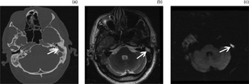
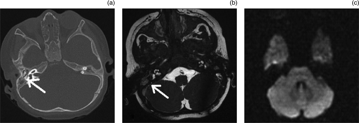
Echo-planar diffusion-weighted MRI accurately predicted the presence or absence of cholesteatoma in 90 per cent of group one patients (37/41) and 76 per cent of group two patients (13/17). Of these correct diagnoses, 23 were true positive diagnoses and 14 were true negatives in group one, while seven were true positives and six true negatives in group two. Examples of scans from patients with true positive and true negative diagnoses are shown in Figures 1 and 2, respectively.
Imaging scans for a 35-year-old man with diffuse cholesteatoma in the epitympanum, antrum and mastoid cells at surgery. (a) Axial computed tomography scan showing a soft tissue density (arrow) in the antrum and epitympanum. (b) Axial, T2-weighted magnetic resonance imaging (MRI) scan at the same level as (a), showing a soft tissue density (arrow). (c) Axial, echo-planar diffusion-weighted MRI showing a flare (arrow) in the same location as the lesion seen in (b); this ‘shine-through’ effect was radiologically interpreted as cholesteatoma.

Imaging scans for a 21-year-old woman with granulation tissue and dark secretions in the middle ear and mastoid air cells at surgery. (a) Axial computed tomography scan and (b) axial, T2-weighted magnetic resonance imaging (MRI) scan, both showing a soft tissue density (arrow) in the middle ear and mastoid air cells. (c) Axial, echo-planar diffusion-weighted MRI scan showing a lesion in the same location as that seen in (b); no ‘shine-through’ effect is observed, and the lesion was radiologically interpreted as not representing cholesteatoma.
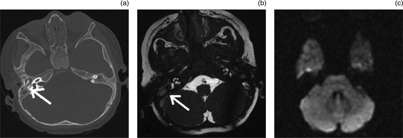
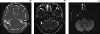
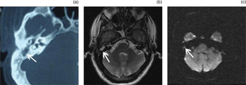
In both group one and group two, three patients had false negative diagnoses and one patient had a false positive diagnosis. The three false negative diagnoses in group one were attributed variously to a cholesteatoma smaller than 5 mm, a dry, hollow retraction pocket, and an air–bone artefact at the base of the skull. The three false negative diagnoses in group two were attributed variously to cholesteatomas smaller than 5 mm (two patients) and a dry, hollow retraction pocket (one patient). The one false positive diagnosis in group one was attributed to dense tympanosclerosis found at surgery, while the one false positive in group two was attributed to motion artefact. Examples of scans from false positive and false negative patients are shown in Figures 3 and 4, respectively.
Imaging scans for a 47-year-old woman with a 4 mm cholesteatoma limited to the antrum, with granulation tissue surrounding the cholesteatoma. (a) Axial computed tomography scan showing sclerotic mastoid cells and a soft tissue density in the antrum (arrow). (b) Axial, T2-weighted magnetic resonance imaging (MRI) scan showing soft tissue density (arrow) in the region of the mastoid cells. (c) Axial, echo-planar diffusion-weighted MRI scan showing no ‘shine-through’ effect in the same location as the lesion seen in (b); this was radiologically interpreted as not representing cholesteatoma.

Imaging scans for a 47-year-old man with dense tympanosclerosis in the middle ear and antrum at surgery. (a) Axial computed tomography scan showing a soft tissue density (arrow) in the mastoid antrum and epitympanum. (b) Axial, T2-weighted magnetic resonance imaging (MRI) scan at the same level as (a), showing soft tissue densities (arrow). (c) Axial, echo-planar diffusion-weighted MRI scan showing a flare (arrow) in the region of the lesion seen in (b); this ‘shine-through’ effect was radiologically interpreted as cholesteatoma.

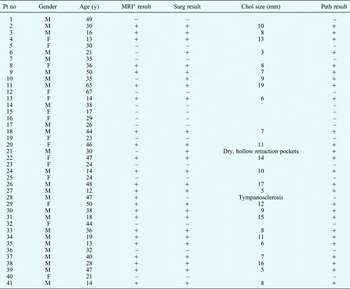
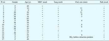
Detailed patient information is given in Tables I and II.
Primary acquired cholesteatoma patients: summary

*Echo-planar diffusion-weighted magnetic resonance imaging. Pt no = patient number; y = years; surg = surgery; chol = cholesteatoma; path = histopathology; M = male; F = female; – = incompatible with chol; += compatible with chol
Second-look or revision surgery patients: summary
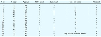
*Echo-planar diffusion-weighted magnetic resonance imaging. Pt no = patient number; y = years; surg = surgery; chol = cholesteatoma; path = histopathology; F = female; M = male; – = incompatible with chol; += compatible with chol
The sensitivity, specificity, and positive and negative predictive values of echo-planar diffusion-weighted MRI for cholesteatoma detection were respectively 88, 93, 95 and 82 per cent in group one, and 70, 85, 87 and 66 per cent in group two.
Discussion
Many clinicians favour high-resolution CT scans for cholesteatoma imaging. Cholesteatoma is suspected in the presence of CT findings such as ossicular erosion, lateral semicircular canal fistula or tegmen destruction. However, on CT scans it is difficult to differentiate whether a soft tissue density in the middle ear or mastoid air cells is due to cholesteatoma, granulation tissue, mucoid secretion or other causes.Reference Tierney, Pracy, Blaney and Bowdler4, Reference Blaney, Tierney, Oyarazabal and Bowdler7–Reference Plouin-Gaudon, Bossard, Fuchsmann, Ayari-Khalfallah and Froehlich9 However, the absence of soft tissue density in the middle ear or mastoid air cells on the CT scan is considered sufficient in itself to exclude a cholesteatoma, as CT has a very high negative predictive value in such cases.Reference Thomassin and Braccini10, Reference Vercruysse, De Foer, Pouillon, Somers, Casselman and Offeciers11
In recent years, MRI has gained importance in cholesteatoma imaging. Although MRI distinguishes cholesteatoma from other tissues better than CT, it is difficult for routine MRI to differentiate cholesteatoma from other soft tissues and mucoid secretions. The sensitivity of routine MRI for cholesteatoma detection varies between 57 and 79 per cent, while its specificity varies between 63 and 71 per cent.Reference Tierney, Pracy, Blaney and Bowdler4–Reference Kimitsuki, Suda, Kawano, Tono and Komune6 These results suggest that classical MRI alone is insufficient when assessing the radiological differential diagnosis of cholesteatoma. Therefore, recent studies have emphasised the use of echo-planar diffusion-weighted MRI, delayed post-contrast T1-weighted MRI and non-echo-planar diffusion-weighted MRI techniques for cholesteatoma imaging. Since echo-planar diffusion-weighted MRI was routinely used in our hospital practice, we preferred this method for cholesteatoma imaging in our study.
During echo-planar diffusion-weighted MRI of the temporal bone, only cholesteatoma gives a high signal intensity, whereas other tissues (e.g. granulation tissue, fibrous tissue, mucoid secretions and cholesterol granuloma) give a low signal intensity.
The sensitivity and specificity of echo-planar diffusion-weighted MRI for cholesteatoma detection have been respectively reported as 80 and 100 per cent by Aikele et al. (in whose study secondary surgery was performed on all patients) and 86 and 100 per cent by Stasolla et al. (likewise, secondary surgery was performed on all patients).Reference Aikele, Kittner, Offergeld, Kaftan, Hüttenbrink and Laniado12, Reference Stasolla, Magliulo, Parrotto, Luppi and Marini13 Vercruysse and colleagues' study found respective rates of 81 and 100 per cent in a group undergoing primary surgery, and 12.5 and 100 per cent in a group undergoing secondary surgery.Reference Vercruysse, De Foer, Pouillon, Somers, Casselman and Offeciers11 Venail et al. found respective rates of 60 and 72 per cent.Reference Venail, Bonafe, Poirrier, Mondain and Uziel1 Our study found that echo-planar diffusion-weighted MRI had a cholesteatoma detection sensitivity and specificity of 88 and 93 per cent in the primary surgery group and 70 and 85 per cent in the secondary surgery group, respectively.
The reported specificity values for diffusion-weighted MRI cholesteatoma detection are consistent with each other, except for Venail and colleagues' findings.Reference Venail, Bonafe, Poirrier, Mondain and Uziel1 In this latter study, two of the three false positive diagnoses were attributed to a Silastic® strip used in the previous operation, of which the radiologist was unaware.
The reported sensitivity values range from 12.5 to 86 per cent. This wide range has been attributed to the large number of small cholesteatomas encountered, rather than to differences of interpretation between investigators.
In Vercruysse and colleagues' study, 29 per cent of all patients in the secondary surgery group had cholesteatomas larger than 3 mm.Reference Vercruysse, De Foer, Pouillon, Somers, Casselman and Offeciers11 Therefore, this study reported a sensitivity of 12.5 per cent. If the studies of Aikele et al., Vercruysse et al., Stasolla et al. and Venail et al. had considered only those cholesteatomas larger than 5 mm, then their sensitivity rates would have approached 100 per cent.Reference Venail, Bonafe, Poirrier, Mondain and Uziel1, Reference Vercruysse, De Foer, Pouillon, Somers, Casselman and Offeciers11–Reference Stasolla, Magliulo, Parrotto, Luppi and Marini13 However, in Plouin-Gaudon and colleagues' study of 21 patients, all false negative diagnoses were found to involve a cholesteatoma smaller than 5 mm.Reference Plouin-Gaudon, Bossard, Fuchsmann, Ayari-Khalfallah and Froehlich9 In our study, a cholesteatoma smaller than 5 mm was found in one of the three false negative patients in group one and two of the three false negative patients in group two. When these patients were removed from our calculations, the sensitivity of echo-planar diffusion-weighted MRI for cholesteatoma detection increased from 88 to 92 per cent in group one and from 70 to 90 per cent in group two.
One reason why cholesteatomas smaller than 5 mm were less frequently found in previous echo-planar diffusion-weighted MRI studies may have been the long time period between MRI scanning and surgery. Therefore, echo-planar diffusion-weighted MRI scanning should be performed as close as possible to the operation date.Reference Vanden Abeele, Coen, Parizel and Van de Heyning5
Despite sensitivity values approaching 100 per cent for the detection of cholesteatomas larger than 5 mm in patients who had previously undergone intact canal wall surgery, recurrent and/or residual cholesteatomas smaller than 5 mm may be missed by echo-planar diffusion-weighted MRI. A more appropriate approach for these patients may be follow up with repeated echo-planar diffusion-weighted MRI, rather than second-look surgery, as cholesteatomas smaller than 5 mm have little effect on the short-term prognosis. The use of repeated MRI as follow up is superior to secondary surgery, as it has lower costs, no complications and no risk of radiation exposure.
Another, previously reported reason for false negative diagnosis with echo-planar diffusion-weighted MRI is dry, hollow retraction pockets.Reference Venail, Bonafe, Poirrier, Mondain and Uziel1 In our study, the false negative diagnoses of one patient each in groups one and two were due to this condition. Therefore, the clinician is recommended to clean the contents of the retraction pockets during examination, before echo-planar diffusion-weighted MRI. The occurrence of an air–bone artefact in the skull base may be another reason for a false negative diagnosis, as seen in one patient in our study.Reference Vercruysse, De Foer, Pouillon, Somers, Casselman and Offeciers11, Reference Aikele, Kittner, Offergeld, Kaftan, Hüttenbrink and Laniado12
Of our two false positive diagnoses, one was attributed to motion artefact and the other to dense tympanosclerosis observed during surgery. There are no previously published data suggesting that tympanosclerosis can cause false positive results. However, if we consider that diffusion-weighted MRI enables better imaging of avascular tissue, compared with CT, it can be understood why tympanosclerosis caused a flare in these images. Although a greater number of cases is needed to adequately investigate this association, it should be borne in mind that tympanosclerosis can cause false positive results during MRI scanning for cholesteatoma. The radiological images and features of this particular patient are given in Figure 4.
Post-contrast T1-weighted MRI is another imaging method used for cholesteatoma detection. The reported sensitivity, specificity, and positive and negative predictive values of the use of this MRI modality for cholesteatoma detection range from 56 to 90 per cent, 54 to 100 per cent, 78 to 100 per cent and 27 to 100 per cent, respectively.Reference Venail, Bonafe, Poirrier, Mondain and Uziel1, Reference Williams, Ayache, Alberti, Héran, Lafitte and Elmaleh-Bergès14–Reference De Foer, Vercruysse, Bernaerts, Meersschaert, Kenis and Pouillon16 In De Foer and colleagues' study, which included the greatest patient number (120) of all the reviewed studies, the sensitivity, specificity, and positive and negative predictive values were 56, 67, 88 and 27 per cent, respectively.Reference De Foer, Vercruysse, Bernaerts, Meersschaert, Kenis and Pouillon16 When we consider this, the most extensive series in the literature, we see that post-contrast T1-weighted MRI is not superior to echo-planar diffusion-weighted MRI for cholesteatoma imaging. In addition, the former modality has major disadvantages, such as greater expense, the need for contrast medium, a longer scanning time (therefore requiring general anaesthesia in children) and the need for more experience during interpretation.
• Echo-planar diffusion-weighted magnetic resonance imaging (MRI) identifies primary and recurrent acquired cholesteatoma with high sensitivity
• This MRI modality can be used instead of ‘second-look’ surgery, with high sensitivity and specificity
In recent years, non-echo-planar diffusion-weighted MRI has been investigated as an alternative imaging method for cholesteatoma. There are currently insufficient reports comparing the results of echo-planar and non-echo-planar diffusion-weighted MRI within the same study. In general, the various studies of echo-planar and non-echo-planar diffusion-weighted MRI have been compared with each other. In Kasbekar and colleagues' study, the results of echo-planar versus non-echo-planar diffusion-weighted MRI were compared with each other on the same patients, although cholesteatomas smaller than 4 mm could not be seen using either of these two imaging methods.Reference Kasbekar, Scoffings, Kenway, Cross, Donnelly and Lloyd17 In De Foer and colleagues' study, the smallest cholesteatoma size detectable by non-echo-planar diffusion-weighted MRI was 2 mm; these authors reported that non-echo-planar diffusion-weighted MRI was superior to echo-planar diffusion-weighted MRI for imaging small cholesteatomas.Reference De Foer, Vercruysse, Bernaerts, Meersschaert, Kenis and Pouillon16, Reference De Foer, Vercruysse, Bernaerts, Maes, Deckers and Michiels18, Reference De Foer, Vercruysse, Bernaerts, Deckers, Pouillon and Somers19 However, De Foer et al. reached that conclusion by comparing the results of different studies using echo-planar diffusion-weighted MRI and non-echo-planar diffusion-weighted MRI. The varying results obtained by these different studies may have been due to differences in the technical properties of the non-echo-planar diffusion-weighted magnetic resonance imaging used, including cross-section thickness.Reference Clark, Westerberg and Fenton20 De Foer et al. used a 1.5 Tesla non-echo-planar diffusion-weighted MRI with 2 mm slice thickness and a single-shot turbo spin-echo sequence, while Kasbekar et al. used a 1.5 Tesla non-echo-planar diffusion-weighted MRI with 3 mm slice thickness and a periodically rotated overlapping parallel lines with enhanced reconstruction sequence (known as ‘PROPELLER’). There are currently no published data comparing these two imaging modalities.Reference Clark, Westerberg and Fenton20 Nevertheless, if non-echo-planar diffusion-weighted MRI comes to be preferred for cholesteatoma imaging, it may well be the single-shot turbo spin-echo sequence which is favoured.
Conclusion
Echo-planar diffusion-weighted MRI is a valuable imaging technique for the detection of primary and recurrent acquired cholesteatoma, with high sensitivity. In the follow up of patients who have undergone intact canal wall surgery, echo-planar diffusion-weighted MRI can be used instead of second-look surgery, with high sensitivity and specificity. Because cholesteatomas smaller than 5 mm have little effect on short-term prognosis, small cholesteatomas which are missed on the initial echo-planar diffusion-weighted MRI can be re-evaluated on repeated scans. This management approach could prevent unnecessary second-look surgery, with its potential complications, in patients who do not have cholesteatoma.