Absent pulmonary valve syndrome is a rare congenital cardiac malformation with absent, dysplastic, or rudimentary pulmonary valve leaflets. The characteristic feature of absent pulmonary valve syndrome is pulmonary arterial dilation, which leads to compression of the tracheobronchial tree and respiratory distress. Most cases of absent pulmonary valve syndrome are associated with tetralogy of Fallot. Double-outlet left ventricle is also a rare congenital anomaly associated with right ventricular outflow obstruction and pulmonary stenosis. There are no reports of patients with both absent pulmonary valve syndrome and double-outlet left ventricle.
Case report
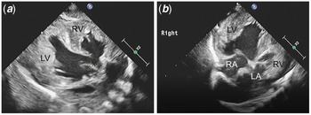
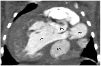
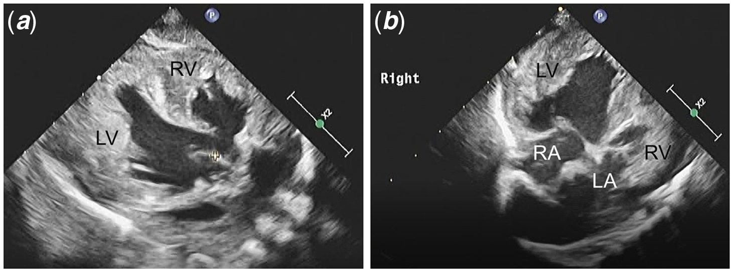
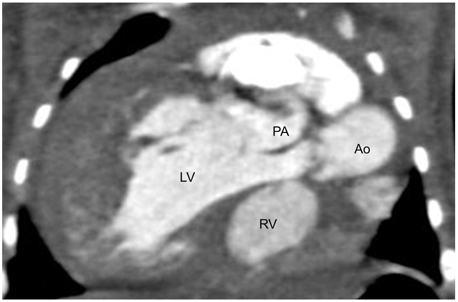
A female infant (birth weight: 2231 g) was delivered vaginally at a gestational age of 37 weeks and 6 days. After birth, she was tachydyspneic and cyanotic with a loud “to-and-fro” murmur over the left sternal border. Hepatomegaly was also observed. Chest radiography revealed a cardiothoracic ratio of 80%. Echocardiography showed the absence of the pulmonary valve with pulmonary insufficiency and stenosis (maximum velocity, 3.8 m/s), double-outlet left ventricle, dextrocardia, hypoplastic right ventricle (70% compared with normal), small tricuspid valve (annulus dimension, 49% compared with normal), valvular aortic stenosis (maximum velocity, 3.8 m/s), subaortic ventricular septal defect, no conus, atrial septal defect, and patent ductus arteriosus with a bidirectional shunt (Fig 1 and Video 1). Segmental anatomy was situs solitus with dextrocardia, l-loop of the ventricles, and corrected transposition of the great arteries. Atrioventricular connection was discordant, and ventriculoarterial connection was double-outlet left ventricle. CT confirmed double-outlet left ventricle (Fig 2), a severe hypertrophic left ventricle connected to the left displaced and dilated main pulmonary artery, with dilation of both branches. The compression of the left main bronchus by the dilated pulmonary artery was diagnosed as bronchomalacia. The left lung was compressed by the dilated pulmonary artery and was suspected to be hypoplastic. It had a less vascular shadow than the right lung.
Pre-operative echocardiography. Unbalanced ventricle ( a ) and discordant atrioventricular connection ( b ) were confirmed. LA, left atrial; LV, left ventricle; RA, right atrial; RV, right ventricle.
Pre-operative CT. Both great arteries originate from LV. Ao, aorta; LV, left ventricle; PA, pulmonary artery; RV, right ventricle.
A tracheal tube was placed immediately after birth because spontaneous breathing effort was severe, and the left lung sounds could not be heard on auscultation. Complex bronchomalacia, severe cardiac failure, and hypoplastic lung seemed to be the cause of respiratory distress. Pulseless ventricular tachycardia requiring adrenaline infusion immediately after birth indicated severe heart failure. Six hours after delivery, the patent ductus arteriosus was ligated because of increased pulmonary insufficiency and exacerbated heart failure. Considering pulmonary stenosis, pulmonary artery banding cannot be performed to regulate pulmonary blood flow. On postnatal day 5, percutaneous balloon aortic valvuloplasty was performed for aortic stenosis. This reduced the peak pressure gradient from 58 to 21 mmHg on echocardiography. At 4 months of age, catheter examination revealed a mean pulmonary artery pressure of 30 mmHg, pulmonary vascular resistance of 3.0 Wood units, and Qp:Qs of 2.1:1. Left ventricle-to-pulmonary artery shunt using an 8-mm expanded polytetrafluoroethylene bicuspid-valved shunt with bulging sinuses (Stretch Vascular Graft, W.L. Gore & Associates, Inc., Flagstaff, AZ, USA) and pulmonary artery plication were performed considering that the Glenn procedure was risky due to the lung condition. Although pulmonary insufficiency could be completely controlled surgically, the peak inspiratory pressure could decrease from 22 to 18 cmH2O postoperatively. Unfortunately, bronchomalacia of the left main bronchus persisted. Tracheostomy was performed at the age of 5 months. There was poor post-operative weight gain and repeated necrotising enterocolitis. Catheterisation showed that Qp:Qs was 1.6:1, and aortopulmonary collaterals were observed in the right intrathoracic artery. Embolisation of the aortopulmonary collateral was performed to decrease pulmonary blood flow at the age of 9 months. Shunt was clipped at the age of 10 months to decrease pulmonary blood flow as an emergency operation because repeated necrotising enterocolitis causes septic shock and Glenn procedure could carry a high risk due to infectious lung condition. Despite recurrent respiratory tract infections, the patient was discharged at the age of 14 months in a stable condition while waiting for the Glenn procedure.
Discussion
Absent pulmonary valve syndrome is a rare variant of Tetralogy of Fallot, accounting for 3–6% of patients with Tetralogy of Fallot. Reference Nair, Haranal, Elkhatim, Dillon, Hew and Sivalingam1 Double-outlet left ventricle is a rare congenital cardiac malformation wherein both great arteries originate entirely or predominantly from the anatomical left ventricle Reference Menon and Hagler2 ; 90% of double-outlet left ventricle cases have pulmonary stenosis. Reference Menon and Hagler2
Although most absent pulmonary valve syndrome cases could have Tetralogy of Fallot with biventricular physiology (1), our patient had a univentricle physiology, which is extremely rare. Reference Futuki, Fijuwara, Yoshizawa and Sakazaki3 Our patient had bronchomalacia, hypoplastic left lung, and high pulmonary vascular resistance, which made it difficult to perform the Glenn procedure. Therefore, under her difficult condition to perform Glenn procedure, we consider that the left ventricle-, similar to the right ventricle, to-pulmonary artery shunt could be optimal to control pulmonary overcirculation because the right ventricle-to-pulmonary artery shunt had a better survival rate than the modified Blalock–Taussig shunt in univentricular cases. Reference Ohye, Sleeper and Mahony4
Conclusion
To the best of our knowledge, this is the first reported case of a patient with absent pulmonary valve syndrome and double-outlet left ventricle who underwent left ventricle-to-pulmonary artery shunt, pulmonary artery plication, and tracheostomy. The patient was discharged in a stable condition while waiting for the Glenn procedure.
Acknowledgements
None.
Authors’ contribution
Wataru Sakai and Hidetsugu Asai: Concept, interpretation, drafting article, critical revision of the article, and approval of article.
Noriyoshi Ebuoka and Junichi Oba: Concept, interpretation, drafting article, and approval of article.
Financial support
This research received no specific grant from any funding agency, commercial, or not-for-profit sectors.
Conflicts of interest
None.
Ethical standards
This study was approved by the Ethics Committee of the Hokkaido Medical Center for Child Health and Rehabilitation.