Introduction
Age-related hearing loss is the most common sensory disorder in the older population, yet it is often underdiagnosed and undertreated. Approximately one-third of individuals aged 65–74 years and nearly half of those over 75 experience hearing loss.Reference BS, Bush, Weinreich, Schwartz, Anne and Adunka1 Based on Dr Schuknecht’s temporal bone studies, age-related hearing loss can be classified into four subtypes: sensory, neural, strial and/or metabolic and conductive.Reference Schuknecht and Gacek2 Among these, outer hair cell loss stands out as one of the fundamental pathophysiological mechanisms of the disorder.Reference Wu, O’Malley, de Gruttola and Liberman3
The cochlea has the capacity to decompose and analyse sounds in real time according to their frequencies. In this process, the travelling wave, which propagates along the basilar membrane from the base toward the apex, reaches its maximum amplitude at frequency-dependent regions, where neural transmission is supported through the activity of outer hair cells.Reference He, Burwood, Fridberger, Nuttall and Ren4 Estimation of travelling wave delay is of great importance for understanding cochlear physiology and evaluating structural-mechanical changes associated with ageing or pathology.Reference Polak, Lorens, Walkowiak, Furmanek, Skarzynski and Skarzynski5 Although direct assessment of travelling wave delay is technically challenging, indirect evaluation can be achieved through methods such as derived auditory brainstem responses (ABRs), tone-burst ABR, otoacoustic emissions and recently electrocochleography.Reference Murray, Cohn, Harker and Gorga6–Reference Harte, Pigasse and Dau9
Electrocochleography is a technique used to record the electrical potentials generated by cochlear hair cells and the auditory nerve, and the extracochlear electrocochleography method has been applied in clinical practice for more than 80 years.Reference Eggermont10, Reference Chertoff, Lichtenhan and Willis11 During these recordings, the electrical signal reflecting the synchronous firing of numerous auditory nerve fibres is referred to as the action potential.Reference Chertoff, Lichtenhan and Willis11 The basal portion of the cochlea is sensitive to high frequencies, while the apical portion is sensitive to low frequencies, therefore high-frequency components are transmitted through the cochlea faster than low-frequency components.Reference Verhulst, Bharadwaj, Mehraei, Shera and Shinn-Cunningham12 This physiological property leads to frequency-specific differences in action potential latencies and results in a natural latency gap. Comparing responses at these two distinct frequency regions therefore provides a meaningful opportunity to evaluate the timing of neural transmission across different cochlear segments.
Satar et al.Reference Satar, Yildirim, Karahatay and Sen8 demonstrated that the action potential latency difference measured between 0.5 and 4 kHz in a frequency-specific electrocochleography setting in normal-hearing individuals is significant and reflects the cochlear tonotopic organisation.Reference Satar, Yildirim, Karahatay and Sen8 However, these findings have mostly been limited to young adults without hearing loss. More recent studies, including those by Harris et al.Reference Harris, Ahlstrom, Dias, Kerouac, McClaskey and Dubno13 and Qian et al.Reference Qian, Wang, Yang, Wang, Hu and Li14, Reference Qian, Chen, Wang, Zhou, Chen and Huang15, have reported age-related reductions in action potential amplitudes and impaired neural synchrony using electrocochleography, suggesting early auditory nerve dysfunction. Notably, while Qian et al. conducted their analyses in strictly normal-hearing individuals, Harris et al. included older participants with a broader range of hearing thresholds, up to 65 dB HL below 4 kHz.Reference Harris, Ahlstrom, Dias, Kerouac, McClaskey and Dubno13–Reference Qian, Chen, Wang, Zhou, Chen and Huang15
Despite these valuable contributions, none of these studies assessed travelling wave delay using action potential latency differences across different frequencies. These findings nonetheless underscore the sensitivity of frequency-specific action potential measurements in detecting early neural degeneration and highlight the potential of electrocochleography to reveal age-related cochlear transmission delays. In fact, existing travelling wave delay assessments have predominantly focused on overtly pathological cochlear conditions, with limited attention to its relevance in physiological ageing and subtle hearing loss.Reference Claes, Wyndaele, De Valck, Claes, Govaerts and Wuyts16–Reference Sen, Satar and Yetiser18 The present study aims to address this gap in the literature and represents one of the few human investigations evaluating how travelling wave delay changes in the context of ageing and hearing loss through frequency-specific action potential latency differences. It is hypothesised that age- and hearing-related prolongation of action potential latencies would result in a concomitant deceleration of travelling wave delay.
Methods
Participants
A total of 49 participants (26 males, 23 females) were included in the study, and data from 85 ears from these individuals were analysed. Measurements were obtained from both ears in 36 participants and from one ear in 13 participants. The mean age for all groups was 52.37 ± 17.35 years. Participants were divided into four groups according to age and hearing status. Those aged 65 years and older with sensorineural hearing loss were assigned to the older adults with hearing loss group (n = 11, 22 ears), those aged 65 years and older with normal hearing were assigned to the older adults with normal hearing group (n = 9, 18 ears), those aged 18–60 years with sensorineural hearing loss were assigned to the younger adults with hearing loss group (n = 12, 19 ears) and those aged 18–60 years with normal hearing were assigned to the younger adults with normal hearing group (n = 17, 26 ears).
The audiological profiles of the ears in the older adults with hearing loss group were consistent with features typically associated with the ‘sensory’ type in Schuknecht’s classification of age-related hearing loss, such as high-frequency sloping audiograms and relatively preserved speech discrimination. Participants with middle-ear pathology, those younger than 18 years and those with a diagnosis of neurological or psychiatric disorders were excluded from the study.
This study was conducted at University of Health Sciences Gülhane Training and Research Hospital (General Dr Tevfik Sağlam St No. 1 Etlik/Ankara, Turkey). The study was conducted in accordance with the principles of the Declaration of Helsinki and was approved by the Gülhane Scientific Research Ethics Committee (Registration number: 2025/31, 7 January 2025). Written informed consent was obtained from all participants prior to their inclusion in the study.
Procedure
All participants underwent pure-tone audiometry and speech audiometry, and the groups were determined based on the results obtained. Hearing loss was classified according to the pure tone average (PTA) at 0.5, 1, 2 and 4 kHz, with thresholds greater than 25 dB HL considered indicative of sensorineural hearing loss. Assessments were performed with an AC-40 audiometer (Interacoustics, Assens, Denmark) using TDH-39 supra-aural headphones.
Before the electrocochleography test, the ear to be tested was cleaned with a 3 per cent hydrogen peroxide solution. A non-invasive extratympanic electrode (TM-electrode; Sanibel, Assens, Denmark) was placed in the ear canal and secured with the foam tip of the insert earphone. The skin was cleaned to ensure that electrode impedances remained below 5 kΩ. Participants were instructed to remain as still as possible or to rest quietly during the recording. Tone-burst stimuli at two different frequencies (0.5 and 4 kHz) were presented at an intensity of 90 dB nHL and a stimulation rate of 19.7 per second. The action potential latencies recorded at 0.5 and 4 kHz were determined, and the latency difference between them was analysed to estimate the travelling wave delay. Latency measurements were manually marked by two experienced audiologists to ensure accuracy.
All recordings and analyses were performed using the Interacoustics Eclipse EP25 software system. The test parameters used in this study followed the same protocol as our previous work.Reference Bozkurt, Karaçaylı and Satar19 Briefly, the active electrode was placed on the tympanic membrane, with the reference on the contralateral mastoid and the ground at frontopolar zero (Fpz). Insert earphones (ER-3A; Etymotic Research, Inc., Elk Grove Village, IL, USA) delivered monaurally presented 2-0-2 ms tone bursts. Signals were bandpass filtered between 10 and 3000 Hz, with an analysis window of −1.0 to 10 ms, and 1000 sweeps were averaged per condition.
Statistical analysis
The normality of the data distribution was assessed with the Shapiro–Wilk test and variance homogeneity with Levene’s test. Because Levene’s test indicated a violation of homogeneity (p < 0.05), Welch one-way analysis of variance (ANOVA) was used instead of standard ANOVA. For post hoc analysis, the Games–Howell test was applied because of unequal variances. Adjusted eta squared (η 2adj) was calculated for Welch ANOVA and interpreted following Cohen’s thresholds: approximately 0.01 small, approximately 0.06 medium and greater than 0.14 large. Cohen’s d was computed for all pairwise comparisons using pooled standard deviations, with values ∼0.2, 0.5 and 0.8 considered small, medium and large, respectively.Reference Cohen20
When normality assumptions were not met, the Kruskal–Wallis H test was applied. If significant, pairwise comparisons were run with the Mann–Whitney U test and Bonferroni correction was applied to control type I error. Epsilon squared (ε 2) was calculated as an effect size for Kruskal–Wallis: 0.01–0.08 small, 0.08–0.26 moderate and greater than 0.26 large. Effect sizes for pairwise comparisons were also reported using rank-biserial correlation (r), with 0.10, 0.30 and 0.50 interpreted as small, medium and large effects.Reference Tomczak and Tomczak21
Results
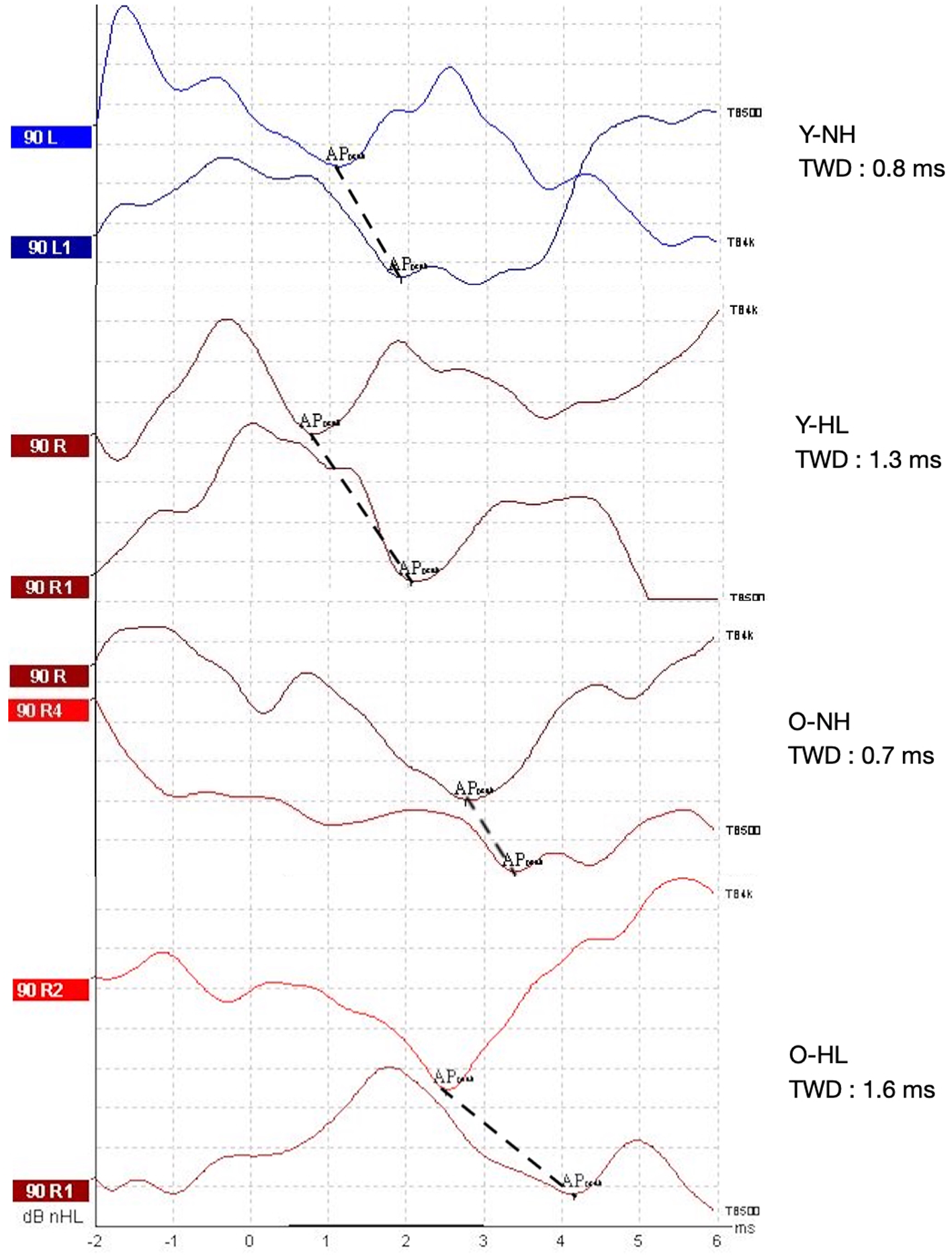
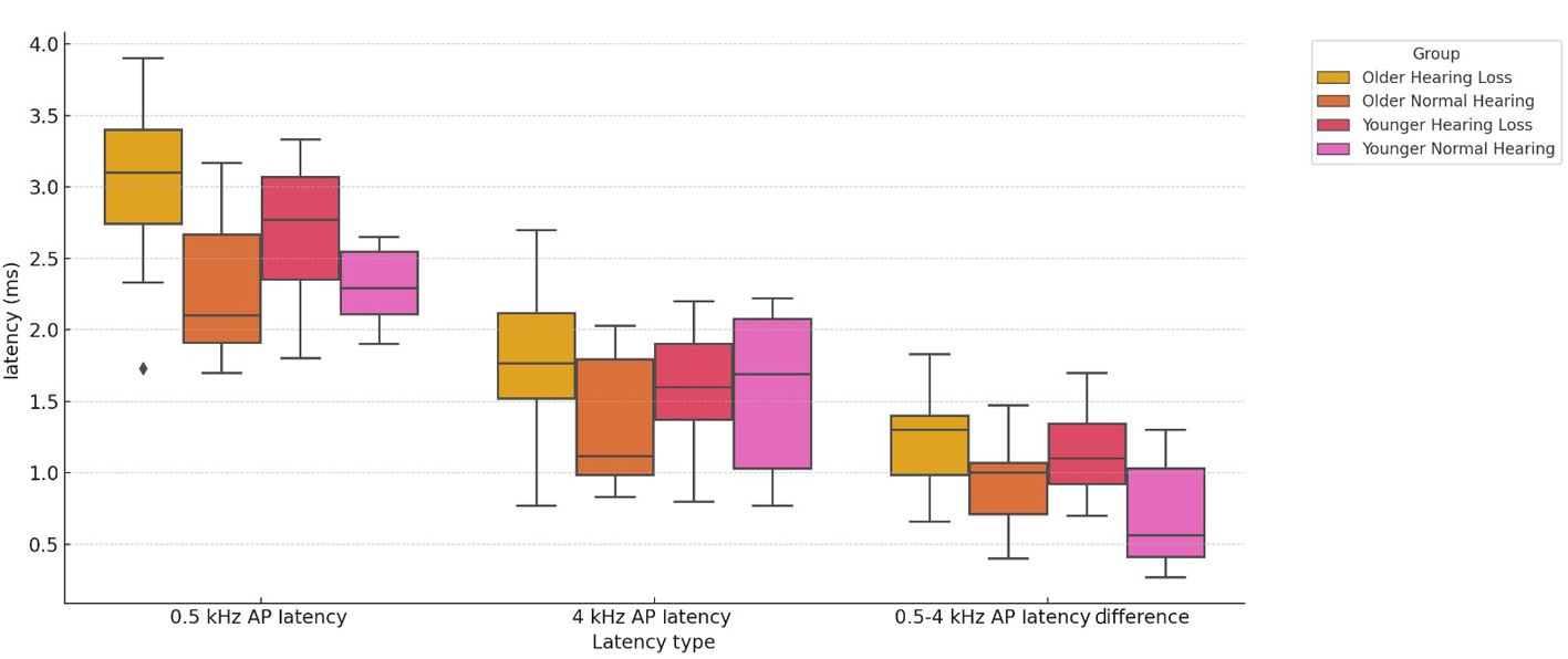
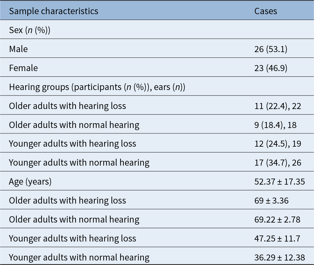
A total of 49 participants (85 ears) were included in the study. The overall mean age was 52.37 ± 17.35 years (range, 20–75). Participants were divided into four groups: older adults with hearing loss (69 ± 3.36 years, 22 ears), older adults with normal hearing (69.22 ± 2.78 years, 18 ears), younger adults with hearing loss (47.25 ± 11.7 years, 19 ears) and younger adults with normal hearing (36.29 ± 12.38 years, 26 ears). Sex distribution was nearly balanced, with 23 females (46.9 per cent) and 26 males (53.1 per cent). Descriptive statistics are summarised in Table 1 and latency values are presented in Table 2. Figure 1 shows representative electrocochleography waveforms, demonstrating that younger adults with normal hearing exhibited the shortest travelling wave delay, while older adults with hearing loss showed the longest. These group differences are also illustrated in the box plot (Figure 2) to improve visual clarity.
Representative electrocochleography waveforms recorded at 0.5 kHz and 4 kHz for each hearing and age group. APpeak = action potential peak; Y-NH = younger adults with normal hearing; TWD = travelling wave delay; Y-HL = younger adults with hearing loss; O-NH = older adults with normal hearing; O-HL = older adults with hearing loss.

Latency distributions by group. AP = action potential.

Sample demographics and hearing group distribution

Group comparisons for 0.5, 4 and 0.5–4-kHz action potential latency measures across hearing and age groups

Superscripts indicate significant group differences (p < 0.05). Groups that do not share the same letter are significantly different. For 0.5-kHz action potential latency, post hoc comparisons were based on Welch analysis of variance (ANOVA) and letters are shown next to means. For 4 and 0.5–4-kHz latency differences, Mann–Whitney U tests were conducted following Kruskal–Wallis and letters are shown next to medians. Effect sizes are reported as η 2adj for Welch ANOVA and ε 2 for Kruskal–Wallis tests, respectively (displayed in parentheses). SD = standard deviation
A Welch one-way ANOVA was conducted to assess differences in 0.5-kHz action potential latencies across groups because Levene’s test indicated a violation of homogeneity of variances (F(3, 81) = 3.431, p = 0.021). The analysis showed a significant group effect, F(3, 81) = 16.891, p < 0.001, with a large overall effect size (η 2adj = 0.368). Post hoc Games–Howell comparisons revealed that older adults with hearing loss had significantly higher 0.5-kHz action potential values than older adults with normal hearing (p < 0.001, d = 1.612), younger adults with hearing loss (p = 0.002, d = 0.754) and younger adults with normal hearing (p < 0.001, d = 1.939). Additionally, older adults with normal hearing differed significantly from younger adults with hearing loss (p = 0.014, d = −0.928), whereas no significant difference was found between older adults with normal hearing and younger adults with normal hearing (p = 0.999, d = −0.105).
A Kruskal–Wallis H test was used for 4-kHz action potential latencies, revealing a significant group effect (H(3) = 8.283, p = 0.041) with a moderate effect size (ε 2 = 0.0986). Post hoc Mann–Whitney U tests with Bonferroni correction showed that older adults with hearing loss had significantly higher 4-kHz action potential values than older adults with normal hearing (p = 0.027), with a large effect size (Cohen’s d = −1.02, r = 0.45). The rank-biserial correlation (r) indicates a moderate-to-large effect, as values of approximately 0.10, 0.30 and 0.50 are conventionally interpreted as small, medium and large effects, respectively.Reference Tomczak and Tomczak21 No other pairwise comparisons were significant.
A Kruskal–Wallis H test was conducted to evaluate group differences in the latency difference (0.5–4 kHz action potential values), revealing a significant group effect (H(3) = 22.952, p < 0.001) with a large overall effect size (ε 2 = 0.277). Post hoc Mann–Whitney U tests with Bonferroni correction showed that younger adults with normal hearing exhibited significantly smaller 0.5–4-kHz action potential latency differences compared with both younger adults with hearing loss (p = 0.005, Cohen’s d = −1.215, r = 0.501) and older adults with hearing loss (p < 0.001, Cohen’s d = −1.494, r = 0.643). These findings indicate a large effect for the younger adults with normal hearing versus younger adults with hearing loss comparison and a very large effect for the younger adults with normal hearing versus older adults with hearing loss comparison. No other pairwise comparisons reached statistical significance after Bonferroni adjustment.
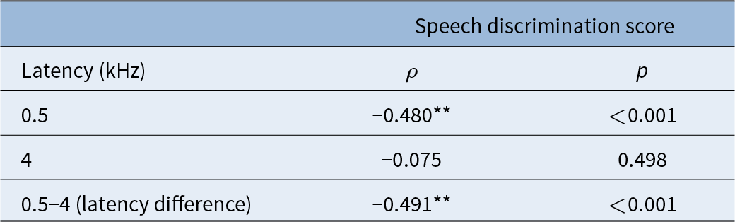
To further examine the functional implications of the latency findings, we analysed the relationship between electrocochleography latency measures and speech discrimination scores. As shown in Table 3, speech discrimination scores were significantly negatively correlated with 0.5-kHz latency (ρ = −0.480, p < 0.001) and the 0.5–4-kHz latency difference (ρ = −0.491, p < 0.001). No significant correlation was observed for 4-kHz latency (ρ = −0.075, p = 0.498). These results suggest that increased latency at low frequencies and greater latency differences are associated with poorer speech discrimination ability.
Correlations between speech discrimination scores and electrocochleography latencies at 0.5, 4 and 0.5–4 kHz

p < 0.05 indicates statistical significance. ρ = Spearman’s rank correlation coefficient
Discussion
This study aimed to compare the action potential latencies of four groups differing in age and hearing status at 0.5 and 4 kHz, and to investigate the latency difference between these two frequencies (0.5–4-kHz latency difference). The primary objective was to evaluate the effects of ageing and sensorineural hearing loss on travelling wave delay and to explore the potential interaction between these two factors. To the best of our knowledge, this is the first study to directly investigate travelling wave delay in individuals with age-related hearing loss.
The findings obtained from our study revealed that age and hearing loss exert significant effects on action potential latencies, both independently and in combination. Notably, at the low frequency of 0.5 kHz, individuals who were older and had hearing loss exhibited the longest and statistically significant delays in action potential latencies. Furthermore, the significant difference observed between the older adults with normal hearing and younger adults with hearing loss groups suggests that older individuals may retain more physiological (within expected limits) neural transmission compared with younger individuals with hearing loss, provided that their hearing thresholds remain normal.
At 4 kHz, a significant difference was observed only in the older groups, suggesting that ageing, when combined with hearing loss, more prominently prolongs action potential latency. The finding that the older adults with hearing loss group exhibited significantly longer 4-kHz action potential values compared with the older adults with normal hearing group indicates that age-related hearing loss may impose an additional burden on cochlear transmission. In contrast, the absence of a significant difference between younger hearing-impaired and younger normal-hearing individuals suggests that hearing loss occurring at a younger age may not, by itself, lead to a meaningful alteration in travelling wave dynamics.
One of the most noteworthy findings of our study is that the 0.5–4-kHz action potential latency difference significantly varied as a function of age and hearing loss. The observation of the shortest latency difference in young normal-hearing participants indicates that cochlear transmission in this group is the fastest and most efficient. In contrast, the longest latency difference observed in the older group with hearing loss suggests that ageing and hearing impairment together markedly slow travelling wave delay.
Post hoc analyses further revealed that young normal-hearing individuals exhibited significantly shorter latency differences than both young hearing-impaired and older hearing-impaired participants. This pattern suggests that the effects on travelling wave delay may not be solely attributable to ageing, but may also be associated with additional cochlear alterations that arise in relation to hearing loss. Indeed, various histopathological findings related to the basilar membrane have been reported in the literature.
Temporal bone sections from older individuals have revealed a marked thickening and calcification of the basilar membrane.Reference Nelson and Hinojosa22 Similarly, studies on aged rat models have demonstrated collagen fibre loss and structural alterations in both the basilar and tectorial membranes.Reference Ishii, Murofushi and Takeuchi23 More recent molecular-level investigations have indicated that miR-29a deficiency may lead to an accumulation of type IV collagen and laminin, thereby contributing to basilar membrane thickening.Reference Ma, Wang, Geng, Gong, Li and Xie24 Furthermore, a recent experimental study has suggested that cochlear wave propagation is not limited to the travelling wave along the basilar membrane, but may also involve a ‘cortilymph wave’ occurring within the cortilymph fluid. This novel mechanism is thought to arise from outer hair cell vibrations and may enhance cochlear transmission efficiency by supporting travelling wave amplification.Reference Guinan25
Taken together, these findings suggest that the slowing of travelling wave delay observed in our study may not only be attributable to delays in neural transmission but also to histological and molecular-level alterations occurring in the basilar membrane and the micromechanical structure of the cochlea.
In addition to these physiological and micromechanical alterations, our findings indicate that the slowing of cochlear transmission may have functional consequences for auditory performance. The negative correlation between speech discrimination scores and electrocochleography latency measurements suggests an impairment of auditory processing at the suprathreshold level. This implies that, even when hearing thresholds are similar, variations in cochlear transmission speed may influence speech perception. In line with the literature, pure-tone thresholds do not fully represent auditory nerve integrity, and it has been demonstrated that deafferentation particularly affecting low-spontaneous-rate fibres, as well as age-related spiral ganglion cell loss, contributes to reduced speech discrimination performance.Reference Wu, O’Malley, de Gruttola and Liberman3, Reference Verhulst, Bharadwaj, Mehraei, Shera and Shinn-Cunningham12, Reference Nelson and Hinojosa22
Moreover, age-dependent changes in the micromechanical structure of the basilar membrane and cochlea have been shown to slow cochlear transmission.Reference Ishii, Murofushi and Takeuchi23, Reference Ma, Wang, Geng, Gong, Li and Xie24 The decline in speech discrimination performance observed in our study may therefore reflect these transmission-related and structural alterations. Among the limitations of our study, the cross-sectional design restricts the ability to evaluate the longitudinal course of age-related changes. In addition, the lack of an objective distinction between the underlying pathophysiological types of hearing loss limits a more detailed examination of their specific effects on travelling wave delay.
• Cochlear travelling wave delay reflects tonotopic transmission speed
• Age-related hearing loss alters neural transmission at the cochlear level
• Frequency-specific action potential latency differences (0.5–4 kHz) may provide additional insight into cochlear transmission integrity beyond pure-tone thresholds
• Travelling wave delay analysis may complement conventional audiometry by revealing early cochlear transmission deficits that are not detected in threshold measurements
Conclusion
The assessment of travelling wave delay in age-related hearing loss holds promise as an objective and complementary tool for detecting early cochlear transmission impairments. Particularly in older adults, travelling wave delay analysis may provide valuable insights into supra-threshold auditory difficulties that are not fully accounted for by pure-tone thresholds alone. However, we do not propose travelling wave delay as a replacement for conventional audiometry, but as a complementary metric that may reveal aspects of auditory dysfunction not captured by threshold-based testing. Future studies are warranted to further investigate the clinical relevance and validity of travelling wave delay measurements, rather than their immediate use as a routine diagnostic marker, with the potential to enhance the understanding and management of auditory dysfunction in ageing populations.
Acknowledgements
We sincerely thank all the patients who took part in this study; their valuable contributions made this research possible.
Data availability statement
Data are available on request from the authors.
Competing interests
None declared.