Introduction
Cysticercosis is an infestation caused by the larval stage of the tapeworm, Taenia solium in which humans are intermediate hosts. A case of solitary cysticercosis of the right parotid gland in a young female is reported here, highlighting its clinical presentation, diagnosis and management.
Case report
A 24-year-old female from a good socioeconomic background presented with a swelling below the right ear to the Department of Otorhinolaryngology of St John's Medical College Hospital, a tertiary referral hospital in Bangalore, South India. It was of insidious onset, gradually enlarging over the previous six months. She noted a sudden increase in size associated with pain for about a week prior to seeking medical attention.
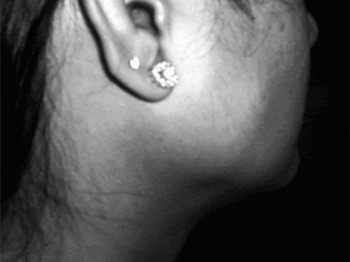
A warm, tender, immobile and firm swelling was apparent on palpation over the right parotid region. It measured approximately 40 mm in diameter (Figure 1).
Clinical photograph of the swelling in the right parotid region.

The differential diagnoses considered ranged from acute on chronic suppurative parotitis, acute on chronic lymphadenitis, benign conditions like pleomorphic adenoma, to granulomatous conditions like Sjögren's syndrome, sarcoidosis and tuberculosis with superadded acute infection.
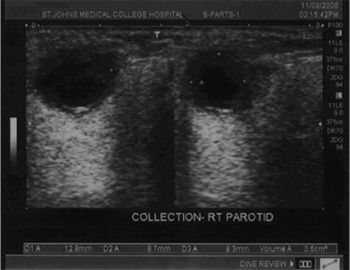
All haematological investigations including the eosinophil counts were normal. The ultrasound examination of the right parotid region showed a fluid-filled cyst in the parotid gland measuring 13 mm × 8 mm × 9 mm (volume of 0.5 cc) (Figure 2). The radiologist noted that the swelling was tender when touched with the probe.
Ultrasonography showing fluid-filled cyst in the right parotid gland.

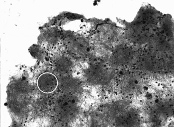
A fine needle aspiration cytology (FNAC) study showed stromal fragments with spindle cells, aggregates of histocytes (some with epithelioid transformation) and many macrophages with phagocytosed karyorectic nuclear debris. Many neutrophils – some with degenerative changes – were seen, along with a few lymphocytes, eosinophils, plasma cells and multinucleated giant cells. Occasional fibrillary fragments with basophilic granules and nuclear dust-like material were seen in the midst of the infiltrate (Figure 3). On the basis of these findings, a diagnosis of cysticercosis with a granulomatous host response was made.
FNAC smear shows cellular fibrillary fragments with scattered nuclear material characteristic of the body wall of a cysticercus (H&E; ×200).

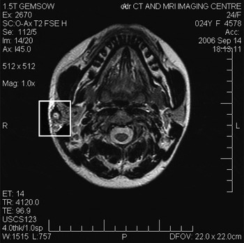
A multiplanar, multisequence magnetic resonance imaging (MRI) scan of the parotid gland without contrast showed classic features of a cysticercus characterised by a well-defined, hyperintense lesion on T2 weighting, measuring 10 mm × 8 mm, with irregular margins and areas of cystic signal intensity. A focal hyperintense speck suspected to be a scolex (the adult head) was also noted (Figure 4). There was mild thickening of the adjacent parotid fascia. The deep portion of the gland, internal carotid artery and adjacent muscles were normal.
MRI scan showing cysticercus in the right parotid gland. Note the hyperintense speck, possibly a scolex.

Further questioning did not elicit any history of headaches, seizures, orbital symptoms or any nodular swellings over the body suggestive of lesions elsewhere. An MRI scan of the brain was performed, which ruled out neurocysticercosis. A stool examination was negative for parasites.
The patient was hospitalised for medical management. She was treated with albendazole (400 mg three times a day) as a larvicidal and tramadol hydrochloride/acetaminophen tablets (Ultrazac) as an analgesic. The swelling regressed markedly within two days. She was discharged and asked to continue medication at home for two weeks. The swelling has not recurred. She is asymptomatic after a period of six months.
Discussion
Cestodes, or tapeworms are segmented worms. Their hosts are termed definitive hosts when the adult tapeworms live in their gastrointestinal tract, or intermediate hosts when only larval stage parasites are present in the tissues. Pigs are the usual intermediate hosts of the tapeworm Taenia solium. Human beings can act as both definitive hosts and intermediate hosts. When humans are definitive hosts, the condition is called taeniasis. If they act as intermediate hosts, the disease caused is called cysticercosis.Reference White, Weller, Kasper, Braunwald, Fauci, Hauser, Longo and Jameson1
Cysticercosis is endemic in India, China, Mexico, Latin America, Africa and other parts of the developing world. It is transmitted to humans by eating infected pork and through the faeco-oral route by ingesting food or water contaminated with eggs or segments of the body of the parasite called proglottids. Internal autoinfection is rare.Reference Kung, Lee and Yu2
The ingested eggs or proglottids form embryos which leave the intestine via the hepatoportal system, are carried passively by the bloodstream, and dispersed throughout the tissues and organs of the body.Reference Pushker, Bajaj and Balasubramanya3 Viable cysticerci elicit a granulomatous inflammatory response with foreign body giant cells. From an interplay of immunological and chemical mechanisms, the larva dies, provoking acute inflammation and necrosis and ultimately undergoes resorption with calcification within the affected organ.Reference Arora, Gupta, Singh and Bhatia4
The brain is most often affected and is involved in 60 to 92 per cent of all patients with cysticercosis. Other common locations for cysticercosis include subcutaneous tissue (17.8 per cent), skeletal or heart muscle (8.5 per cent) and the eye (5.1 per cent). Other reported sites include the liver, lung, kidney, pancreas, intestine, diaphragm, spleen, peritoneum, mouth and peripheral nervous system.Reference Sheehan, Sheehan, Lopes and Jane5 To the best of our knowledge there is no case of cysticercosis of the parotid gland reported in the indexed literature.
The disease is relatively difficult to diagnose as there are no specific manifestations. A history of residence or extended travel in an endemic area may be obtained. An asymptomatic, isolated cyst may remain undetected, until it enlarges, migrates or dies and produces symptoms.Reference Ogilvie, Kasten, Rovinsky, Workman and Johnston6 The clinical manifestations depend on the size and site of cyst lodgement, and its stage of evolution.Reference Pushker, Bajaj and Balasubramanya3 Our patient presented with a warm, tender, immobile and firm swelling, mimicking an acute inflammation.
Plain radiographs rarely show cysticerci, except as calcified lesions in advanced cases. Multiple fusiform, cigar shaped calcifications of thigh and calf muscle on plain radiographs are highly indicative of cysticercosis and multiple punctate soft tissue calcifications have been reported.Reference Ogilvie, Kasten, Rovinsky, Workman and Johnston6
Computed tomography (CT) and MRI are the preferred methods for imaging brain and orbital cysticerci, showing their location, identifying areas of atrophy and oedema, and assessing the degree of ventricular dilatation.Reference Ogilvie, Kasten, Rovinsky, Workman and Johnston6 The age of the parasite and the nature of the inflammatory host response markedly influence the radiological appearance of cysticercosis. Viable cysticerci usually appear as 1 mm to 35 mm hypodense images. They may be isodense with brain tissue and radiologically occult, even in contrast studies. Dying or the dead cysticerci, surrounded by the oedema from the inflammatory reaction, are visible as spherical hypodensities, often with the protoscolex of the parasite appearing as an eccentric dot of calcium. Contrast studies demonstrate the inflammatory response surrounding the cysticercus and give the cyst a radiographic image of an enhanced ring structure. Later, obliteration of the cyst cavity by the inflammatory process may produce a solid enhanced image.Reference Richards and Schantz7
Serological tests play a role in diagnosis when CT or MRI findings are inconclusive since palpable subcutaneous nodules are scarce and demonstrable calcified cysticerci may take years to develop. A significantly positive titre is strong corroborating evidence though negative results do not rule out the diagnosis.Reference Loo and Braude8 The enzyme-linked immunoelectro transfer blot is the most effective assay, with a documented specificity of 100 per cent and a sensitivity varying from 60 per cent to 98 per cent depending on the number of lesions.Reference Ogilvie, Kasten, Rovinsky, Workman and Johnston6 The sensitivity of antibody testing tends to be high with multiple cysts but is substantially lower with single cysts or calcified cysts.Reference Proano-Narvaez, Meza-Lucas, Mata-Ruiz, Garcia-Jeronimo and Correa9 Serological tests were not performed in our case as a diagnosis of cysticercosis had already been reached based on radiological investigations and cytological findings.
• Cysticercosis is an infestation caused by the larval stage of the tapeworm, Taenia solium in which humans are intermediate hosts
• This paper describes the case of a young female patient presenting with features resembling acute parotitis
• Solitary cysticercosis of the parotid gland was diagnosed, based on fine needle aspiration cytology and radiological investigations
• Medical therapy was effective, consequently surgery was deferred
When within the salivary glands, cysticercosis should be differentiated from pleomorphic adenoma, adenolymphoma, chronic suppurative parotitis, chronic lymphadenitis, sarcoidosis, tuberculosis, Sjögren's syndrome, malignant lymphoma and metastatic carcinoma. FNAC helps confirm the diagnosis in such a patient. The diagnosis of cysticercosis is made when fragments of the body wall, composed of a fine protoplasmic network of eosinophilic material studded with several small nuclei are aspirated. Scolices may or may not be found.Reference Kini, Shariff and Nirmala10
Del Brutto et al. have proposed definite criteria for the diagnosis of human cysticercosis based on information from imaging studies, clinical manifestations, serological tests and epidemiological data in which the demonstration of cysticercosis by microscopic examination of aspirate and the visualisation of the scolex in the parotid gland by MRI are both considered absolute criteria.Reference Del Brutto, Rajshekhar, White, Tsang, Nash and Takayanagui11
Therapy is indicated for patients with symptoms with live cysticerci but will not benefit patients with dead calcified cysts. Neurocysticercosis is commonly treated with praziquantel (50 mg/kg per day for 15–20 days) or albendazole (10–15 mg/kg per day for 8–15 days). Albendazole is reported to have better response rates than praziquantel. Central nervous system inflammation may be reduced by concurrent administration of corticosteroids. For symptomatic cysts outside the central nervous system, the optimal approach is surgical resection. Medical therapy also should be used if the number or the location of the lesions makes surgical removal of all cysticerci technically unfeasible. Patients with cysticercosis should be examined for possible concomitant intestinal taeniasis and must be treated if intestinal disease is present.Reference Ogilvie, Kasten, Rovinsky, Workman and Johnston6 In our case, response to medical therapy was effective. Hence surgical excision was not required.
Measures for the prevention of intestinal Taenia solium infection consist of adequate cooking, freezing or salting of food for long periods. Other preventive measures involve minimising the opportunities for ingestion of faecally derived eggs by means of good personal hygiene, effective faecal disposal, and treatment and prevention of human intestinal infections.Reference White, Weller, Kasper, Braunwald, Fauci, Hauser, Longo and Jameson1
Conclusion
A case of solitary cysticercosis of the parotid gland is presented for its rarity, with a review of relevant literature. This lesion responded to medical treatment, hence surgical excision was not performed. A high index of suspicion for parasitic infestations of this nature is essential as frequent travel across endemic areas has increased the risk of isolated cases presenting in non-endemic areas as well.