Introduction
Desmoid tumour (DT) is a neoplasm also previously named ‘aggressive fibromatosis’, that originates from superficial or deep musculo-aponeurotic structures. DT can occur intra- or extra-abdominally. It is a rare tumour, the incidence is 2–4 per million. It occurs twice more often in women than in men and is more likely to arise in the third and the fourth decade of life, however it can occur in children.
DTs are slow-growing mesenchymal tumours with a high predilection for local recurrence. Surgery, radiotherapy (RT), chemotherapy, hormonal therapy, anti-inflammatory therapy, vitamin C or a combination of all these modalities are used to treat DT.Reference Karabulut, Keskin and Ekenel 1 Adjuvant RT is a treatment aimed at increasing local control, especially for patients with high-grade tumour and positive microscopic surgical margin. RT significantly decreases local relapse when compared with patients who receive only surgical interventions. According to Nuyttens compilation of 22 articles, the local control rate is 78% among patients who receive both surgical operation and RT, however the local control rate is 68% among patients who receive surgery only.Reference Nuyttens, Rust and Thomas 2
Case
A 16-year-old female had repeated operations for DT located under the right toe, 4 years ago. Subsequently, the distal aspect of the left foot was amputated and the patient administered two cycles of chemotherapy. The chemotherapy was stopped at the patient’s own request. On follow up, the patient received further surgery to remove a bulky tumour located in the right femur. Pathology demonstrated this was a DT, and the surgical borders were tumour positive. It was decided to treat the patient with RT.
Materials and Method
Before starting the adjuvant intensity-modulated radiotherapy (IMRT) planning, the patient was immobilised in the supine position using a vacuum board with legs apart. For treatment planning a computed tomography scan was obtained using 3 mm. The target volume and organs at risk (OaRs) were defined by the physician. To cover all tumour extension, the planning target volume (PTV) was extended from the right inguinal fossa to the distal end of the femur. The femur, femoral head, ovaries, bladder, rectum, uterus were all delinated as OaRs by using the velocity planning software.
The treatment was planned to deliver 54 Gy in 30 fractions. The PTV received 95% of the 54 Gy. Tomotherapy-based IMRT plan was optimised on the TomoHi-art work station. Optimisation parameters were as follows; field width was 5 cm, pitch was 0·25, planning modulation factor was 2·00, plan calculation grid was normal (0·610×0·610 cm).
OaRs doses were evaluated using RTOG (Radiation Therapy Oncology Group) protocols 3 and quality of PTV was evaluated with parameters of homogeneity index (HI) and the conformity index (CI).
CI was calculated using the following formula for PTV:
 $${\rm CI}\,{\equals}\,{{\left( {{\rm TV}_{{{\rm PIV}}} } \right)^{2} } \mathord{\left/ {\vphantom {{\left( {{\rm TV}_{{{\rm PIV}}} } \right)^{2} } {\left( {{\rm TV}{\times}{\rm PIV}} \right)}}} \right. \kern-\nulldelimiterspace} {\left( {{\rm TV}{\times}{\rm PIV}} \right)}}{\rm }$$
TVPIV is the target volume covered by the prescription isodose volume, TV is target volume and PIV is the prescription isodose volume in the Formula.Reference Paddick 4
The HI formula was as follows:
 $${\rm HI}\,{\equals}\,{{\left( {D_{{\rm 5}} {\minus}D_{{{\rm 95}}} } \right)} \mathord{\left/ {\vphantom {{\left( {D_{{\rm 5}} {\minus}D_{{{\rm 95}}} } \right)} {D_{p} }}} \right. \kern-\nulldelimiterspace} {D_{p} }}$$
D 5 and D 95 represent the doses received by the 5 and 95% volumes of PTV, respectively, and D p is the prescribed dose.Reference Pathak and Vashisht 5
Results
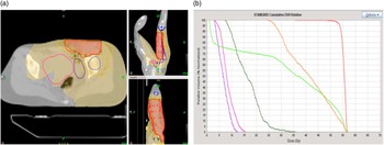
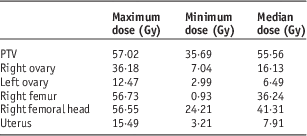
Coronal, sagittal and transverse isodose distributions of the planning and dose volume histograms (DVH) including PTV and critical organs are showed in Figures 1a and 1b. PTV and critical organs maximum, minimum and median doses are demonstrated in Table 1. Because the patient is so young, it is important that the PTV dose and the dose to the ovary is kept to a minimum so as not to impair fertility.
(a) Isodose distribution for coronal, sagital and transverse planes (Dark Orange colour: 54 Gy, light orange colour: 51.3 Gy, dark yellow: 27 cGy, light yellow: 5.4 cGy—red: planning target volume (PTV), pink: uterus, light blue: right femoral head, dark blue: right femur, purple: right ovary) (b) Dose volume histogram (DVH) distribution (red: PTV, orange: right femoral head, light green: right femur, dark green: right ovary, pink: uterus, purple: left ovary).

Doses of planning target volume (PTV) and Organ’s at Risk (OaR’s) (right and left ovaries, right femur, right femoral head and uterus)

Maximum, minimum and median doses of PTV were 57·02 Gy, 35·69 Gy and 55·56 Gy, respectively (Table 1). In all, 5% of the femoral head received ~55 Gy, 10% of the femoral head receives 53 Gy and the femur receives an average of 36·24 Gy. The uterus receives a maximum of 15·49 Gy and an average of 7·91 Gy. The right ovary receives a maximum of 36·8 Gy, and an average of 16·13 Gy. The left ovary receives a maximum of 12·47 Gy and an average of 6·49 Gy. The HI, the CI and the treatment time were 0·8, 1·056 and 413·4 MU, respectively.
Discussion
Reducing the dose to the critical organs especially the ovary dose was problematic given the extent of the area requiring RT. Ionising radiation has adverse effects on the ovary at all ages. The degree and persistence of the damage to the ovary is dependent on the dose, irradiation field and the patient’s age when irradiated. It is necessary to shield the ovaries using beam shaping blocks or by placing the treatment fields to avoid direct irradiation to the ovaries, but in some treatments treating the ovaries cannot be avoided.Reference Meirow and Nugent 6 – Reference Stroud, Mutch and Rader 8
Modern RT techniques such as image-guided radiotherapy (IGRT) and IMRT are more effective than classical RT techniques and allow for the reduction of the dose to OaRs. In this study, the doses of the femur, femoral head and uterus as defined as OaR’s were acceptable. Right ovary was very close to PTV therefore decreasing the dose to the ovary was not an option. The average dose to the right ovary was decreased to 16·13 Gy. Dose to the left ovary was decreased to 6·49 Gy. Meirow describes the dose of radiation required to destroy 50% of primordial follicles (LD50) has previously been judged to be <4 Gy—an estimate later deemed to be an over-simplification.Reference Meirow and Nugent 6 Although doses of >6 Gy consistently cause permanent primary hypogonadism in women over age 40 years, the impact of lower doses varies, with reports of conceptions in women under 20 years of age who have received up to 30 Gy.Reference Green, Yakar and Brecher 9
Using the best available model for follicle decline, Wallace et al.Reference Wallace, Thomson, Saran and Kelsey 10 reported their calculation of the radio-sensitivity of the human oocyte in terms of LD50. According to Wallece et al., for the a 16-year-old patient, the effective (ovarian failure in 97·5%) sterilising dose is about 18 Gy (upper) and the mean (ovarian failure in 50%) sterilising dose is about 16·5 Gy (lower) of radiation for a given treatment age. According to this study, the dose to the left ovary in this patient is within limits, however the maximum value to the right ovary is out of limits. Dose to the left ovary within limits, however, this does not necessarily indicate that ovarian failure will not occur. According to Wallace, for our patient and the dose to the left ovary being ~6·49 Gy, the predicted age of ovarian failure, with 95% confidence limits of 25·4 (low), 29·3 (mean) and 33·2 (high).Reference Wallace, Thomson and Kelsey 11
The patient is followed up with estradiol, follical stimulating hormone and leutenising hormone tests periodically. In the last follow-up menstrual irregularity was noted in the patient.
Conclusion
The use of RT is likely to improve local control in DT. However, RT is more likely to result in long-term complications and an impact on fertility, therefore individual decision making and special attention to OaR is warranted. The patient tolerated the RT well and only grade 1 skin toxicity occurred.
Acknowledgements
The authors would like to thank Eda Kaya Pepele.
Conflicts of Interest
None.