Introduction
The freshwater shark Wallago attu (local name: Lanchi) is widely distributed in the Ganga River and its tributaries in the state of Uttar Pradesh, India. This fish is an excellent food fish, famous for rapid growth and rich in nutritional quality of its flesh (Goswami & Devraj, Reference Goswami and Devraj1992; Lilabati & Viswanath, Reference Lilabati and Viswanath1996). Much information is available on the helminth parasite fauna of W. attu but, due to inadequate descriptions, most species were assigned to species inquirendae by Lim et al. (Reference Lim, Timofeeva and Gibson2001) as there was no possibility of further evaluating the status of many of these monogenean species parasitizing W. attu.
The genus Mizelleus (Monogenea: Ancyrocephalidae) was erected by Jain (Reference Jain1957) for the species Mizelleus indicus Jain (Reference Jain1957), a gill parasite of the freshwater shark W. attu at Lucknow, India. Gusev (Reference Gusev1976) included Mizelleus in Ancylodiscoidinae, subsequently transferred to the Ancyrocephalinae by Lim & Lerssutthichawal (Reference Lim and Lerssutthichawal1996) and later to the Ancylodiscoididae by Lim et al. (Reference Lim, Timofeeva and Gibson2001). Various workers from India reported different species of this genus on the basis of morphology (Agrawal & Pandey, Reference Agrawal and Pandey1981; Venkatanarsaiah & Kulkarni, Reference Venkatanarsaiah and Kulkarni1981; Singh & Sharma, Reference Singh and Sharma1992; Singh et al., Reference Singh, Kumari and Agrawal1992a, Reference Singh, Kumari, Sharma and Agrawalb; Agrawal & Sharma, Reference Agrawal and Sharma2002; Pandey et al., 2003) but most of these species were poorly described on the basis of morphology only. Hence, identification of species and a clear differentiation between closely related species is not possible. Therefore, re-description of these species using molecular approaches is warranted, correct species identification being well supported by the study of DNA of the species in question.
The aim of the present study was to re-describe two species of the genus Mizelleus parasitizing W. attu, i.e. M. indicus Jain (Reference Jain1957) and Mizelleus longicirrus (Tripathi, Reference Tripathi1959) Venkatanarasaiah & Kulkarni, 1981, using light microscopy. Besides this, both species were also characterized based on the sequences of 18S ribosomal DNA in order to validate their phylogenetic position.
Materials and methods
Morphology
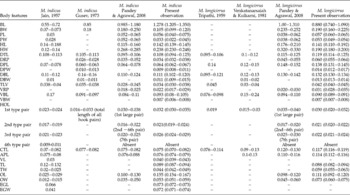
Fifty-one specimens of W. attu (Bloch & Schn. 1801) identified according to Froes & Pauly (Reference Froese and Pauly2010), were collected from the local markets of Meerut (28°59′0″N 77°42′0″E), Uttar Pradesh, India, during the period 2011–2012. Specimens were transported live to the laboratory of the Department of Zoology, Chaudhary Charan Singh University, Meerut, and held until observation. The gill filaments were checked for monogenean infection with a Motic SMZ-168 series stereomicroscope (Xiamen, People's Republic of China). Monogeneans were removed with the help of triangular surgical needles and some were immediately mounted in glycerin for the preparation of semi-permanent slides. Others were stained with acetocarmine, dehydrated through ascending grades of alcohol and mounted in Canada balsam for preparation of permanent slides. Some samples of monogeneans were fixed in 95% ethanol in vials for molecular biological examination. Photos of monogeneans were taken with an Olympus CH30 (Shinjuku, Tokyo, Japan) fitted with an AxioCam ERc 5s. Measurements were made according to the guidelines suggested by Gusev (Reference Gusev and Bauer1985) for Dactylogyridae. In the description of species, different morphotypes were used to characterize anchors, bars and hooks (Gusev, Reference Gusev and Bauer1985): Wunderoid-type anchors with sharply recurved point and comparatively small roots diverging, both roots rarely developed equally, outer root smaller than the inner one; Juvenile-type anchors with recurved point but without roots; Anchoratoid–Wegeneri-type anchors with very long inner root but almost without outer root; Vastator-type transverse bar having a rough massive piece; and Definitive-type hooks with a thick end and oblong spindle shaped handle. The type and voucher specimens of helminths were deposited in the Museum d'Histoire naturelle, Geneva, Switzerland and the Museum of the Department of Zoology, Chaudhary Charan Singh University, UP, India, as indicated in the respective taxonomic accounts. Abbreviations of the measurements are given in table 1.
A comparison of the morphometrics (measurements given in mm unless otherwise stated) of Mizelleus indicus Jain (Reference Jain1957) and M. longicirrus (Tripathi, Reference Tripathi1959) Venkatanarasaiah & Kulkarni 1981 in the freshwater shark W. attu from India; ranges given in parentheses.

BL: body length; BW: body width; PL: pharynx length; PW: pharynx width; HL: haptoral length; HW: haptoral width; DTL: total length of dorsal anchor; DRP: recurved point of dorsal anchor; DPL: dorsal patch length; DPW: dorsal patch width; DBL: dorsal bar length; DBW: dorsal bar width; TLV: total length of ventral anchor; VRP: recurved point of ventral anchor; VBL: ventral bar length; VBW: ventral bar width; HOL: hook length; CTL: copulatory tube length; APL: accessory piece length; VL: vaginal length; TL: testis length; TW: testis width; OL: ovary length; OW: ovary width; EGL: egg length; EGW: egg width.
Molecular analysis
DNA was extracted from preserved samples using QIAGEN DNeasyTM tissue kit according to manufacturer's instructions (Qiagen, Hilden, Germany). The 18S rDNA was amplified in a 25-μl reaction mixture using the following primers: Worm A, GCGAATGGCTCATTAAATCAG; 1270R, CCGTCAATTCCTTTAAGT; 930F, GCATGGAATAATGGAATAGG; Worm B, CTTGTTACGACTTTTACTTCC (Littlewood & Olson, Reference Littlewood, Olson, Littlewood and Bray2001); and S1, ATTCCGATAACGAACGAGACT; IR8, GCTAGCTGCGTTCTTCATCGA (Šimková et al., Reference Šimková, Plaisance, Matějusová, Morand and Verneau2003). The reaction mix consisted 3 μl DNA, 2.5 μl of 10 × Taq buffer (Biotools, Madrid, Spain), 1 μl of Taq polymerase (1 U; Biotools), 3.4 μl of deoxyribonucleoside triphosphates, 0.9 μl of each primer and 13.3 μl of water. The polymerase chain reaction (PCR) was performed in an AG22331 Thermocycler (Eppendorf, Hamburg, Germany). The PCR cycle comprised the following steps: an initial denaturation for 3 min at 94°C; followed by 40 cycles of 94°C for 40 s, 57°C for 45 s and 72°C for 1 min; with a final extension of 10 min at 72°C, then stored at 4°C. An aliquot (5 μl) of each amplicon was examined on 1.5% agarose-TAE (Tris–acetic acid–EDTA) buffer gel, stained with ethidium bromide, and then purified with PurelinkTM Quick Gel Extraction and PCR purification Combo Kit (Invitrogen, Löhne, Germany) following the manufacturer's instructions. Purified PCR products were sequenced (in both directions) using the Big Dye Terminator v. 3.1 cycle sequencing kit in an ABI 3130 genetic analyser (Applied Biosystems, Foster City, California, USA) with the primers as cited above.
Amplified forward and reverse sequences were aligned in Bioedit (Hall, Reference Hall1999) and ambiguous bases were clarified using corresponding ABI chromatograms. A BLAST homology search was performed to evaluate the relationship of both Mizelleus spp. to other monogeneans. For molecular phylogenetic analysis, the species have been chosen that were found to be closely related (maximum identity 90–93%) with the 18S rDNA sequence of both Mizelleus spp., including species of Euryhaliotrematoides, Euryhaliotrema, Pseudodactylogyrus and Dactylogyridae. Monogenean sequences were downloaded after a search resulting in the selection on the basis of BLAST matches. Nucleotide sequences were aligned using CLUSTAL W (Thompson et al., Reference Thompson, Higgins and Gibson1994) and the alignment was manually corrected using alignment editor in MEGA v. 6 (Tamura et al., Reference Tamura, Stecher, Peterson, Filipski and Kumar2013). The dataset was tested for the nucleotide substitution model of best fit. The best fitting model Akaike Information Criterion (AIC) was chosen for each partition. The Kimura two-parameter (K2P) model was used to calculate DNA pairwise distances in MEGA 6. Subsequent phylogenetic analyses were conducted in MEGA 6 employing the maximum likelihood (ML). The ML method was performed using the GTR +G+I model. The bootstrap values based on 1000 resampled datasets were generated. Bayesian inference (BI) analysis was computed by TOPALI 2.5 (Milne et al., Reference Milne, Lindner, Bayer, Husmeier, McGuire, Marshall and Wright2008) and was based on the GTR +G+I model. The ML tree was visualized using MEGA 6. Pseudodactylogyroides apogonis (AB065115) was selected as the out-group species.
Results
Based on morphological examination, two species of genus Mizelleus were identified as M. indicus and M. longicirrus.
Mizelleus indicus Jain (Reference Jain1957)
- Host.
-
Freshwater shark, Wallago attu (Bloch & Schn. 1801).
- Locality.
-
District Meerut, Uttar Pradesh (UP), India.
- Site of infection.
-
Gill lamellae.
- Specimens studied.
-
Fifteen.
- Material deposited.
-
The slides were deposited in the Museum d'Histoire naturelle, Geneva, Switzerland (MHNG-INVE-91 847) and the Museum, Department of Zoology, Chaudhary Charan Singh University, UP, India, under the voucher number HS/monogenea/2015/01.
- Synonyms.
-
Mizelleus linorchis Kulkarni, Reference Kulkarni1969; M. inglishi Gupta & Sharma, Reference Gupta and Sharma1984; M. ramalingami (Agrawal & Singh, Reference Agrawal and Singh1984) Pandey et al., 2003; M. hindanensis Tewari & Agrawal, Reference Tewari and Agrawal1986; M. lucknowensis (Agrawal & Sharma, Reference Agrawal and Sharma1986) Pandey et al., 2003; M. kherai (Pandey & Mehta, Reference Pandey and Mehta1986) Pandey et al., 2003; M. wallagonius (Singh et al., Reference Singh, Kumari, Sharma and Agrawal1992b) Pandey et al., 2003
Re-description
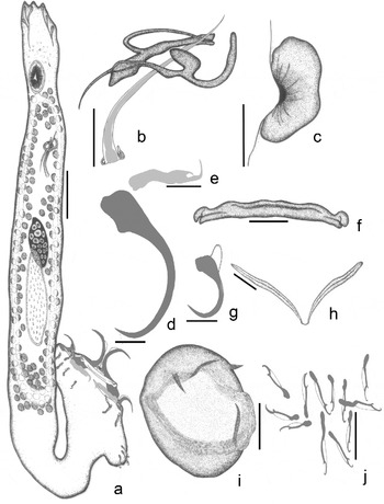
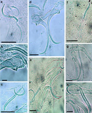
Measurements are given in table 1. Body elongated, fusiform with well-demarcated haptor, moderate to large-sized worms, maximum width attained at the level of gonads (fig. 1a). Cephalic region bilobed, equipped with four pairs of well-developed head organs. Eyespots may or may not be compact, numerous pigmented granules present in cephalic region. Pharynx oval in outline, muscular structure (fig. 1a). Intestinal caeca simple, bifurcated and confluent posteriorly, anterior to haptor (fig. 1a). Testis single, oval, intercaecal, pre-equatorial (fig. 1a). From anterior border of testis, a fine duct vas deferens arises that runs anteriorly, loops around left intestinal caecum and dilates to form a dactylogyrid-type seminal vesicle. Male copulatory organ comprises a cirrus proper and an accessory piece. Cirrus proper long, double-walled tube with narrow proximal part and broad distal part (figs 1b and 2a). Accessory piece chitinoid and tape-like going from the initial part and transforming into wide curved processes (fig. 1b and fig. 2a). Ovary post-equatorial, intercaecal, elliptical, post-testicular (fig. 1a). Egg spherical, smooth in outline (fig. 1i). A spur-like polar filament present at broad end of egg. Vagina sinistral (fig. 1c).
The morphology of Mizelleus indicus to show: (a) the entire worm, ventral view; (b) copulatory complex; (c) vaginal armature; (d) dorsal anchor; (e) dorsal patch; (f) dorsal bar; (g) ventral anchor; (h) ventral bar; (i) egg; (j) hooks. Scale bars: (a) 0.02 mm; (b–j) 0.005 mm.

Morphology of (a–d) Mizelleus indicus to show (a) the copulatory complex, (b) dorsal patch, (c) hooks and ventral anchor with ventral bar, (d) dorsal anchor with dorsal bar; and (e–h) M. longicirrus to show (e) haptor armature, (f) copulatory complex, (g) dorsal anchor with patch, (h) ventral anchor with ventral bar. Scale bars: (a, c–h) 0.05 mm; (b) 0.01 mm.

Opisthohaptor markedly set off from body proper. Dorsal anchors Wunderoid–Juvenile-type with strong recurved point but without roots, armed with articulated finger-like protuberance on inner side of basal part, supported by sleeve sclerites (fig. 1d and fig. 2d). Forked supporting dorsal patch with long shaft and thin process at end (fig. 1e and fig. 2b). Dorsal transverse bar Vastator-type, thin, roughly straight, curved by its medial part backward with distinctly marked articulate heads (fig. 1f and fig. 2d). Ventral anchors Wunderoid-type with smoothly curved point, base without root (fig. 1g and fig. 2c). Ventral connective bar two pieces, connected together by a thin ligament, stick-like, wide at the middle portion with narrow outer ends (fig. 1h and fig. 2c). Marginal hooks seven pairs, Definitive-type, unequal sizes, consist of blade and handle. The differences between the lengths of hooks are due to size of handle, as shaft and blade are similar in length (fig. 1j and fig. 2c).
Remarks
Jain (Reference Jain1957) described M. indicus. Later, Kulkarni (Reference Kulkarni1969) described a second species, M. linorchis, which was apparently found to be identical with M. indicus. The present form is characterized by the presence of a protuberance at the basal part of dorsal anchors; a median piece of ventral bar present between two halves of the bar; dorsal patches forked, with a typical thin process at the end; absence of ventral patches; absence of any side connectives; cirrus and accessory piece non-articulate at the base; massive accessory piece having a wide, curved process; and vagina sinistral.
Mizelleus longicirrus (Tripathi Reference Tripathi1959) Venkatanarasaiah & Kulkarni 1981
- Host.
-
Freshwater shark, Wallago attu (Bloch & Schn. 1801).
- Locality.
-
District Meerut, Uttar Pradesh (UP), India.
- Site of infection.
-
Gill lamellae.
- Specimens studied.
-
Eleven.
- Material deposited.
-
The slides were deposited in the Museum d'Histoire naturelle, Geneva, Switzerland (MHNG-INVE-91 848) and the Museum, Department of Zoology, Chaudhary Charan Singh University, UP, India under the voucher number HS/monogenea/2015/02.
- Synonyms.
-
Mizelleus chauhani (Agrawal & Pandey, Reference Agrawal and Pandey1981) Dubey et al., Reference Dubey, Gupta and Agarwal1992; M. indicus (Singh & Sharma, Reference Singh and Sharma1992) Pandey et al., 2003; M. orientalis (Yadav et al., Reference Yadav, Singh and Yadav2000) Pandey et al., 2003
Re-description
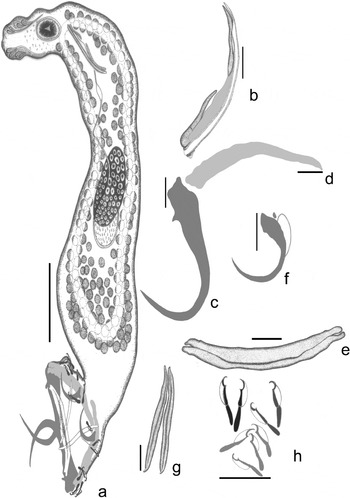
Measurements are given in table 1. Body elongated with bluntly lobed anterior end and well-demarcated haptor (fig. 3a). Cephalic region well lobed, having four pairs of glandular head organs. Eyespots found to be dispersed. Pharynx elongated and oval (fig. 3a). Oesophagus short. Intestinal caeca simple, bifurcate and confluent posteriorly above haptor (fig. 3a). Testis single, oval, intercaecal, post-equatorial (fig. 3a). From anterior end of testis, a fine, long vas deferens arises, it extends anteriorly, sometimes looped around left intestinal caeca and dilates to form an oval seminal vesicle. Male copulatory organ simple and comprises a cirrus proper and an accessory piece. Cirrus proper long, double-walled chitinoid tube with narrow distal part (figs 2f and 3b). Accessory piece ribbon-like, running parallel to cirrus tube (fig. 2f and fig. 3b). Ovary intercaecal, equatorial, elongated (fig. 3a). Vitelline follicles follicular and extending between confluent end of intestinal caeca. Vaginal armature not found.
The morphology of M. longicirrus to show: (a) the entire worm, ventral view; (b) copulatory complex; (c) dorsal anchor; (d) dorsal patch; (e) dorsal bar; (f) ventral anchor; (g) ventral bar; (h) hooks. Scale bars: (a) 0.02 mm; (b–h) 0.005 mm.

Opisthohaptor fairly set off from body proper by narrow peduncle (fig. 2e). Dorsal anchors Wunderoid–Juvenile-type, having strong recurved point and without roots (figs 2g and 3c). At base of dorsal anchor, a small protuberance is present that helps in articulation with the dorsal transverse bar. Dorsal patch very long and tusk-like (figs 2g and 3d). Dorsal transverse bar Anchoratoid-Wegeneri-type, rounded ends having small socket and a slit in middle (figs 2e and 3e). Ventral anchors small, Wunderoid-type with smooth, recurved point and base without roots (figs 2h and 3f). Ventral bar V-shaped, two pieces/bar, each joined together by a thin ligament of length 0.007 (0.006–0.007) (figs 2h and 3g). Marginal hooks: seven pairs, Definitive-type. Handle distinguished into two portions, a thin, small shaft and a swollen, large, bag-shaped handle at distal end (fig. 2e and fig. 3h). Filament loop attached at distal part of blade on ventral side.
Remarks
Tripathi (Reference Tripathi1959) described M. longicirrus for the very first time. Later, the same species was re-described by Venkatanarasaiah & Kulkarni (1981). The present form was found to be very similar to Wallagotrema chauhani reported by Agrawal & Pandey (Reference Agrawal and Pandey1981). But it differs from W. chauhani by the number of head organs and marginal hooks; the presence of eyespots and absence of a pair of accessory pieces joining the dorsal bar with the dorsal patch; cirrus long but with an accessory piece, mentioned as absent in W. chauhani. Furthermore, it differs from M. indicus in the shape of the copulatory complex and in the presence of a ventral patch at the base of the ventral anchor. By taking into account all the above considerations, M. longicirrus is well-characterized morphologically by the dorsal patch being tusk-like and very long; the shape of the cirrus tube and accessory piece, both running parallel; and the copulatory tube being a long, double-walled chitinoid structure.
Molecular analyses
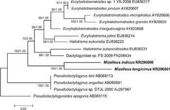
The consensus 18S rDNA sequences of M. indicus and M. longicirrus were composed of 2084 and 2200 bp, respectively. Both sequences were deposited in GenBank (accession numbers: KR296800 and KR296801, respectively). On submission to BLAST, the sequenced sequences showed high similarity scores (90–93%). After a BLAST search, the two newly generated sequences of the partial 18S rDNA fragment of M. indicus and M. longicirrus were aligned together with 13 published sequences, including Euryhaliotrematoides sp. 1, E. pirulum, E. microphallus, E. grandis, E. triangulovagina, Euryhaliotrema johni, Haliotrema eukurodai, H. subancistroides, Dactylogyridae sp., Pseudodactylogyrus bini, P. anguillae, Pseudodactylogyrus sp. and Pseudodactylogyroides apogonis (see accession numbers in fig. 4). Maximum likelihood (ML) and Bayesian inference (BI) analyses of 18S rDNA sequences placed the sequences of M. indicus and M. longicirrus within the same clade alongside the species of Dactylogyridae and Haliotrema with a bootstrap support value of 100% for ML and 1.00% for BI (fig. 4). The generated ML and BI trees, exhibited an identical topology. Therefore, we present here only the ML tree along with the posterior probabilities for BI (fig. 4). However, no other sequence of the genus Mizelleus was available in GenBank to be included in the analysis. Mizelleus indicus and M. longicirrus share a common clade and are found to be closely related to each other.
Phylogenetic tree generated by maximum likelihood analysis of the 18S rDNA sequences of monogeneans, rooted at Pseudodactylogyroides apogonis. Bootstrap confidence values (1000 replications) and posterior probabilities are shown at nodes. Accession numbers are given in parentheses. The Mizelleus species examined in this study are indicated in bold.

The phylogenetic tree clearly exhibits that there are no other taxa common to a node branch with sequences of Mizelleus, hence it persists as a separate genus from other groups of Dactylogyrids. The DNA sequence of M. indicus is most closely related to the other Indian species, i.e. M. longicirrus (90%), and in the phylogenetic tree both species were placed on the same branch supported by high bootstrap values (fig. 4). The clade of M. indicus and M. longicirrus was 100% supported in both trees constructed by ML and BI methods, showing that both species exhibited a close relationship to each other as related sister species with strong bootstrap values. However, due to the lack of sequences available from this group of parasites, no closely related species were shown with similarities in 18S rDNA sequences.
Discussion
The taxonomic status of the genus Mizelleus has been far from clear (Lim et al., Reference Lim, Timofeeva and Gibson2001). In India, since the work of Jain (Reference Jain1957) and Tripathi (Reference Tripathi1959) there have been few systematic studies for this group (Kulkarni, Reference Kulkarni1969; Venkatanarsaiah & Kulkarni, Reference Venkatanarsaiah and Kulkarni1981; Agrawal & Singh, Reference Agrawal and Singh1984; Pandey & Mehta, Reference Pandey and Mehta1986; Tewari & Agrawal, Reference Tewari and Agrawal1986; Agrawal & Sharma, Reference Agrawal and Sharma1989; Singh et al., Reference Singh, Kumari and Agrawal1992a, Reference Singh, Kumari, Sharma and Agrawalb). The majority of Indian monogenean species were described by relying on only the morphological methods, which are not as informative as phylogenetic studies. In the present study, using morphological tools as well as molecular methods, the taxonomic and phylogenetic status of two species of Mizelleus has been re-examined.
Mizelleus indicus and M. longicirrus are strictly specific parasites of silurid fishes, especially W. attu. Jain (Reference Jain1957) established the genus Mizelleus from W. attu at Lucknow, India, with type species M. indicus. Mizelleus longicirrus was first described by Tripathi (Reference Tripathi1959) from the same fish; later it was re-described by Venkatarasaiah & Kulkarni (1981) from Manair River, Hyderabad, India. The genus Mizelleus was included in Ancylodiscoidinae by Gusev (Reference Gusev1976), in the Ancyrocephalinae by Lim & Lerssutthichawal (Reference Lim and Lerssutthichawal1996) and in the Ancylodiscoididae by Lim et al. (Reference Lim, Timofeeva and Gibson2001). This genus was restricted to W. attu and had been found only in India until Lim & Lerssutthichawal (Reference Lim and Lerssutthichawal1996) described M. siamensis from Thailand. At present, 11 species of Mizelleus Jain (Reference Jain1957) have been reported, i.e. M. indicus Jain Reference Jain1957; M. linorchis Kulkarni, Reference Kulkarni1969; M. postorchidis Kulkarni, Reference Kulkarni1969; M. hindanensis Tewari & Agrawal, Reference Tewari and Agrawal1986; M. kherai Pandey & Mehta, Reference Pandey and Mehta1986; M. longicirrus (Tripathi, Reference Tripathi1959) Venkatanarsaiah & Kulkarni, Reference Venkatanarsaiah and Kulkarni1981; M. ramalingami Agrawal & Singh, Reference Agrawal and Singh1984; M. chauhani Agrawal & Sharma, Reference Agrawal and Sharma1989; M. lucknowensis Agrawal & Sharma, 1989; M. wallagonius Singh et al., Reference Singh, Kumari and Agrawal1992a from India and M. siamensis Lim & Lerssutthichawal (Reference Lim and Lerssutthichawal1996) from Thailand. With regard to the Indian species, it is still difficult to determine the validity of most of the species due to the lack of proper morphological descriptions and molecular evidence.
The existence of Mizelleus species was reported from India where they were first identified. In the present study, the worms were recovered from the gills of W. attu in the Ganga River flowing through Meerut, UP. Although both species were first recovered from the Gomti River, Lucknow, records are also available from other localities, such as Raipur, Hyderabad, Sultanpur, Faizabad, Gorakhpur and Deoria, showing their widespread existence in India. Morphologically, the two forms are undoubtedly members of the suborder Tetraonchinae, as they show four pairs of anchors and an absence of squamodiscs on the peduncle. At first sight, both forms look very much similar in morphology. Both share similarities: the presence of protuberances in the basal part of dorsal anchor; the presence of the wing of the ventral anchor but absence of wings of the dorsal anchor; the shape of the dorsal as well as the ventral bar; the shape of hooks; the absence of a patch on ventral anchor. However, both forms differ by significant characters: the different shape of dorsal patches of dorsal anchors (in M. indicus it is forked in shape but in M. longicirrus it is tusk-like); the different shape of the copulatory complex. These two differences cannot be neglected as they are significant characters for species differentiation. All these considerations led us to consider the two forms as belonging to two different species, i.e. M. indicus and M. longicirrus.
In addition to morphology, phylogenetic analysis using 18S rDNA genes has become a significant tool for strengthening the differentiation between species. Now molecular tools have become important criteria for species identification and for re-description of an inadequately described species. 18S rDNA genes are particularly useful for elucidating molecular identification and phylogenetic analysis (Littlewood & Olson, Reference Littlewood, Olson, Littlewood and Bray2001). The phylogenetic trees obtained in the present study, constructed by ML and BI, revealed the same topology and showed that both Mizelleus species are distinguishable and strongly supported by molecular data, though close in morphological characteristics. These results prompt us to interpret that both species show individual existence as their nucleotide sequences show interspecific distance (0.09%) within the same genus. This is further supported by morphological observation mentioned in the present communication. We could not compare the 18S rDNA sequences with other members of the genus Mizelleus due to the unavailability of data in GenBank. Hence, this phylogenetic analysis does not allow us to draw detailed conclusions. The confirmation of the existence of all the species will likely require a comprehensive phylogenetic analysis of Mizelleus utilizing both morphological and molecular data.
In conclusion, to understand the more definite species identification and phylogeny of Indian monogenean species, new sequences from different species are needed for further analysis and comparison. For the first time in this study, the use of molecular methods for the Mizelleus group has clearly been shown to be complementary to traditional morphological techniques and accurately confirmed the validation of species.
Acknowledgements
The authors thank the Head of the Department of Zoology, Chaudhary Charan Singh University, Meerut, for providing laboratory facilities.
Financial support
This study was partially supported by grants from the UGC (University Grants Commission) India, under the Senior Research Fellowship [RGNF; F.14-2(SC)/2010 (SA-III)] to C.V. and Post-doctoral Fellowship [(F.15-191/2012 (SA-II)] to A.C.
Conflict of interest
None.