Left ventricular twist results from the movement of two orthogonally oriented muscular bands of the helical myocardial structure, with a consequent clockwise rotation of the left ventricular base and counterclockwise rotation of the left ventricular apex.Reference Sengupta, Tajik, Chandrasekaran and Khandheria 1 , Reference Nemes, Kalapos, Domsik and Forster 2 A three-dimensional speckle-tracking echocardiography seems to be a feasible method for the non-invasive evaluation of left ventricular rotational mechanics.Reference Zhou, Ashraf and Hu 3 , Reference Nemes, Kalapos, Domsik and Forster 4 This case report presents alterations in left ventricular twist in a patient with palliated hypoplastic right-heart syndrome.
Case report
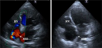
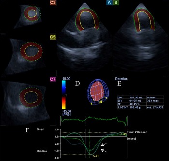
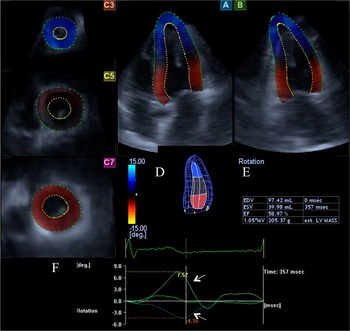
A 19-year-old girl with surgically palliated hypoplastic right-heart syndrome with intact ventricular septum, ostium secundum atrial septal defect, and small right ventricular cavity is presented. The patient had undergone palliation with a superior caval vein to pulmonary artery shunt – bidirectional Glenn – at 3 years of age. The patient proved to be compensated without palpitations. She was involved in the MAGYAR-Path Study – Motion Analysis of the heart and Great vessels bY three-dimensionAl speckle-tRacking echocardiography in Pathological cases. A complete two-dimensional Doppler echocardiography was carried out with a commercially available Toshiba ArtidaTM (Toshiba Medical Systems, Tokyo, Japan) echocardiography equipment with a PST-30SBP (1–5 MHz) phased-array transducer (Toshiba Medical Systems, Tokyo, Japan), confirming right ventricular dysplasia and atrial septal defect (Fig 1). The movement of the septum was found to be dyskinetic. Following two-dimensional echocardiographic study, three-dimensional data acquisition was performed using a PST-25SX matrix-array transducer (Toshiba Medical Systems) with three-dimensional speckle-tracking echocardiographic capability. During a constant RR interval and within a single breath-hold, six wedge-shaped subvolumes were acquired from an apical window to create a full-volume three-dimensional data set. The chamber quantification was performed off-line using 3D Wall Motion Tracking software version 2.5 (Toshiba Medical Systems). The multiple long- (A, B) and short-axis views (C3, C5, C7) extracted from three-dimensional echocardiographic data sets of the patient with hypoplastic right-heart syndrome and that of a healthy control are demonstrated in Figures 2 and 3. Visual information on left ventricular rotational mechanics are shown in colour overlay superimposed on grey-scale images in both cases.
Transthoracic Doppler echocardiography images from a 19-year-old girl with palliated hypoplastic right-heart syndrome. The images demonstrate an intact ventricular septum, atrial septal defect (ASD), and small right ventricular (RV) cavity.

In a patient with palliated hypoplastic right-heart syndrome, apical four-chamber ( A ) and two-chamber ( B ) views and short-axis views ( C3, C5, C7 ) at different levels of the left ventricle extracted from the three-dimensional echocardiographic data set are presented. A three-dimensional cast of the left ventricle ( D ) and calculated volumetric and functional left ventricular parameters are also demonstrated ( E ). Left ventricular basal and apical rotations proved to be in the same direction with near absence of left ventricular twist ( F ). EDV=end-diastolic volume, EF=ejection, ESV=end-systolic volume.

In a healthy patient, apical four-chamber ( A ) and two-chamber ( B ) views and short-axis views ( C3, C5, C7 ) at different levels of the left ventricle extracted from the three-dimensional echocardiographic data set are presented. A three-dimensional cast of the left ventricle ( D ) and calculated volumetric and functional left ventricular parameters are also demonstrated ( E ). Counterclockwise rotation of the left ventricular apex (white arrow, positive value) and clockwise rotation of the left ventricular base (dashed arrow, negative value) are shown demonstrating normal rotational directions ( F ). EDV=end-diastolic volume, EF=ejection, ESV=end-systolic volume.

Left ventricular end-diastolic and end-systolic volumes, ejection fraction, mass and right ventricular outflow tract, and tricuspid annular plane systolic excursion of the patient proved to be 107 ml, 64 ml, 40%, 198 g, 17 mm, and 14 mm, respectively. Results of the present case with hypoplastic right-heart syndrome suggest that all left ventricular regions move in almost the same clockwise direction (Fig 2f, dashed arrow, negative value). Global left ventricular radial, circumferential, longitudinal, three-dimensional and area strain parameters, and the range for all segments proved to be 5.1±4.3, −12.2±8.0, −14.1±8.5, 6.0±4.8, and −24.3±13.8%, respectively. Quantitative data of left ventricular rotation of the control patient demonstrate adequate rotational directions with counterclockwise motion of the left ventricular apex (white arrow, positive value) and clockwise motion of the left ventricular base (dashed arrow, negative value; Fig 3F).
Discussion
To the best of the authors’ knowledge, this is the first time that left ventricular basal and apical rotations could be in the same clockwise direction, with near absence of left ventricular twist as called “rigid body rotation” of the left ventricle in hypoplastic right-heart syndrome, has been demonstrated. In a recent study using two-dimensional speckle-tracking echocardiography, left ventricular “rigid body rotation” was demonstrated in patients with non-compaction cardiomyopathy.Reference Van Dalen, Caliskan and Soliman 5 The near absence of left ventricular twist was found to be an objective, quantitative, and reproducible functional criterion with good predictive value for the diagnosis of left ventricular non-compaction.Reference Van Dalen, Caliskan and Soliman 5 , Reference Van Dalen, Caliskan and Soliman 6 This phenomenon was found in 53–100% of non-compaction cardiomyopathy patients by other working groups.Reference Peters, Khandheria and Libhaber 7 , Reference Kalapos, Domsik, Forster and Nemes 8 Similar alterations in left ventricular rotational mechanism could be demonstrated in a patient with univentricular heart with left ventricular characteristics as well.Reference Nemes, Havasi, Domsik and Forster 9 Hypoplastic right-heart syndrome is a congenital heart defect in which the right ventricle of the heart fails to develop appropriately. However, rotational mechanics of the left ventricle has never been assessed by speckle-tracking echocardiography in this disease.
It is known that two-dimensional speckle-tracking echocardiography based on frame-by-frame tracking of speckle patterns created by interference of the ultrasound beam within the myocardial tissue in one plane is likely to be affected by geometric assumptions and the use of foreshortened apical views.Reference Nemes, Kalapos, Domsik and Forster 4 – Reference Van Dalen, Caliskan and Soliman 6 The three-dimensional speckle-tracking echocardiography detects speckles in the three-dimensional space, and therefore is able to merge benefits of three-dimensional and speckle-tracking echocardiographies, allowing clinicians to visualise the heart as it is: a three-dimensional organ.Reference Nemes, Kalapos, Domsik and Forster 4 It was found to be a quantitave way to assess left ventricular rotation and twist deformations.Reference Zhou, Ashraf and Hu 3 , Reference Nemes, Kalapos, Domsik and Forster 4 , Reference Kalapos, Domsik, Forster and Nemes 8 – Reference Nemes, Kalapos, Domsik and Forster 10 “Rigid body rotation” of the left ventricle was confirmed by this methodology in a non-compaction cardiomyopathy, as well.Reference Kalapos, Domsik, Forster and Nemes 8 , Reference Nemes, Kalapos, Domsik and Forster 10
In conclusion, left ventricular “rigid body rotation” was identified in a single patient with hypoplastic right-heart syndrome. This finding could be partially explained by the impairment in ventricle-to-ventricle interaction. Alterations in the anatomic myocardial fibre orientation, which is a left-handed helix in the epicardium and a right-handed helix in the endocardium in normal situation, could be altered in this disease. However, other reasons could not be excluded as well. Further studies with larger number of hypoplastic right-heart syndrome patients are warranted to confirm our findings with deeper insights into pathophysiology.
Acknowledgement
Many thanks to linguistic corrections by Dr Péter Hausinger.
Financial Support
Attila Nemes, MD, holds a János Bolyai Research Fellowship (Budapest, Hungary).
Conflicts of Interest
None.
Ethical Standards
The authors assert that all procedures contributing to this work comply with the ethical standards of the relevant national guidelines on human experimentation and with the Helsinki Declaration of 1975, as revised in 2008, and has been approved by the institutional committee of the University of Szeged.