
I. INTRODUCTION
The past few decades have witnessed burgeoning exploitation of bimetallic nanoparticles (NPs) from both academic and technological point of view. Various composite bimetallic nanostructures have been tailored because of their wide spectrum of applications in controlling energy of plasmon adsorption band, electrochemical, magnetic, catalytic, and biomedical applications, tuneable optical behavior and play a crucial role in bio-sensing. Reference Reddy, Mohanaraju and Reddy1,Reference Valodkar, Modi, Paland and Thakore2 Generally, they exhibit unique properties compared to their pure individual metallic nanodimensional particles. Reference Tao3 Bimetallic nanomaterials of various size and morphology synthesized via diverse techniques like vapor deposition, ionic liquid, micro emulsions, electrochemical method, solvothermal reduction, thermal decomposition of organic complexes, radiolytic reduction, environmental benign methods, alcohol reduction etc. are well documented in the literature. Reference Ferk, Stergar, Makovec, Hamler, Jaglicic, Drofenik and Ban4–Reference Lui, Cai and Liang11 Even though chemical methods have the advantages of ease to synthesis and precise control of the size and morphology, all these methods involved usage of huge amount of chemicals, consumed energy, and used environmentally hazardous substances. In spite of the fact that there are numerous routes for the preparation of composite bimetallic nanoparticulate system of different morphologies, green approaches have drawn must attention, unambiguously, owing to the growing need for development of eco-friendly environmentally technologies, its high atom economy, less time consumption, simplicity for the experimental procedures, versatility etc. Several biosynthesis routes of nanoscopic particles were developed using plants (leaves, roots, stems etc.), plant products and microorganism, which is a huge contribution to the progress of development for green synthesis. Such biosynthesis of nanomaterials has been providing an alternative solution to chemical and physical methods. Reference Li, Wang, Zhang, Li and Liu12,Reference Wu and Chen13 Although preparation of bimetallic NPs employing various techniques have gained maturity, still there is a need to envisage a novel, facile route of tailoring bimetallic nanostructure of different compositions utilizing nontoxic, biocompatible, and abundantly available resources.
In this work, we have manifested a simple, down-to-earth biocompatible green approach for the preparation of NiO, Ag monometallic and NiO–Ag bimetallic NPs of alloy type utilizing tannic acid (TA) in alkaline medium at room temperature. In literature many works were reported in synthesis of NPs using TA. Reference Ahmad14,Reference Çakar and Ozacar15 TA served a dual purpose by acting both as an excellent reducing and capping agent. The aforementioned particles are remarkably stable. Water was used as green solvent throughout the synthesis procedure. The utilization of toxic solvent and harsh chemicals is completely ignored during the synthesis process. Insofar, we believed this is the first report of tailoring NiO–Ag bimetallic alloy type NPs using TA at alkaline pH. The fabricated bimetallic system displayed significant catalytic behavior during photodegradation and chemical degradation of dye; methyl violet (MV) was used as a model dye for degradation. Synergistic electronic effect due to composition of the bimetallic nanocatalyst preponderantly plays a typical part during the degradation of the MV dye. Organic pollutants include dyes, pesticides, polynuclear aromatic hydrocarbons, polychlorinated and polybrominated biphenyls, phenols, and drug residues. Reference Mittala, Mittala, Malviyaa and Gupta16–Reference Mittal, Mittal, Malviya and Gupta18 Organic pollutants and contaminants like heavy metals are one of the major environmental issues and should be treated well before discharging in the environment. Reference Saleh and Gupta19–Reference Gupta, Jain, Nayak, Agarwal and Shrivastava21 Nanocatalysis might be a solution of such environmental issues. Reference Saleha and Gupta22–Reference Saleh and Gupta25
II. MATERIALS AND EXPERIMENT DETAILS
A. Materials
All the chemicals were of AR grade. Silver nitrate (AgNO3) and nickel nitrate hexahydrate Ni(NO3)2·6H2O from were purchased from Merk Pvt. Ltd. While, TA and MV 10B (MV) were obtained from HIMEDIA Laboratories Pvt. Ltd. NaOH was procured from Merk Pvt, Ltd. and their solutions were prepared in double distilled water (DDW).
B. Synthesis of NiO, Ag monometallic and NiO–Ag bimetallic NPs
0.004 M of Ni(NO3)2·6H2O and AgNO3 solutions in DDW were prepared by weighing out the required amount of salts (0.01163 g of Ni salt and 0.00678 g of AgNO3 in 10 mL of DDW respectively). The fabrication of monometallic NPs was accomplished by taking 1.6 mL of 0.04 M TA in 10 mL of each metal salt solution. An alkaline pH of the reaction mixture was established by introducing 400 µL of 0.02 M NaOH solution. The reaction mixtures were allowed to stir uniformly for 2 h at room temperature and the whole reaction mixture was left undisturbed for one night. The generation of NiO–Ag bimetallic nanoscale particles was performed by maintaining the same reaction condition and procedure except instead of 10 mL of salt solution; 5 mL each of Ni and Ag salt solutions (0.004 M) were taken. The pH of the reaction system was also maintained by employing 0.02 M NaOH solution. The ensuing particles are markedly stable and used as nanocatalysts in the degradation of MV dye.
III. CHARACTERIZATIONS OF THE SYNTHESIZED NANOSCOPIC PARTICLES
A. Ultraviolet visible (UV–vis) spectroscopy
All UV–vis spectra were recorded on Perkin Elmer UV–vis spectrophotometer (Perkin Elmer, Wokingham, United Kingdom) fitted with Petlier System. The absorption spectra of the prepared NPs were recorded by taking the aqueous dispersion of the NPs and scanned in the range of 200–800 nm.
B. Scanning electron microscopy (SEM) and energy dispersive x-ray (EDX) analysis
SEM and EDAX data of the so-synthesized NPs were obtained by employing SEI Quanta 250 model (FEI Company, Hillsboro, Oregon) of SEM instrument. An adequate amount of dried powder NiO, Ag monometallic and NiO–Ag bimetallic NPs were used for the measurement.
C. Transmission electron microscopy (TEM) and selected area electron diffraction (SAED)
TEM and SAED micrographs of the particles were taken with JEM-2100 transmission electron microscope (JEOL Ltd., Freising, Germany) operating at 60–200 kV STEPS. After preparation, the particles were centrifuged at 12,000 rpm for 10 min, washed with DDW for three times and redispersed in it by sonication for 4–5 min. A drop of dilute aqueous dispersion of the NiO, Ag, and NiO–Ag bimetallic NPs were put on carbon coated copper grids and the grids were dried at room temperature. After complete drying of the grids, TEM and SAED patterns of the particles were taken.
D. Dynamic light scattering (DLS) and zeta potential (ζ)
To measure size using DLS, sample preparation has to be carried out very carefully as this measurement is very sensitive to foreign particles like dust. Sample was prepared by diluting the required amount of sample in double distilled water and sonicated for 1–2 min. A minimum of 10 mm of sample solution was taken and put in a glass. This cuvette is placed in the cell holder and scanned in range of 1–100 nm at room temperature. All the size and zeta potential measurement in this work has been carried out using Zeta Sizer Nano ZS90 (Malvern Instruments Ltd., Worcestershire, United Kingdom).
E. X-ray diffraction (XRD) analysis
X-ray diffraction patterns of the samples were taken by Philips XRD instrument (Philips, Woodlands, Texas); properly dried powder samples were utilized to carry out the diffraction experiment. After preparation, the samples were washed effectively with DDW and finally with ethanol. The samples were then allowed to dry at room temperature.
F. Fourier transformed-infra red (FTIR) spectroscopy
Perkin Elmer Spectrum-2 FT-IR instrument (Perkin Elmer, Waltham, Massachusetts) was utilized to record FT-IR spectra of TA and the so-synthesized NPs. The particles after fabrication were washed thoroughly, collected and then finally redispersed in DDW. Liquid FT-IR spectra of TA and the synthesized NiO, Ag monometallic and NiO–Ag bimetallic NPs were recorded in the range 400–4000 cm−1. The transmission of the solvent, water was finally omitted from the readings of the samples recorded.
IV. NANOCATALYTIC ACTIVITY OF THE SYNTHESIZED PARTICLES
Reduction of MV in aqueous medium was monitored with the help of UV–vis spectrophotometer at λmax = 584 nm, a prominent characteristic peak of MV. To 10 µL of 1 × 10⁻³ M MV, 20 µL of 0.0033 M NPs (i.e., NiO, Ag monometallic and NiO–Ag bimetallic NPs) were added separately in a 3.5 mL of opti-quartz cuvette followed by the addition of 10 µL of 1 × 10⁻² M NaBH4. The total volume for the chemical degradation reaction system was fixed at 3 mL by adding DDW. The progress of the reaction was monitored by noting the fall in absorbance at 584 nm in room temperature. Similar procedure was also repeated for the uncatalysed reaction, instead of introducing metallic nanoscaled particles, 10 μL of DDW was added to maintain a consistent total volume of the reaction system. Kinetic dependences of the reaction on the pH of the solution were carried out for the catalyzed reaction by adjusting the pH of the reaction mixture either by the addition of NaOH or HCl solution.
Moreover, a comparative study for photocatalytic degradation of MV was also performed spectrophotometrically at the same characteristic maximum wave length of MV i.e., 584 nm. The reaction was monitored by taking 10 µL of 1 × 10⁻³ M MV and 20 µL of 0.0033 M each of the aforementioned NPs at natural pH medium. The total volume of the reaction mixture was also maintained at 3 mL by adjusting with 2970 μL of DDW. Photocatalytic degradation of MV was also carried out in the absence of nanocatalyst by fixing the total volume of the reaction mixture at 3 mL with DDW. All the kinetic studies reported in this work were carried out at an ambient temperature of 25 °C.
V. RESULT AND DISCUSSION
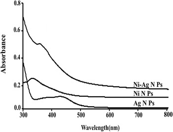
NiO, Ag monometallic, and NiO–Ag bimetallic NPs were synthesized successfully by utilizing TA in alkaline medium; the formation of NiO–Ag bimetallic particles was accomplished by co-reduction of Ni2+ to Ni0 and Ag1+ to Ag0 at room temperature. The formation of nanoscale NiO, Ag, and NiO–Ag particles is directly confirmed by UV–vis spectroscopy. Figure 1 displays the UV–vis spectra of NiO and Ag monometallic NPs with distinct surface plasmon resonance (SPR) bands at 330 and 422 nm respectively. Such SPR band of nanoscopic NiO particles is well documented in the literature. Reference Gupta, Jain, Mittal, Saleh, Nayak, Agarwal and Sikarwar26–Reference Li and Komarneni30 While, NiO–Ag bimetallic NPs exhibit SPR band at 363 nm as presented in Fig. 1 this sudden disappearance of the characteristic peaks of individual components of the composite bimetallic system along with shifting of peak in the bimetallic system reveal that the bimetallic system is not just a physical mixture of the constituent metals. Reference Dutta and Dolui31–Reference Toshima and Yonezawa34 Physical mixture of two metals in a solution is expected to show two SPR bands where as an alloy or core shell shows a single plasmon resonance band. Reference Lahiri, Bunker, Mishra, Zhang, Maisel, Doudna, Bertino, Blum, Tokuhiro, Chattopadhyay, Shibata and Terry35,Reference Boote, Byun and Kim36 However, from the UV–vis spectra alone it is difficult to conclude whether the bimetallic system is an alloy or of core shell in nature. But co-reduction or simultaneous reduction technique, generally, results to the formation of alloy type of bimetallic system.
UV–vis spectra of NiO, Ag and NiO–Ag bimetallic NPs.

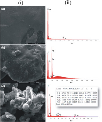
Figure 2(i) highlights SEM pictures of the prepared monometallic and bimetallic nanoscopic particles. The images indicate the formation of particles; however, the size and morphology of the particles cannot be determined subtly under the measured scale. EDAX provides elemental information regarding the constituents of multi-components metallic system. The inclusion of metallic Ni and Ag in EDAX pattern reflects the bimetallic nature of NiO–Ag composite NPs, shown in Fig. 2(ii). The individual metals, Ni and Ag, are present almost in the ratio of 1:1.7 (weight ratio) within the bimetallic system. The EDAX patterns of the individual monometallic NPs are also provided for better comparison. EDAX results rule out the presence of C and Na in the prepared particles, author believes that this elements come from the tannic acid and NaOH.
(i) SEM images and (ii) EDAX data of (a) NiO (b) Ag and (c) NiO–Ag bimetallic NPs.

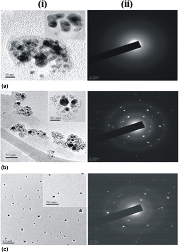
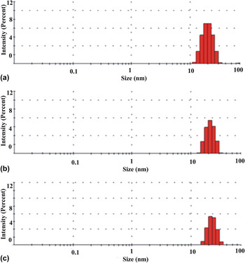
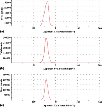
The size and morphology of these mono and bimetallic NPs were studied by using transmission electron microscopic technique. Fig. 3 shows TEM and SAED images of NiO, Ag monometallic and NiO–Ag bimetallic NPs. The prepared particles are spherical in shape with an average diameter ranging from 7 nm to 10 nm in both mono and bimetallic system as estimated via HRTEM [Fig. 3(i)]. The SAED patterns of the synthesized NPs display well dotted ring structures indicating crystalline behavior of the tailored nanoscale particles and are presented in Fig. 3(ii). On the other hand DLS data's rule out that the size of both mono and bimetallic particles of Ni and Ag range in between 19 and 22 nm as given in Fig. 4. The size of particles determined using DLS are slightly larger than size measure from TEM which may be due the smelling of particles in aqueous medium. The size of particles measured from DLS is actually the hydrodynamic size of particles so result of TEM and DLS usually show some variation. Reference Singh, Gupta, Sharma and Sharma37 The stability of the particles is characterized by measuring the zeta potential of the particles in the dispersion. Particles seem to be very stable as its zeta potential is higher than ±30 mV as given in Fig. 5. Zeta potential of Ag NPs is −42.6 mV that of Ni NPs is −42.4 mV and bimetallic NPs is of −42 mV.
(i) TEM and (ii) SAED micrographs of (a) NiO (b) Ag and (c) NiO–Ag bimetallic NPs.

Size distribution plot of (a) Ag (b) NiO and (c) NiO–Ag NPs.

Plot of total counts versus zeta potential (mV) of (a) Ag (b) Ni and (c) NiO–Ag NPs.

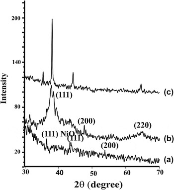
XRD patterns of the fabricated NiO, Ag monometallic and NiO–Ag bimetallic NPs are shown in Fig. 4, further substantiate their crystalline nature. NiO NPs shows diffraction peaks at 2θ = 37.2°, 44.3°, and 53° which may be indexed to (111) and (200) planes of FCC crystal structure as displayed in Fig. 6(a). Reference Dharmaraj, Prabu, Nagarajan, Kim, Park and Kim38,Reference Park, Kang, Son, Park, Lee, Kim, Kim, Noh, Park, Bae, Park and Hyeon39 The authors strongly believed that the formation of NiO is due to the occurrence of aerial oxidation during drying of the particles at room temperature, since first row transition metals are very prone to oxidation. On contrary, Ag NPs exhibit three characteristic obvious peaks at 2θ = 38.7°, 47.5°, and 65.8° corresponding to (111), (200), and (220) FCC crystal planes as displayed in Fig. 6(b). Reference Swamy, Sudipta, Jayanta and Balasubramanya40–Reference Kim, Lim and Shin42 Figure 6(c) displays XRD pattern of the bimetallic system showing sharp peaks at 2θ = 38.11°, 44.35°, and 64.59°. In the bimetallic system, the characteristic peaks of NiO and Ag are absent instead their peaks got shifted. This result further supports the fact that bimetallic NiO–Ag NPs is an alloy not a core shell or a physical mixture of individual metals. Reference Tao3
XRD patterns of (a) NiO, (b) Ag and (c) NiO–Ag bimetallic NPs.

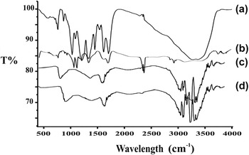
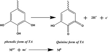
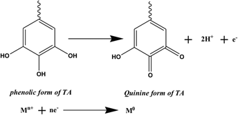
Besides, Fig. 7 show liquid FT-IR spectra of the synthesized metallic NPs and TA. In the spectra the broad peak of OH radical (group) (3400 cm−1) were shrunken and it is almost reduced in case of metallic NPs indicating that the hydroxyl groups of TA are utilized in reduction of metal ions and stabilizing the particles. Bands at 1490 cm−1 and 1300 cm−1 correspond to aromatic C=C and phenolic C–O stretching vibrations from which it can be concluded that majority of TA is present in its oxidized form. Reference Swamy, Sudipta, Jayanta and Balasubramanya40 IR spectra vividly indicate that TA is well coated over NiO, Ag and NiO–Ag bimetallic NPs hereby acting both as reducing as well as capping agent. Both the monometallic and bimetallic NPs exhibit excellent stability as we observed that particles remained unsettle for more than 6 months. Thus, it can be anticipated that identical size and morphology of the prepared NPs might be due to similar reaction conditions and effective capping capability of TA. The possible mechanism for the formation of NPs through reduction of metal salts by TA is given below in Scheme 1. Reference Yi, Li, Xu, Luo, Luo, Wu, Yi and Tang43,Reference Sylvia, Rajmuhon and David44
IR spectra of (a) tannic acid, (b) Ag NPs (c) NiO NPs and (d) NiO–Ag bimetallic NPs.

Probable reaction mechanism for the formation of NPs.

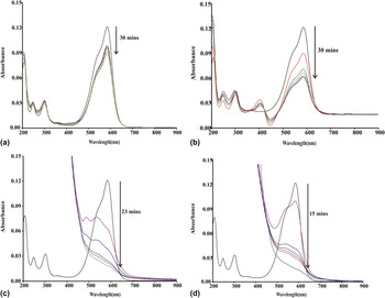
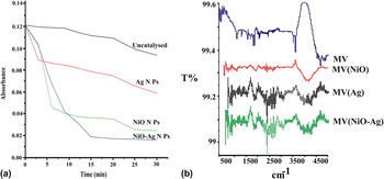
The fabricated metallic NPs were then utilized as nanocatalysts in reduction and photocatalytic degradation of MV at room temperature. MV exhibits prominent characteristic peak at 584 nm and surfacing of this absorption peak indicates the degradation/reduction of MV. Reduction of MV dye with NaBH4 in presence and absence of nanocatalyst were monitored spectrophotometrically at 584 nm, presented in Fig. 8. Figure 8(a) displays reduction of MV by NaBH4 in the absence of nanocatalyst; rate of decrease in absorbance of the dye is very slow. With the addition of nanocatalyst, the rate of reduction is greatly enhanced as there is tremendous decrease in absorbance of MV as given in Figs. 8(b)–8(d). From this result it can be concluded that the monometallic and bimetallic NPs act as catalyst but the process of reduction is more effective in the presence of bimetallic nanoscopic particles as compared to monometallic. Kinetic studies for the reduction of MV were also assayed in absence and presence of NPs given in Fig. 9 at room temperature in natural pH medium of 6.4. NiO–Ag bimetallic NPs completely transform MV into its leuco form in 15 min, while monometallic particles took more time. Degradation process followed pseudo first order reaction at the optimized concentration of NaBH4 and rate constant follows the order: k NiO–Ag bimetallic NPs > k NiO NPs > k Ag NPs > k uncat at room temperature. Catalytic mechanism for reduction of MV can be explained as: NaBH4 being a nucleophile donate electron to the metallic NPs which serve an electron relays in the redox reaction. From the surface of metal particles MV capture electrons and get reduces to its colourless leuco form. Reference Mallick, Witcomb and Scurell45
Reduction of MV dye (a) uncatalysed (b) catalyzed with NiO NPs, (c) catalyzed with Ag NPs and (d) catalyzed with NiO–Ag bimetallic NPs.

(a) Kinetics studies of uncatalysed and catalyzed reduction of MV and (b) IR spectra of MV and MV treated with NPs.

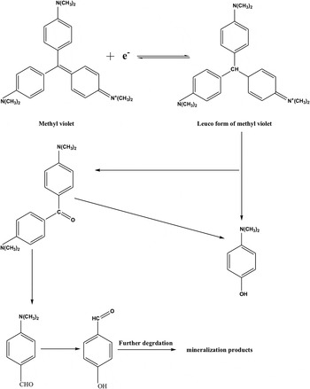
IR spectra of MV composed of band at 3467 cm−1 which is attributed to N–H stretching, bands at 2921 cm−1 and 2856 cm−1 are of asymmetric and symmetric stretching of C–H, bands at 1605 cm−1 and 1473 cm−1 can be attributed to C=C stretching usually occurred in pair, band at 1583 cm−1 is of N–H bending, C–N stretching in range of 1176–1015 cm−1. Reference Pavia, Lampman, Kriz and Vyvyan46 Significant changes in the spectra of MV are observed after treating with NPs as shown in the spectra Fig. 9(b). MV treated with NiO NPs shows IR stretching/bands at 3334, 2528, 1652, 1343, 1156, and 1012 cm−1. After treating MV with Ag NPs IR spectra of MV are observed at 3439, 2873, 1853, 1587, 1354, and 1157 cm−1. Likewise in case of MV (NiO–Ag) bands are obtained at 3486, 2749, 1849, 1579, 1362, and 1189 cm−1. Most of the significant IR bands of MV where shifted indicating the strong interaction of NPs with MV and formation of a new compound different from original substrate. Possible mechanism for the reduction of MV is given in Scheme 2. Reference Gan, Cheng, Palanisami, Chen, Megharaj and Naidu47–Reference Retnamma, Novais and Rangel49
Possible mechanism for the reduction of MV using NPs.

Moreover, pH also play important role in degradation of MV and so degradation of MV was also studied at a stronger acidic and basic pH of 2 and 10 respectively. The rate constants for the different types of nanocatalyst at different pH conditions are summarized in Table I(a). With increase in pH of the reaction mixture from 2 to 6.4, rate of degradation increase to certain point but it again decrease when pH is alkaline i.e., pH = 10. It can be explained as with increase in pH, the stability of borohydride in aqueous solution increases and this make the hydrolysis reaction difficult causing hindrance to the reduction process so the rate of degradation decreases in alkaline medium. In contrast, at low pH the half-life of borohydride is very small and so the release of hydrogen by borohydride occurs quickly to be adsorbed on the surface of metal NPs which consequently results in decrease of reduction process of MV. Reference Kaufman and Sen50 From this observation it can be concluded that degradation of MV is favorable in natural medium.
Calculated rate constants for the (a) chemical and (b) photocatalytic degradation of MV at room temperature.

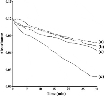
Photocatalytic degradation of MV was also studied in presence of metallic NPs at room temperature in pH medium of 6.4, shown in Fig. 10. Photocatalysis degradation of dye in aqueous medium involves generation of electron (e−) and hole (h+) pair leading to the formation of hydroperoxyl radicals, superoxide radicals anions, and hydroxyl radicals. These radicals act as oxidizing species in photo oxidation of dye by scavenging the electrons in conduction band which indirectly prevent the recombination of e− and h+. Electron in the conduction band can be utilized by dye molecules to generate dye radicals anions and leading to the degradation of dye. Reference Hoffman, Martin, Choi and Bahnemann51 The possible mechanism for the degradation of MV dye can be explained as. Reference Houas, Lachheb, Ksibi, Elaloui, Guillard and Herrmann52,Reference Ameta, Vardia, Punjabi and Ameta53
 $${}^{\rm{1}}{\rm{M}}{{\rm{V}}_{\rm{0}}}\mathrel{\mathop{\kern0pt\longrightarrow}\limits_{{\rm{ISC}}}^{h\nu }} {}^{\rm{1}}{\rm{M}}{{\rm{V}}_{\rm{1}}}$$
 $${}^{\rm{1}}{\rm{M}}{{\rm{V}}_{\rm{1}}} \to {}^{\rm{3}}{\rm{M}}{{\rm{V}}_{\rm{1}}}$$
 $${\rm{Photocatalyst}}\left( {{\rm{PC}}} \right) \to {\rm{PC}}\left( {{{\rm{h}}^{\rm{ + }}}} \right) + {\rm{PC}}\left( {{{\rm{e}}^ - }} \right)$$
 $${\rm{PC}}\left( {{{\rm{e}}^ - }} \right) + {{\rm{O}}_2}\left( {{\rm{dissolved}}} \right) \to {\rm{PC}} + {\rm{O}}_2^{ \cdot - }$$
 $${{\rm{H}}^{\rm{ + }}} + {\rm{O}}_2^{ \cdot - } \to {\rm{HO}}_{\rm{2}}^{ \cdot - }$$
 $${\rm{2HO}}_{\rm{2}}^{ \cdot - } \to {{\rm{H}}_2}{{\rm{O}}_2} + {{\rm{O}}_2}$$
 $${{\rm{H}}_2}{{\rm{O}}_2} \to 2{}^ \cdot {\rm{OH}}$$
 $${{\rm{H}} \mathord{\left/ {\vphantom {{\rm{H}} {{\rm{HO}}_{\rm{2}}^{ \cdot - }}}} \right. \kern-\nulldelimiterspace} {{\rm{HO}}_{\rm{2}}^{ \cdot - }}} + {}^3{\rm{M}}{{\rm{V}}_{\rm{1}}} \to {\rm{Leuco\ form}}$$
 $${\rm{Leuco\ form}} \to {\rm{Product}}$$
Photocatalytic degradation of MV (a) uncatalysed, (b) catalyzed with NiO NPs, (c) catalyzed with Ag NPs and (d) catalyzed with NiO–Ag bimetallic NPs.

The photocatalytic degradation follows first order kinetics and the rate constants determined from the plot of ln A versus time are also listed in Table I(b). In this case also the rate of degradation of MV is maximum for NiO–Ag bimetallic NPs followed by NiO and Ag NPs even though the variation is less significant. This result confirms the higher catalytic efficacy of bimetallic NPs over the monometallic NPs. Such dominating catalytic behavior of bimetallic NPs in both the reactions may be attributed to synergistic effect with arises due to uneven distribution of electrons. Basically, this synergistic electronic effect is contributes from composition effect of multicomponent system. The electronic interaction between different components may lead to significant modification of the electronic structure of NPs, which leads to an enhancement of catalytic activity. Reference Valodkar, Modi, Paland and Thakore2,Reference Ameta, Vardia, Punjabi and Ameta53 At least our work have directed a rational way to design NiO–Ag bimetallic NPs via an environmental friendly approach using TA for the first time and facilitates in the study of composition effect on nanocatalysis. Such preparation route of bimetallic system may find its way in tuning the properties of composite nanoparticulate structures for a wide range of applications.
VI. CONCLUSION
Monometallic and bimetallic NiO and Ag NPs were fabricated through an environmental friendly method using TA in alkaline medium. The synthesized nanodimensional particles were stable for more than six months in aqueous medium. Co-reduction of Ni and Ag metal salt solutions by TA enables successfully in the formation of alloy particles having excellent catalytic efficiency as compared to monometallic particles. Chemical degradation of MV dye was studied at different pH using the synthesized NPs and it was found that the favourable environment for the degradation of MV was in natural medium. Rate constants calculated from the plot of ln A versus time reveals that NiO–Ag bimetallic NPs possess greater catalytic property as compared to its constituent monometallic particles. The rate constant follows the order k NiO–Ag bimetallic NPs > k NiO NPs > k Ag NPs at room temperature. This result was further supported by the information obtained from the photocatalytic degradation of MV by monometallic and bimetallic NPs. Introduction of a second component to a metal system results in the alternation of electronic property of the composite system hereby enhancing catalytic property of the bimetallic particles as compared with individual metal particles. The significant catalytic potential of the NiO–Ag bimetallic NPs is attributed to the synergistic effect which arises due to uneven distribution of electrons contributing to the effect of composition in nanocatalysis.
ACKNOWLEDGMENTS
Authors are also grateful to SAIF, NEHU for allowing us to take TEM-SAED images and Physic Department, Manipur University for recording SEM, EDAX. Authors are thankful to Dr Raghumani Ningthoujam of BARC, Trombay for technical supports and S. Sanatombi Devi, Deparment of Physics, National Institute of Technology for her supports.