Introduction
Many abnormalities of the otic capsule which result in sensorineural hearing loss (SNHL)Reference Schuknecht1 are visible on computed tomography (CT) of the temporal bone.Reference Valvassori, Mafee and Carter2 We present a child with SNHL whose temporal bone CT showed bilateral widening of the superior vestibular nerve canal, with bilateral distortion of the nerve's entry site into the vestibule.
Case report
A 10-month-old male infant failed his newborn hearing screening test.
Physical examination was unremarkable. Auditory brainstem response testing showed no waves detectable below 70 dB on the left and none detectable below 80 dB on the right.
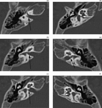
Temporal bone CT (Figure 1) showed bilaterally enlarged superior vestibular nerve canals and bilateral distortion of the vestibules at the nerve entry site. The superior vestibular nerve canal length and width at the most medial portion (i.e. their exit from the internal auditory canal) were measured from the temporal bone CT using the Centricity Dicom viewer software. The canal depth in the z axis was estimated from the number of serial sections in which it appeared. The right canal's length was 2.28 mm, its width was 2.24 mm and its depth was approximately 3 mm. The left canal's length was 2.74 mm, its width was 1.82 mm and its depth was approximately 2 mm. The bony wedge between the superior vestibular nerve and inferior vestibular nerve canals was not well seen on CT, so our depth estimate remains crude. The remaining structures appeared normal, including the facial nerve and the internal auditory canals.
Axial, noncontrast, bone window computed tomography scans of the right and left temporal bones of an infant with congenital sensorineural hearing loss. The only visible malformation is the bilateral enlargement of the superior vestibular nerve canal and the distortion of the vestibule at the nerve entry site. Arrows indicate the superior vestibular nerve canals at their most medial portion, where width measurements were taken. Auditory nerve canals are indicated by asterisks directly adjacent. Small white arrows indicate inferior vestibular nerve canals.

Discussion
This radiographic finding of isolated enlargement of the canal for the superior vestibular nerve has not previously been reported in ears with SNHL, nor has it been reported in normal hearing ears. Schuknecht's 1993 pathological analysis of the temporal boneReference Schuknecht1 identified many inner-ear malformations, but no enlargements of the superior vestibular nerve canal. A careful survey of this author's photomicrographs of temporal bones from patients with congenital hearing loss identified five which clearly show a normal superior vestibular nerve canal. Another eight photomicrographs of temporal bones from patients with congenital hearing loss show areas of temporal bone directly adjacent to the superior vestibular nerve canal into which an enlarged canal – similar in size to that seen in our patient – would have intruded, had it been present.
A 1995 radiographic review of inner-ear malformationsReference Valvassori, Mafee and Carter2 also gave no evidence of this particular malformation.
Two recent studies of vestibular canal measurements in temporal bones found the average length of the superior vestibular nerve canal to be 2.30 mmReference Goebel, O'Mara and Gianoli3 and 1.944 mm.Reference Gianoli, Goebel, Mowry and Poomipannit4 The second study also calculated the average width of the superior vestibular nerve canal at its most medial portion, which is its widest portion, to be 1.04 mm. Our patient's superior vestibular nerve canals were 2.28 and 2.74 mm in length – close to these previously measured averages. Our patient's superior vestibular nerve canal widths were 2.24 and 1.82 mm, approximately twice the normal width. Direct coronal images could not be obtained in this paediatric patient; therefore, the depth of the superior vestibular nerve canal was estimated from the number of sections in which it was visible on axial imaging. In addition, careful review of the temporal bone CT did not clearly identify a bony wedge between the superior and inferior vestibular nerves. The vestibular canals may have been confluent in this patient.
Unfortunately, magnetic resonance imaging could not be obtained for this child. Therefore, the superior vestibular nerve canal may be filled with nerve, cerebrospinal fluid or a combination of the two.
• Pathological analysis of the temporal bone has identified many inner-ear malformations, but enlargement of the superior vestibular nerve canal has not previously been reported
• This paper describes a 10-month-old male with sensorineural hearing loss together with bilaterally enlarged superior vestibular nerve canals
• This inner-ear malformation may be a marker for sensorineural hearing loss
This anomaly should be added to the canon of inner-ear malformations, and further study may establish its role, if any, in SNHL.