Introduction
Malignant triton tumour is a rare, aggressive soft tissue sarcoma first described by MassonReference Masson1 in 1932. It is a subtype of malignant peripheral nerve sheath tumour comprising rhabdomyoblasts and malignant Schwann cells.Reference Woodruff, Chernik, Smith, Millett and Foote2 The name ‘triton tumour’ derives from Locatelli's experiment in which a sciatic nerve implanted in the back of a triton salamander caused the growth of supernumerary limbs consisting of neural and muscular tissue.Reference Locatelli3 Although malignant peripheral nerve sheath tumours may consist of many types of differentiated tissues (such as glandular epithelium, squamous cell, cartilage and bone), those which contain only differentiated skeletal muscle are referred to as malignant triton tumours. The biological behaviour of this tumour is more aggressive than the usual malignant schwannoma, and the prognosis is grim.Reference Matsunou, Shimoda, Kakimoto, Yamashita, Ishikawa and Mukai4–Reference Bhatt, Graeme-Cook, Joseph and Pilch7
Malignant triton tumour can occur as a sporadic tumour. Approximately half to two-thirds of cases occur in association with neurofibromatosis type one, usually affecting individuals at a relatively young age.Reference Woodruff and Perino8, Reference Brooks, Freeman and Enterline9 Of the reported cases, nearly one-third appear to arise in the head and neck. A recent review by Victoria et al. Reference Victoria, McCulloch, Callagan and Bauman5 included 27 malignant triton tumours localised in the head and neck; however, none of these involved the parapharyngeal space. A Medline- and PubMed-based search indicated that the current case is the second malignant triton tumour of the parapharyngeal space to be reported, and the first case arising from the cervical sympathetic nerve.
In this article, we present this case and review the previously reported case, in order to discuss the difficulties in the diagnosis and management of parapharyngeal malignant triton tumours, and to emphasise the importance of adjuvant therapy in addition to complete resection in the treatment of malignant triton tumour in this rare location.
Case report
A 40-year-old man presented with a history of a foreign body sensation in his throat and an enlarging, painless mass below the right mandible for four years. His past history was otherwise negative, and the family history was negative for neurofibromatosis type one.
Physical examination showed widespread café au lait spots on the skin of the trunk and extremities. The patient was considered to have von Recklinghausen's neurofibromatosis. Bimanual palpation of the neck revealed a mobile, firm, nontender, approximately 7 × 4 × 3 cm mass in the right parapharyngeal space. The rest of the physical examination was normal.
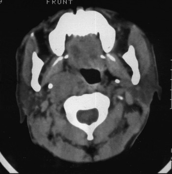
A computerised tomographic scan revealed a soft tissue mass occupying the right parapharyngeal space. The lesion measured approximately 4 × 3.5 cm (Figure 1).
Axial computed tomography scan showed a soft tissue density mass occupying the right parapharyngeal space.

An excisional biopsy of the mass was performed under general anaesthesia. The tumour appeared to be encapsulated and arose from the right cervical sympathetic trunk. The entire mass was resected.
The patient developed Horner's syndrome after the operation.
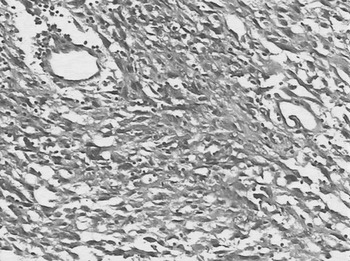
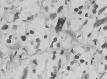
Microscopically, the tumour was composed of fascicles of compact spindle cells. Large cells with abundant, eosinophilic cytoplasm were identified as rhabdomyoblasts (Figure 2). Immunohistochemistry demonstrated that the spindle cells were positive for S-100 protein and vimentin, suggesting Schwann cell origin. The large cells showed strong positivity for desmin (Figure 3) and vimentin, supporting skeletal muscle differentiation. The diagnosis of malignant triton tumour was therefore confirmed. All surgical margins were negative.
Photomicrograph showing rhabdomyoblasts scattered between fascicles of spindle-shaped cells with abundant eosinophilic cytoplasm (H&E; ×100, original magnification).

Photomicrograph showing immunohistochemical staining. The rhabdomyoblasts are positive for desmin (×400, original magnification).

The suggested adjuvant therapies were refused by the patient.
In the third post-operative month, the patient found a 3 × 2.5 cm mass in his neck. An excisional biopsy of the lesion was carried out. Histopathological examination showed it to be a metastatic lymph node. However, the patient still refused further surgery and adjuvant therapies.
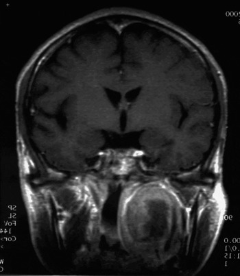
Three months later, the patient developed progressive hoarseness and dysphagia. Magnetic resonance imaging demonstrated a mass in the parapharyngeal space, extending to the skull base (Figure 4). One month later, the patient succumbed to his disease and died.
Coronal magnetic resonance imaging scan demonstrated a mass in the parapharyngeal space, extending to the skull base.

Discussion
Malignant triton tumour is a very rare clinicopathological entity, with about 90 cases reported in the literature. Because of their rarity and a lack of standardised diagnostic criteria, these tumours are difficult to diagnose.
The diagnostic criteria, suggested by Woodruff and Perino,Reference Woodruff and Perino8 are as follows. Firstly, the tumour arises from the course of a peripheral nerve and occurs with von Recklinghausen's disease. Secondly, it consists predominantly of spindle tumour cells resembling Schwann cells. Thirdly, it contains a rhabdomyoblast that arises within the body of the peripheral nerve tumour and is neither an extension nor a metastasis from an extrinsic rhabdomyosarcoma.
The current case met these criteria. Although the biphasic appearance of a pale, pleomorphic, spindle cell tumour with scattered rhabdomyoblasts is highly suggestive of malignant triton tumour, immunohistochemical staining helps to prove the origin of the cell. In the current case, a panel of antibodies to vimentin, desmin and S-100 protein was utilised; these were all positive. Nerve sheath differentiation was confirmed by S-100 protein, and rhabdomyoblastic differentiation was confirmed by positivity to desmin and vimentin.Reference Jong, van Vark, Albus-Lutter, van Raamsdonk and Voute10
Brooks et al. classified malignant triton tumours into two types.Reference Brooks, Freeman and Enterline9 Those with von Recklinghausen's neurofibromatosis constituted over 70 per cent in a review of 36 cases and showed a male predominance with a relatively young age (average 35 years), and usually presented in the head and neck.Reference Brooks, Freeman and Enterline9 Those without von Recklinghausen's neurofibromatosis had a female predominance with an older age, and frequently involved the trunk.Reference Brooks, Freeman and Enterline9 The current case and the previously reported case of malignant triton tumour of the parapharyngeal space both occurred in men with a mean age of 33 years and were associated with von Recklinghausen's neurofibromatosis.Reference Oysu, Aslan, Bilgic and Yazicioglu11 However, a recent review of cases arising in the head and neck showed no difference in the age of onset between patients with von Recklinghausen's neurofibromatosis and sporadic cases.Reference Victoria, McCulloch, Callagan and Bauman5
The prognosis of malignant triton tumour is poor, and it is defined as a highly malignant and deadly tumour.Reference Woodruff, Chernik, Smith, Millett and Foote2 Brooks et al. described the malignant triton tumour as more aggressive than the usual malignant schwannoma.Reference Brooks, Freeman and Enterline9 Local recurrence and metastasis are found in nearly 43 and 48 per cent, respectively.Reference Brooks, Freeman and Enterline9 The average two- and five-year survival rates for malignant triton tumour in all regions are 33 and 12 per cent, respectively.Reference Brooks, Freeman and Enterline9 Lesions that occur in the head and neck have a better outcome than those arising in the retroperitoneum, buttocks or trunk.Reference Victoria, McCulloch, Callagan and Bauman5 The overall survival rate for patients with head and neck malignant triton tumour is 26 per cent.Reference Victoria, McCulloch, Callagan and Bauman5 However, the previously reported case localised in the parapharyngeal space, unlike other malignant triton tumours of the head and neck, and showed a more aggressive course.Reference Victoria, McCulloch, Callagan and Bauman5, Reference Oysu, Aslan, Bilgic and Yazicioglu11 Despite complete resection, our patient died after three months with a devastating intracranial recurrence.Reference Oysu, Aslan, Bilgic and Yazicioglu11 The current case also showed very aggressive behaviour. These findings may be due to this tumour's location. The parapharyngeal space is a potential and relatively inaccessible space. By the time patients present for medical treatment, tumours in this location may have already reached a large size. Also, the parapharyngeal space contains several vital neurovascular structures. To avoid severe operative complications, the extent of excision is limited. Therefore, parapharyngeal malignant triton tumours may show more aggressive behaviour compared with those elsewhere in the head and neck.
• Malignant triton tumour is a rare, aggressive soft tissue sarcoma, a subtype of malignant peripheral nerve sheath tumour
• Malignant triton tumour of the parapharyngeal space is extremely rare, with only one case previously reported
• This case report describes an additional case of malignant triton tumour originating from the parapharyngeal space and arising from the cervical sympathetic nerve
• This paper discusses the difficulties in the diagnosis and management of malignant triton tumour of the parapharyngeal space and emphasises the importance of adjuvant therapy
Because of the rarity of malignant triton tumours, no treatment guidelines have thus far been established. However, as with any other sarcoma, resection of the tumour with a wide margin, followed by radiotherapy, is generally recommended. The role of chemotherapy has not been clearly defined. After reviewing 26 reported cases of malignant triton tumour occurring in the head and neck and analysing their own case, Victoria et al. Reference Victoria, McCulloch, Callagan and Bauman5 found that survival tended to be improved in patients undergoing complete resection with adjuvant therapy, compared with those undergoing complete resection alone; however, this difference was not statistically significant. Dewit et al. Reference Dewit, Albus-Lutter, de Jong and Voute12 found that cytotoxic drug therapy had no value in the treatment of malignant triton tumour. In both the current and the previously reported cases, the patients underwent complete resection alone, with a poor outcome. This indicates that complete resection may not be enough to treat malignant triton tumour of the parapharyngeal space; adjuvant therapy should be employed in these cases to reduce the risk of local recurrence.
Conclusion
We present a case of malignant triton tumour of the parapharyngeal space. This special location increases the difficulty of early detection and complete resection with a wide margin. The experience of treating patients with parapharyngeal malignant triton tumour, both in the current and previously reported cases, indicates that adjuvant therapy in addition to complete resection may be important in the management of malignant triton tumour of the parapharyngeal space, in order to obtain a better prognosis.