Introduction
Human beings choose to exist in social environments that necessitate countless social interactions on a daily basis. Although these interactions are routine for most people, the social world we live in is highly complex and navigating it successfully requires a set of sophisticated skills. The importance of these skills becomes most obvious when they go awry. This is evident in the phenomenology of various psychiatric disorders and reflected in their diagnostic criteria in the DSM-V (APA, 2013). Autism spectrum disorders (ASD), for example, are characterized by a lack of social or emotional reciprocity and an inability to regulate social interactions. Difficulties in interpersonal relations, social dysfunction and isolation accompany schizophrenia (SZ) and anti-social personality disorder/psychopathy (hereafter PP) is associated with aggressive or deceitful behaviour and a failure to conform to social norms. Obviously, this list is not exclusive and other disorders such as social anxiety disorder, borderline personality disorder and disorders without primary social diagnostic criteria also impact on the ability to engage in social interactions.
There is growing evidence that difficulties in social interactions and poor functional outcomes in patients suffering mental health disorders are underpinned by deficits in social cognitive information processing and that these deficits are also important factors in the instantiation and maintenance of other illness symptoms (Dodge, Reference Dodge1993; Burns, Reference Burns2006; Fett et al. Reference Fett, Viechtbauer, Dominguez, Penn, van Os and Krabbendam2011; Roepke et al. Reference Roepke, Preißler and Dziobek2013). The increasing acknowledgement of the functional and therapeutic significance of social cognition has led to an increased interest in its neurobiological mechanisms (Adolphs, Reference Adolphs1999, Reference Adolphs2003, Reference Adolphs2006; Anckarsäter, Reference Anckarsäter2006; Neuhaus et al. Reference Neuhaus, Beauchaine and Bernier2010; Herpertz, Reference Herpertz2013). Social neuroscience has evolved to take an interdisciplinary approach to unravel these mechanisms (Cacioppo & Berntson, Reference Cacioppo and Berntson1992). Its methods are broad and include techniques from social psychology, anthropology, neuroeconomics, cognitive neuroscience and classical neuropsychology; the application of neurobiological techniques [e.g. functional magnetic resonance imaging (fMRI), positron emission tomography, transcranial magnetic stimulation (TMS) and electroencephalography]; the assessment of bio-signals, such as eye movements; endocrinology, immunology, and animal models. While social neuroscience has employed many ways to investigate the biological substrate of social cognition and functioning, this review will focus on investigations of human social brain mechanisms with fMRI.
Since social neuroscience made its official entry into science in the early 1990s, an ever-increasing rate of studies has been published in the field (for reviews see e.g. Adolphs, Reference Adolphs2003, Reference Adolphs2010; Amodio & Frith, Reference Amodio and Frith2006; Cacioppo et al. Reference Cacioppo, Amaral, Blanchard, Cameron, Carter, Crews, Fiske, Heatherton, Johnson, Kozak, Levenson, Lord, Miller, Ochsner, Raichle, Shea, Taylor, Young and Quinn2007; Lieberman, Reference Lieberman2007; Norman et al. Reference Norman, Hawkley, Cole, Berntson and Cacioppo2011; Frith & Frith, Reference Frith and Frith2012) and has led to the instantiation of dedicated journals such as Social Neuroscience and SCAN. Initially, research focused on social brain mechanisms in healthy individuals, which were mostly investigated by means of so called ‘off-line’ measures that assessed social cognition from a third-person perspective. These studies are the basis of normative models of social brain function. In the last decade more and more research started to use social interactive paradigms, which are superior in the assessment of online mechanisms of social behaviour. In addition, researchers began to explore the underlying biological substrate of social cognitive deficits in psychiatric populations, as well as the biological substrate of social risk factors to identify how they drive psychiatric symptoms and social dysfunction with the ultimate aim to apply this knowledge in the development and evaluation of treatment interventions (Adolphs, Reference Adolphs2010; Mendez & Manes, Reference Mendez and Manes2011; Frith & Frith, Reference Frith and Frith2012; Meyer-Lindenberg & Tost, Reference Meyer-Lindenberg and Tost2012; Pemment, Reference Pemment2013).
Social cognition and its neural substrate
Social cognition refers to information processing within social-emotional contexts. It is essential for the understanding of social stimuli and interpersonal cues and guides actions appropriate for the social environment. This review focuses on social cognitive domains that are directed at understanding others’ social signals and social interactions. However, other lower level (social) cognitive mechanisms, which are not the focus of this review, also contribute to social functioning and have been elegantly described by others (Lieberman, Reference Lieberman2007; van der Meer et al. Reference van der Meer, Costafreda, Aleman and David2010; van Veluw & Chance, Reference van Veluw and Chance2014). Research in psychiatry investigating social cognitive processes aimed at understanding others has mostly focused on emotion recognition (ER) and aspects of perspective-taking (Grady & Keightley, Reference Grady and Keightley2002). ER, the ability to perceive and interpret emotions, is fundamental in deciphering others’ social signals (Gonzalez-Liencres et al. Reference Gonzalez-Liencres, Shamay-Tsoory and Brüne2013). Perspective-taking can be broadly distinguished into two closely related cognitive-affective processes. The first, more cognitive process is the ability to take another person's standpoint into account and to make attributions about their intentions, desires and beliefs. It is most commonly referred to as theory of mind (ToM) or cognitive empathy (Ang & Pridmore, Reference Ang and Pridmore2009) and is highly relevant to normal social interactions and functioning in the social community (Frith, Reference Frith2007, Reference Frith2008). The second, more affective process is the ability to share others’ feelings in absence of emotional stimulation to oneself. It is crucial for altruistic behaviour and referred to as empathy or affective ToM (e.g. see Singer, Reference Singer2006; Decety et al. Reference Decety, Michalska, Akitsuki and Lahey2009). This review focuses on emotional empathy, excluding motor empathy (i.e. the unconscious mirroring of the facial or gestural expressions of others). In the remainder of this article we will use ToM to refer to cognitive perspective-taking processes and empathy to refer to affective perspective-taking processes. The social cognitive processes directed at understanding others are closely related (Ochsner, Reference Ochsner2008; Frith & Frith, Reference Frith and Frith2012). This is reflected in the identification of a surprisingly homogeneous pattern of associated brain activation – which is now frequently referred to as the ‘social brain network’ (Brothers, Reference Brothers1990; Frith, Reference Frith2007; Blakemore, Reference Blakemore2008; Kennedy & Adolphs, Reference Kennedy and Adolphs2012) – through experiments across a variety of social cognitive tasks. This network comprises several prefrontal cortex (PFC) areas including dorsolateral (dlPFC), medial (mPFC), ventral medial, lateral (vm/vlPFC) and orbitofrontal (OFC) regions and temporal lobe regions [temporal poles, posterior superior temporal sulcus (pSTS)]. Closer examination of the involvement of specific regions suggests a preferential role for medial frontal brain areas in ToM. The temporal poles and OFC play a role in the processing of (social) reward-related information and the integration of social conceptual knowledge (van Overwalle, Reference van Overwalle2009; van Overwalle & Baetens, Reference van Overwalle and Baetens2009). The (p)STS has been associated with the perception of biological movement and features of human faces, which have been suggested to be important for ToM (Allison et al. Reference Allison, Puce and McCarthy2000; Pelphrey et al. Reference Pelphrey, Mitchell, McKeown, Goldstein, Allison and McCarthy2003). The ‘social brain network’ also includes other parts of the parietal cortex [e.g. temporo-parietal junction (TPJ), inferior parietal lobule (IPL), precuneus], which have been implicated in change of perspective from self to other and ToM, and several deeper brain structures (i.e. insula, amygdala, striatum), which have been associated with arousal, empathy, emotional processing, and social reward learning (Mitchell et al. Reference Mitchell, Mason, Macrae and Banaji2005; Ross & Olson, Reference Ross and Olson2010). In addition, several brain areas, including the inferior frontal gyrus (IFG), the premotor and parietal cortices (e.g. IPL and postcentral/somatosensory), are implicated in decoding actions and emotional states of others by subserving ‘mirror mechanisms’ in which the neural activation underlying others’ behaviour is thought to be mimicked in the brain of the observer (Rizzolatti & Craighero, Reference Rizzolatti and Craighero2004; Dinstein et al. Reference Dinstein, Hasson, Rubin and Heeger2007; Kilner et al. Reference Kilner, Friston and Frith2007; van der Gaag et al. Reference van der Gaag, Minderaa and Keysers2007; Keysers et al. Reference Keysers, Kaas and Gazzola2010; Molenberghs et al. Reference Molenberghs, Cunnington and Mattingley2012). There is accumulating evidence showing that the brain areas that are typically activated during social-cognitive processing often overlap with brain regions of the default mode network (DMN) that are active during the resting state. This resting state is conventionally considered an index of unconstrained non-task-related cognition. However, given the observed overlap with social cognition, it has been suggested that in the absence of any specific task-related focus, people's thoughts are likely to focus on themselves and others – and that this overlap in brain activation reflects the inherently social nature of humans and their evident predisposition for social cognition (Schilbach et al. Reference Schilbach, Eickhoff, Rotarska-Jagiela, Fink and Vogeley2008, Reference Schilbach, Bzdok, Timmermans, Fox, Laird, Vogeley and Eickhoff2012; Mars et al. Reference Mars, Neubert, Noonan, Sallet, Toni and Rushworth2012; Amft et al. Reference Amft, Bzdok, Laird, Fox, Schilbach and Eickhoff2014).
The current review
In this review we aim to outline the major findings of the body of work covering behavioural and neuroimaging research on social cognitive mechanisms directed at understanding others (specifically ER, ToM and empathy) and social interactions, and implications for treatment and future research. Clinically, the review is focused on a comparative approach of three disorders where most of the work has been done to date; SZ, ASD and PP. Studies focusing on subclinical expressions of these phenotypes were not included. Across the included studies PP is defined using the Psychopathy Checklist – Revised (PCL-R; Hare & Vertommen, Reference Hare and Vertommen2003). We identified the relevant literature by means of PubMed searches including the search terms: schizophrenia, psychopathy, anti-social*, or autism for the three different groups of psychopathology, theory of mind, mentalizing, emotion recognition or empathy for the social cognitive domains, and neuroimaging or fMRI; and through searches of reference lists of the retrieved articles. Studies that have been included in the recent meta-analyses that we review (Li et al. Reference Li, Chan, McAlonan and Gong2010; Sugranyes et al. Reference Sugranyes, Kyriakopoulos, Corrigall, Taylor and Frangou2011; Anticevic et al. Reference Anticevic, van Snellenberg, Cohen, Repovs, Dowd and Barch2012; Taylor et al. Reference Taylor, Kang, Brege, Tso, Hosanagar and Johnson2012) have not been reviewed separately.
We classified studies into the domain of ER if they required passive viewing of facial emotional expressions (implicit tasks) or the identification of emotions from facial expressions (explicit tasks, independent of situational circumstances). Research that required the inference of mental or emotional states of others based on descriptions or graphic illustrations of particular situations has been categorized as ToM. Studies that required seeing or reading about others in pain or other negative emotions or that asked participants to indicate how much they were feeling with others have been classified into the domain of empathy. To allow for comparison between the findings of behavioural studies, we transformed all reported effect sizes to Cohen's d and such that higher positive effect sizes indicate stronger impairments in the patient population. See Supplementary online Tables for the neuroimaging findings in the three social cognitive domains ER, ToM and empathy and for the findings pertaining to social interactive designs. Within the Tables, the findings are sorted according to the nature of the task or fMRI contrast and sample size, with the largest studies and meta-analyses being reported first. To allow for optimal comparability of the neuroimaging literature, we checked all labels of the reported coordinates in the Talairach client (version 2.4.3, http://talairach.org/client.html). We report the Talairach-based labels with the labels as reported by the original research in brackets. Tables and graphical displays of the neuroimaging findings show spatial coordinates in the Montreal Neurological Institute standardized brain template. We used conversion tools icbm_spm2_tal/other and tal2icbm_spm/other (http://brainmap.org and http://noodle.med.yale.edu/~papad/mni2tal/). The graphical displays of the neuroimaging findings have been generated in Matlab by means of the meta-analysis toolbox point plotting function (http://wagerlab.colorado.edu/files/tools/meta-analysis.html) and are shown in the online Supplementary material (Supplements 1–3). The majority of research in the field, and studies that we reviewed here, followed a traditional neurological lesion approach that aims to associate specific brain areas with specific social cognitive functions. However, the findings from neuroimaging research, as in many psychological domains, are more likely to reflect a complex interaction between different brain regions, emphasizing connectivity. Although the heuristic of a social brain is useful for the developing field, it is clear that none of the previously mentioned and reviewed brain areas are specific to social cognition and that social cognitive processes are contingent on widely distributed structure-function networks, rather than particular topographical areas (Menon, Reference Menon2011; Barrett & Satpute, Reference Barrett and Satpute2013).
Social cognitive brain mechanisms of psychiatric illness
Emotion recognition
Behavioural research
ER is fundamental for understanding others’ affective signals. It is typically assessed with tasks that require the identification or discrimination of facial emotions in pictures or the recognition of emotional prosody in speech. The tasks include the emotions regarded as universal, i.e. happiness, sadness, anger, fear, disgust and surprise and also neutral expressions, which in neuroimaging studies are mainly used as control condition. ER has extensively been researched in SZ. There is strong evidence for behavioural impairments from meta-analyses with large effect sizes of d = 0.85 (N = 1820) and 0.89 (N = 3822) (Chan et al. Reference Chan, Li, Cheung and Gong2010; Kohler et al. Reference Kohler, Walker, Martin, Healey and Moberg2010) and deficits that seem particularly pronounced for negative emotions (Hoekert et al. Reference Hoekert, Kahn, Pijnenborg and Aleman2007). ER deficits in SZ are present at all stages of the illness, including at-risk states, but deteriorate further during acute illness phases (Mandal et al. Reference Mandal, Pandey and Prasad1998; Edwards et al. Reference Edwards, Pattison, Jackson and Wales2001; Bediou et al. Reference Bediou, Asri, Brunelin, Krolak-Salmon, D'Amato, Saoud and Tazi2007; Alfimova et al. Reference Alfimova, Abramova, Barhatova, Yumatova, Lyachenko and Golimbet2009; Kohler et al. Reference Kohler, Walker, Martin, Healey and Moberg2010). The evidence for ER impairment is somewhat more equivocal in ASD. This may be partly due to a greater heterogeneity of samples, which included children and adults with classical and high functioning forms of the condition. A meta-analysis of 48 studies on ER in ASD (N = 932) indicated a large effect of d = 0.80 that was reduced to a moderate, although significant effect of d = 0.41 when publication bias was controlled for. Against an emotion-specific account of ER impairment in ASD, the study revealed no significant differences between emotions (Uljarevic & Hamilton, Reference Uljarevic and Hamilton2013). The findings are supported by a recently published meta-analysis that showed an overall ER impairment with an effect size of d = 0.77 (N = 1545; Lozier et al. Reference Lozier, Vanmeter and Marsh2014). Deficits in ER have also been associated with PP. It has been proposed that particularly problems with the recognition of fear and sadness contribute to interpersonal difficulties and a lack of empathic behaviour in the condition. In support of this hypothesis a meta-analysis on children and adults (N = 1244) with different anti-social conditions (including antisocial personality disorder, conduct disorder, externalizing behaviour disorders and psychopathy) showed the strongest impairments in the recognition of fear, followed by sadness. However, impairments were also present in the recognition of surprise, albeit to a lesser degree. Effect sizes ranged from d = 0.22 to 0.72. No consistent ER deficits were found for other emotions (d = 0.20–0.24; Marsh & Blair, Reference Marsh and Blair2008). A meta-analysis (N = 1387), which focused exclusively on psychopathy revealed only weak ER impairments for all emotions (d = 0.14–0.24). However, again the strongest deficits were present in the recognition of fear (d = 0.20) and sadness (d = 0.24; Wilson et al. Reference Wilson, Juodis and Porter2011). More recent work on ER in adults, children and adolescents with PP (N = 1376) revealed an effect of d = 0.17 for facial ER and d = 0.28 for voice ER deficits, once more with the strongest impairments in the recognition of fear and sadness (both d = 0.30). Interestingly, specific analyses by subgroup revealed small effects of d = 0.08 in adults (N = 376), with the only significant deficits in ER of happiness and surprise. Indicative of differential developmental effects, substantially larger impairments (d = 0.36) were found in children and adolescents (N = 343), who had significant impairments in the recognition of anger, fear and sadness; but not disgust, happiness and surprise (Dawel et al. Reference Dawel, O'Kearney, McKone and Palermo2012).
Neuroimaging research
The results of the neuroimaging studies on ER are displayed in Supplementary Table S1. Four meta-analyses have been conducted to summarize the work on the neural mechanisms of ER in SZ. Li et al.'s (Reference Li, Chan, McAlonan and Gong2010) meta-analysis of ER studies (N = 498) that compared SZ patients to healthy controls (HC) showed decreased brain activation in SZ in areas including the bilateral parahippocampal gyrus, amygdala, right superior frontal gyrus and middle occipital gyrus. There were no significant clusters showing higher activation in SZ (Li et al. Reference Li, Chan, McAlonan and Gong2010). A second meta-analysis (N = 414) by Sugranyes et al. (Reference Sugranyes, Kyriakopoulos, Corrigall, Taylor and Frangou2011) showed decreased activation in the right vlPFC and posterior cingulate (PCC), left amygdala, right occipito-temporal regions including the fusiform gyrus (FFG) and the left thalamus in SZ compared to HC (Sugranyes et al. Reference Sugranyes, Kyriakopoulos, Corrigall, Taylor and Frangou2011). In line with Li et al. (Reference Li, Chan, McAlonan and Gong2010) there were no activation clusters showing greater activation in SZ than in HC. A third meta-analysis (N = 872), which only included studies that used whole brain analyses, was conducted by Taylor et al. (Reference Taylor, Kang, Brege, Tso, Hosanagar and Johnson2012). The results also revealed large clusters of decreased activation in SZ compared to HC in the bilateral amygdala/hippocampal region. Reduced activation was also present in occipital regions associated with early visual processing, the anterior cingulate cortex (ACC), dlPFC, mPFC, and several subcortical areas, including the thalamus, caudate and midbrain (Taylor et al. Reference Taylor, Kang, Brege, Tso, Hosanagar and Johnson2012). Other studies which have been conducted since support the general pattern of these meta-analytical findings (Lepage et al. Reference Lepage, Sergerie, Benoit, Czechowska, Dickie and Armony2011; Derntl et al. Reference Derntl, Finkelmeyer, Voss, Eickhoff, Kellermann, Schneider and Habel2012; Mier et al. Reference Mier, Lis, Zygrodnik, Sauer, Ulferts, Gallhofer and Kirsch2014b ). A fourth meta-analysis by Anticevic et al. (Reference Anticevic, van Snellenberg, Cohen, Repovs, Dowd and Barch2012) which focused exclusively on ER in the amygdala, showed reduced amygdala activation in SZ compared to HC when negative emotion was contrasted with neutral expressions, as opposed to baseline. A normal amygdala response when viewing negative emotions and an elevated response to neutral information might reflect a mechanism of aberrant salience of neutral events (Anticevic et al. Reference Anticevic, van Snellenberg, Cohen, Repovs, Dowd and Barch2012). However, other research also reported reduced activation of the amygdala when emotion expressions were compared to the implicit baseline (Mier et al. Reference Mier, Lis, Zygrodnik, Sauer, Ulferts, Gallhofer and Kirsch2014b ). Taken together, the vast amount of imaging research on ER in SZ suggests that a robust and widespread pattern of reduced activation in brain areas that are regarded as part of the ‘social brain network’, might be underlying ER deficits in the disorder. However, some studies also revealed significantly higher activation in SZ than HC, which might indicate alternative or compensatory processing strategies (Taylor et al. Reference Taylor, Kang, Brege, Tso, Hosanagar and Johnson2012; Mier et al. Reference Mier, Lis, Zygrodnik, Sauer, Ulferts, Gallhofer and Kirsch2014b ; Mothersill et al. Reference Mothersill, Morris, Kelly, Rose, Bokde, Reilly, Gill, Corvin and Donohoe2014). A few contradicting findings that warrant further clarification have been made for the superior temporal gyrus (STG), IPL, ACC and medial frontal gyrus (meFG), with some studies showing more activation in HC than SZ and others reporting the opposite activation pattern.
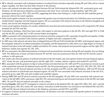
Main neuroimaging findings on ER, ToM and empathy

Sugranyes et al. (Reference Sugranyes, Kyriakopoulos, Corrigall, Taylor and Frangou2011) also meta-analysed ER in ASD (including autism, Asperger's syndrome and pervasive developmental disorder NOS, N = 110). ASD showed more activation than HC in the left STG and less activation in the left postcentral gyrus. Several other studies on ER have been conducted in ASD, but were not included in Sugranyes et al.'s meta-analysis for methodological reasons (see also Harms et al. Reference Harms, Martin and Wallace2010). Here we report studies that employed an ER task in ASD and HC, and either compared ER performance to baseline, a control condition (e.g. scrambled faces) or neutral facial expressions. The majority of studies showed reduced brain activation in ASD compared to HC in the frontal cortex [i.e. middle frontal gyrus (miFG), meFG, superior frontal gyrus (SFG) and IFG], the temporal, parietal and occipital cortex (i.e. middle TG, insula, TPJ, FFG), and subcortical areas, including the putamen and amygdala (Ogai et al. Reference Ogai, Matsumoto, Suzuki, Ozawa, Fukuda, Uchiyama, Suckling, Isoda, Mori and Takei2003; Piggot et al. Reference Piggot, Kwon, Mobbs, Blasey, Lotspeich, Menon, Bookheimer and Reiss2004; Pelphrey et al. Reference Pelphrey, Morris, McCarthy and Labar2007; Kleinhans et al. Reference Kleinhans, Richards, Johnson, Weaver, Greenson, Dawson and Aylward2011; Wicker et al. Reference Wicker, Fonlupt, Hubert, Tardif, Gepner and Deruelle2008; Corbett et al. Reference Corbett, Carmean, Ravizza, Wendelken, Henry, Carter and Rivera2009; Doyle-Thomas et al. Reference Doyle-Thomas, Goldberg, Szatmari and Hall2013). However, in line with differential or compensatory processing strategies in ASD, several studies showed patterns of increased brain activation in inferior, middle and medial frontal, temporal-, parahippocampal- and superior parietal areas and the striatum, amygdala and insula (Corbett et al. Reference Corbett, Carmean, Ravizza, Wendelken, Henry, Carter and Rivera2009; Weng et al. Reference Weng, Carrasco, Swartz, Wiggins, Kurapati, Liberzon, Risi, Lord and Monk2011; Doyle-Thomas et al. Reference Doyle-Thomas, Goldberg, Szatmari and Hall2013; Tottenham et al. Reference Tottenham, Hertzig, Gillespie-Lynch, Gilhooly, Millner and Casey2013).
Sugranyes et al. (Reference Sugranyes, Kyriakopoulos, Corrigall, Taylor and Frangou2011) also directly compared ER in SZ and ASD. In support of differential mechanisms of ER impairment in the two disorders, their findings revealed increased activation in SZ compared to ASD in the IFG, the parahippocampal gyrus, the TPJ, occipital gyrus and cerebellum. By contrast, individuals with ASD showed higher activation in superior temporal regions, the ACC and PCC. Group differences in brain activation were not accounted for by the use of antipsychotics by SZ patients.
To date, imaging research on ER in PP is still scarce and the conducted research yielded mixed findings. Lower activation in PP in the bilateral FFG, the cerebellum and the left pre- and postcentral gyrus was found by an early study during processing of happy and fearful faces (Deeley et al. Reference Deeley, Daly, Surguladze, Tunstall, Mezey, Beer, Ambikapathy, Robertson, Giampietro, Brammer, Clarke, Dowsett, Fahy, Philipps and Murphy2006). However, more recent research did not find reduced, but increased brain activation in PP compared to HC in lateral and medial frontal areas, the FFG and visual cortex (Contreras-Rodríguez et al. Reference Contreras-Rodríguez, Pujol, Batalla, Harrison, Bosque, Ibern-Regàs, Hernández-Ribas, Soriano-Mas, Deus, López-Solà, Pifarré, Menchón and Cardoner2013). Results by Decety et al. (Reference Decety, Skelly, Yoder and Kiehl2013c ) indicated both; extended patterns of decreased activation in the FFG, medial and middle occipital gyrus, IFG, meFG, OFC, dmPFC and ventral PFC, supplementary motor area (SMA), inferior and temporal poles in PP during ER of happy, fearful, painful and sad expressions and increased activation in the amygdala, insula, middle cingulate and the superior temporal poles (Decety et al. Reference Decety, Skelly, Yoder and Kiehl2013c ). Finally, a recent study by Mier et al. (Reference Mier, Haddad, Diers, Dressing, Meyer-Lindenberg and Kirsch2014a ) reported no group differences in neural activation between PP and HC during ER.
In summary, the findings show that SZ is associated with the most profound deficits in ER on the behavioural and neural level, with widespread patterns of decreased brain activation across the social brain network. Behaviourally, ASD has been associated with less severe deficits and imaging research indicates fewer differences in neural processing in comparison to HC. However, the findings in ASD are largely based on small and potentially underpowered studies with more heterogeneous samples and should therefore be regarded with some caution. Behaviourally, PP is associated with mild ER deficits. Here, the differences ER might reflect a processing bias specific to negative emotions, rather than an inability to recognize emotions. The evidence regarding the neural processes of ER in PP is still limited; however, in general the neural activation patterns appear to be less disturbed than in SZ and ASD. Finally, it needs to be noted that the overall findings on the neural mechanisms of ER vary considerably between and, albeit to lesser degree, within disorders. Factors that account for this heterogeneity are sample composition, e.g. including various diagnoses, ability levels and differential age ranges of participants; and methodological differences, such as nature of the ER tasks (e.g. implicit or explicit, different emotions), the utilized analysis strategies (ROI v. whole brain) and the differential contrasts, which compared emotion expressions to either neutral expressions, objects, shapes or the implicit baseline. In addition, the majority of studies included largely male samples, which limits the generalizability of the current findings.
Theory of mind
Behavioural research
The tasks that are used to measure ToM typically require the interpretation of mental states of characters in cartoons or vignettes, ranging from relatively simple false-belief identification or picture sequencing to more intricate measures that require the interpretation of indirect speech (hints), sarcasm, irony or humour. ToM disturbances have been reported in numerous psychiatric illnesses (Ang & Pridmore, Reference Ang and Pridmore2009), but the deficits are probably most profound and most consistently found in SZ and ASD (Baron-Cohen, Reference Baron-Cohen2000; Brune, Reference Brune2005; Sprong et al. Reference Sprong, Schothorst, Vos, Hox and van Engeland2007). Two meta-analyses on ToM in SZ reported large effect sizes of d = 1.21 and d = 1.26 (N = 1181 and 1518, respectively; Sprong et al. Reference Sprong, Schothorst, Vos, Hox and van Engeland2007; Bora et al. Reference Bora, Yucel and Pantelis2009). Meta-analyses on ToM in ASD yielded evidence for somewhat smaller effect sizes of d = 0.88 in children and adolescents (Yirmiya et al. Reference Yirmiya, Erel, Shaked and Solomonica-Levi1998). However, similarly large impairments were found in adults with SZ and ASD (N = 1922, verbal ToM tasks: SZ d = 1.09, ASD d = 1.17; visual ToM tasks: SZ d = 1.07, ASD d = 0.89; Chung et al. Reference Chung, Barch and Strube2014). In both disorders deficits are evident in tasks that assess simple and higher order ToM.
ToM deficits have also been suggested to underlie social deficits in PP. However, the current evidence only supports very mild impairments, if any at all (Richell et al. Reference Richell, Mitchell, Newman, Leonard, Baron-Cohen and Blair2003; Dolan & Fullam, Reference Dolan and Fullam2004; Murphy, Reference Murphy2006; Sharp & Vanwoerden, Reference Sharp and Vanwoerden2014). Dolan et al. (2004) found no differences between HC and PP in simple ToM tasks. But, the PP group performed worse than HC on more complex ToM tasks. Research by Richell et al. (Reference Richell, Mitchell, Newman, Leonard, Baron-Cohen and Blair2003) did not show ToM performance differences between incarcerated men with and without PP (Richell et al. Reference Richell, Mitchell, Newman, Leonard, Baron-Cohen and Blair2003) and recently, Sharp et al. (2014) even presented evidence showing that certain traits of PP are associated with excessive ToM, whereas others are associated with reduced or absent ToM. Research that compared simple and higher-order ToM in ASD, SZ and personality disorders (mainly PP) showed that ASD and SZ performed similarly and significantly worse compared to individuals with personality disorders (Murphy, Reference Murphy2006).
Neuroimaging research
The results of the neuroimaging studies on ToM are displayed in Supplementary Table S2. At the neural level, Sugranyes et al.'s meta-analysis (N = 273), which included a variety of ToM-related tasks that also involved empathy, moral reasoning and certain aspects of ER, found lower activation in the mPFC, temporal cortices and the right PCC in SZ compared to HC. Higher activation in SZ was present in the left PCC and the paracentral lobule. As for ER, other studies also mostly support a pattern of lower activation in SZ than HC including the meFG, STG, temporal poles, IPL and somatosensory areas (Lee et al. Reference Lee, Quintana, Nori and Green2011a ; Das et al. Reference Das, Lagopoulos, Coulston, Henderson and Malhi2012; Pedersen et al. Reference Pedersen, Koelkebeck, Brandt, Wee, Kueppers, Kugel, Kohl, Bauer and Ohrmann2012; Rapp et al. Reference Rapp, Langohr, Mutschler, Klingberg, Wild and Erb2013). In line with the idea of differential or compensatory processing strategies in SZ, research by Varga et al. (Reference Varga, Simon, Tényi, Schnell, Hajnal, Orsi, Dóczi, Komoly, Janszky and Füredi2013) and Pedersen et al. (Reference Pedersen, Koelkebeck, Brandt, Wee, Kueppers, Kugel, Kohl, Bauer and Ohrmann2012) also reported higher activation compared to HC in the miFG and IFG, IPL, STG, precuneus and cerebellum.
Even though ToM has long been regarded as key factor underlying social disturbances in ASD, relatively little research has investigated the neural mechanisms of ToM in the condition. Sugranyes et al.'s meta-analytical work (N = 186) associated ASD with reduced activation in medial, temporal, parietal and limbic brain areas. In contrast to SZ, no areas were significantly more activated in ASD than HC. Kana et al. (Reference Kana, Libero, Hu, Deshpande and Colburn2014) and Lombardo et al. (Reference Lombardo, Chakrabarti, Bullmore and Baron-Cohen2011) also showed patterns of decreased activation in the IPL, IFG, cuneus, TPJ, pSTS, insula and in motor and somatosensory areas. One study, however, that investigated irony comprehension indicated the opposite pattern with increased activation in ASD compared to HC in the precentral gyrus and IFG (Wang et al. Reference Wang, Lee, Sigman and Dapretto2006). A direct comparison between SZ and ASD showed more insula activation in ASD than SZ and greater activation likelihood in the mPFC and somatosensory areas in SZ than ASD. The difference in mPFC activation was partly explained by age differences between the SZ and ASD sample (Sugranyes et al. Reference Sugranyes, Kyriakopoulos, Corrigall, Taylor and Frangou2011). The authors speculated that higher somatosensory activation might reflect possible compensatory mechanisms or a process of ‘overmentalization’ in SZ compared to ASD. The idea that paranoid SZ is associated with the excessive interpretation of others’ behaviour and intentions as malevolent and self-referent (Abu-Akel & Bailey, Reference Abu-Akel and Bailey2000; Frith, Reference Frith2004) has been supported by research showing that specifically positive symptoms were related to ‘overmentalizing’ ToM errors, whereas negative symptoms were more strongly related to a lack of ToM (Montag et al. Reference Montag, Dziobek, Richter, Neuhaus, Lehmann, Sylla, Heekeren, Heinz and Gallinat2011). However, Sugranyes et al. (Reference Sugranyes, Kyriakopoulos, Corrigall, Taylor and Frangou2011) did not specifically investigate the associations between brain activation and symptoms or behavioural performance.
Sommer et al. (Reference Sommer, Sodian, Döhnel, Schwerdtner, Meinhardt and Hajak2010) investigated the neural mechanisms of ToM in PP and found more activation in the OFC compared to HC during mental state attribution in ToM cartoons. Given that the OFC has been associated with more cognitive, as opposed to affective reasoning strategies, the authors suggest that this could reflect more rational, outcome oriented ToM reasoning strategies in PP (Sommer et al. Reference Sommer, Sodian, Döhnel, Schwerdtner, Meinhardt and Hajak2010). By contrast, a recent study by Mier et al. (Reference Mier, Haddad, Diers, Dressing, Meyer-Lindenberg and Kirsch2014a ) found less brain activation in PP compared to HC in the FFG and Declive, but no significant clusters for the reverse contrast during a ToM task that involved intention attribution to facial emotion pictures. Neuroimaging research comparing ToM performance of HC children and adolescents with ASD and PP (research diagnosis conduct problems/high callous-unemotional traits) found no behavioural differences between the groups and no differences in neural activation patterns between PP and HC. ASD, however, was associated with reduced activation in three clusters in the mPFC and vmPFC, compared to HC and PP (O'Nions et al. Reference O'Nions, Sebastian, McCrory, Chantiluke, Happé and Viding2014). Behaviourally, these findings point towards similar, intact ToM mechanisms in children and adolescents with PP traits and adults with a clinical diagnosis of PP. However, the neuroimaging findings suggest differential neural processing in adults, which could be due to a variety of factors, for example clinical characteristics, age or the employed ToM paradigms.
In sum, the current evidence from neuroimaging research indicates partly overlapping neural mechanisms of ToM impairment in SZ and ASD, particularly with reduced activation in the left meFG, middle temporal gyrus (MTG), precuneus, right IFG, IPL and precentral gyrus and increased activation in the left IFG and right meFG. This could imply similar pathological mechanisms, such as changes in neural connectivity, grey-matter volumes and neurotransmitter abnormalities, which have been associated with both disorders (Cheung et al. Reference Cheung, Yu, Fung, Leung, Wong, Li, Sham, Chua and McAlonan2010; Neuhaus et al. Reference Neuhaus, Beauchaine and Bernier2010; Kana et al. Reference Kana, Libero and Moore2011; Coghlan et al. Reference Coghlan, Horder, Inkster, Mendez, Murphy and Nutt2012; Coyle et al. Reference Coyle, Basu, Benneyworth, Balu and Konopaske2012; Fitzsimmons et al. Reference Fitzsimmons, Kubicki and Shenton2013; Greimel et al. Reference Greimel, Nehrkorn, Schulte-Rüther, Fink, Nickl-Jockschat, Herpertz-Dahlmann, Konrad and Eickhoff2013). However, in support of disease specific mechanisms, the current evidence also shows differential patterns of increased and reduced brain activation. Differential neural irregularities in ASD could be explained by the fact that social (brain) mechanisms are disrupted early in life. Individuals with ASD might therefore acquire alternative strategies to process social information. In SZ a relatively normal social development until illness onset, which typically occurs in late adolescence or early adulthood, is likely to be associated with differential mechanisms of impairment. The current evidence with regard to PP is scare, yet points towards relatively unimpaired ToM, with minor behavioural and neural deviations. With respect to the current research it is important to note that the heterogeneity between studies is even higher than for research on ER, due to the nature of ToM tasks, which are more complex and which partly included a conglomerate of related processes, such as ER and empathy.
Empathy
Behavioural research
Empathy is assessed by having participants observe or read about physical or social pain in other people. Other studies use facial expressions of negative emotions and instruct participants to judge the emotional state of self and others. Performance indices reflect the self-reported affective response to distress cues or compare the emotion indicated by the participant with the correct emotion; some studies have taken into account the emotional state as rated by the target (other), which allows the measurement of empathic accuracy (i.e. the congruence between emotions rated by the participant and the target; Zaki & Ochsner, Reference Zaki and Ochsner2011). In SZ, the evidence for abnormal empathy is mostly based on studies using self-report questionnaires on trait empathy. The most consistent finding is that patients report higher personal distress when others are in difficult situations (Montag et al. Reference Montag, Heinz, Kunz and Gallinat2007; Derntl et al. Reference Derntl, Finkelmeyer, Toygar, Hülsmann, Schneider, Falkenberg and Habel2009; Achim et al. Reference Achim, Ouellet, Roy and Jackson2011; Lee et al. Reference Lee, Zaki, Harvey, Ochsner and Green2011b ); some studies also report lower self-reported empathic concern (Lee et al. Reference Lee, Zaki, Harvey, Ochsner and Green2011b ; Smith et al. Reference Smith, Horan, Karpouzian, Abram, Cobia and Csernansky2012). Some studies have used performance-based measures of empathy, suggesting that patients show reduced empathic accuracy (Lee et al. Reference Lee, Zaki, Harvey, Ochsner and Green2011b ) and are less able to indicate the correct affective response to emotional scenarios (Derntl et al. Reference Derntl, Finkelmeyer, Toygar, Hülsmann, Schneider, Falkenberg and Habel2009; Smith et al. Reference Smith, Horan, Cobia, Karpouzian, Fox, Reilly and Breiter2013).
Findings on empathy in ASD are mixed. As in SZ, studies have relied on self-report questionnaires, some showing normal (Rogers et al. Reference Rogers, Dziobek, Hassenstab, Wolf and Convit2007; Dziobek et al. Reference Dziobek, Rogers, Fleck, Bahnemann, Heekeren, Wolf and Convit2008) others reduced (Mathersul et al. Reference Mathersul, McDonald and Rushby2013; Grove et al. Reference Grove, Baillie, Allison, Baron-Cohen and Hoekstra2014) self-reported empathic concern in adults with high-functioning ASD. Similar to SZ, self-reported personal distress may be higher than in HC (Rogers et al. Reference Rogers, Dziobek, Hassenstab, Wolf and Convit2007; Dziobek et al. Reference Dziobek, Rogers, Fleck, Bahnemann, Heekeren, Wolf and Convit2008). Performance-based studies showed normal behavioural (Jones et al. Reference Jones, Happé, Gilbert, Burnett and Viding2010; Schwenck et al. Reference Schwenck, Mergenthaler, Keller, Zech, Salehi, Taurines, Romanos, Schecklmann, Schneider and Warnke2012; Scheeren et al. Reference Scheeren, Koot, Mundy, Mous and Begeer2013; Deschamps et al. Reference Deschamps, Been and Matthys2014) and psychophysiological (Shalom et al. Reference Shalom, Mostofsky, Hazlett, Goldberg, Landa, Faran, McLeod and Hoehn-Saric2006) responses to others’ distress in school-age children with ASD, as well as in adults (Dziobek et al. Reference Dziobek, Rogers, Fleck, Bahnemann, Heekeren, Wolf and Convit2008). There is some evidence, however, for a reduced affective response to parents’ distress in toddlers at risk for ASD (McDonald & Messinger, Reference McDonald and Messinger2012). Little is known about empathic accuracy in ASD; one older study suggested somewhat lower congruence between the affective response of the participant and the emotional state of the protagonist in children with ASD compared to HC (Yirmiya et al. Reference Yirmiya, Sigman, Kasari and Mundy1992).
Deficits in empathy have been suggested to be at the core of PP, and may underlie the instrumental aggression characteristic of this disorder (Blair, Reference Blair2008; Derntl et al. Reference Derntl, Finkelmeyer, Voss, Eickhoff, Kellermann, Schneider and Habel2012; Meffert et al. Reference Meffert, Gazzola, den Boer, Bartels and Keysers2013). Probing the deficit behaviourally, however, has proven difficult, possibly because individuals with PP may use their relatively intact ToM skills to compensate for the lack of affective sharing of emotions. Accordingly, studies using self-report questionnaires showed normal levels of self-reported empathy in individuals with PP (Shamay-Tsoory et al. Reference Shamay-Tsoory, Harari, Aharon-Peretz and Levkovitz2010; Domes et al. Reference Domes, Hollerbach, Vohs, Mokros and Habermeyer2013a ). Male offenders with high levels of PP did not show a reduced affective response to others’ distress when compared to male offenders with low PP; however as a group the offenders scored lower than HC (Domes et al. Reference Domes, Hollerbach, Vohs, Mokros and Habermeyer2013a ). Another study indicated that male violent offenders with high PP traits had similar self-reported affective responses to others’ distress as HC despite lower psychophysiological responses, possibly suggesting that they were able to respond to the distress cues on the basis of cognitive rather than affective processes (Pfabigan et al. Reference Pfabigan, Seidel, Wucherer, Keckeis, Derntl and Lamm2014). Empathic accuracy, however, was reduced as a function of PP in a group of male offenders (Brook & Kosson, Reference Brook and Kosson2013). In children, PP tendencies were associated with lower affective response to distress (Jones et al. Reference Jones, Happé, Gilbert, Burnett and Viding2010); this study showed a double dissociation with empathy being preserved in boys with ASD but impaired in boys with PP tendencies, and ToM impaired in boys with ASD but preserved in boys with PP tendencies. A similar pattern of results was reported by Schwenck et al. (Reference Schwenck, Mergenthaler, Keller, Zech, Salehi, Taurines, Romanos, Schecklmann, Schneider and Warnke2012). Another study reported reduced psychophysiological response to an emotion evocative film, in conjunction with lower self-reported affective response in children with callous-unemotional traits (Anastassiou-Hadjicharalambous & Warden, Reference Anastassiou-Hadjicharalambous and Warden2008). Reduced psychophysiological responses to distress cues have been reported previously both in children (Blair, Reference Blair1999) and in adults (Blair et al. Reference Blair, Jones, Clark and Smith1997) with PP traits. Taken together, the results suggest that PP is associated with a deficit in empathy in the context of relatively intact ToM. In contrast, there is little consistent evidence for a deficit in empathy in ASD. In SZ, evidence for reduced empathy is mostly based on self-report questionnaires, but some studies have shown that empathic accuracy is impaired.
Neuroimaging research
Imaging studies have investigated patterns of brain activation during observation of pain or emotion scenarios compared to neutral scenarios; some studies have further examined the effect of explicit instructions to feel with the other or imagine oneself in the situation.
The results of the neuroimaging studies on empathy are displayed in Supplementary Table S3. Only two studies have investigated the neural correlates of empathy in SZ, suggesting a reduced neural response across cortical and subcortical areas (Derntl et al. Reference Derntl, Finkelmeyer, Voss, Eickhoff, Kellermann, Schneider and Habel2012; Harvey et al. Reference Harvey, Zaki, Lee, Ochsner and Green2013). Derntl et al. (Reference Derntl, Finkelmeyer, Voss, Eickhoff, Kellermann, Schneider and Habel2012) used sentences describing real-life situations; participants were asked to imagine how they would feel if they were experiencing this situation. The study by Harvey et al. (Reference Harvey, Zaki, Lee, Ochsner and Green2013) focused on empathic accuracy, and had participants rate the affective state of another person (target) shown in a series of videos and compared these with the self-rated affect of the target. Given the widespread abnormalities in ER and ToM in this disorder, it can be hypothesized that any abnormal brain response to empathy has to be considered in the context of these other deficits.
Abnormal brain responses to empathy are also seen in individuals with ASD, but the findings are contradictory. Some studies found little evidence for abnormal neural responses to pain scenarios (Bird et al. Reference Bird, Silani, Brindley, White, Frith and Singer2010; Schneider et al. Reference Schneider, Regenbogen, Pauly, Gossen, Schneider, Mevissen, Michel, Gur, Habel and Schneider2013; Hadjikhani et al. Reference Hadjikhani, Zurcher, Rogier, Hippolyte, Lemonnier, Ruest, Ward, Lassalle, Gillberg, Billstedt, Helles, Gillberg, Solomon and Prkachin2014), whereas others did observe reduced activation in areas associated with face processing and empathy (Greimel et al. Reference Greimel, Schulte-Ruther, Fink, Piefke, Herpertz-Dahlmann and Konrad2010; Schulte-Ruther et al. Reference Schulte-Ruther, Greimel, Markowitsch, Kamp-Becker, Remschmidt, Fink and Piefke2011). Fan et al. (Reference Fan, Chen, Chen, Decety and Cheng2014) similarly observed reduced activation in anterior insula and ACC in response to pain scenarios, but an increased response in the somatosensory cortex, possibly indicating heightened empathic arousal in individuals with ASD, which may trigger an increased reliance on cognitive reappraisal (Hadjikhani et al. Reference Hadjikhani, Zurcher, Rogier, Hippolyte, Lemonnier, Ruest, Ward, Lassalle, Gillberg, Billstedt, Helles, Gillberg, Solomon and Prkachin2014). There is also some evidence that atypical processing of one's own emotions (‘alexithymia’) contributes to the empathic deficits in ASD (Bird et al. Reference Bird, Silani, Brindley, White, Frith and Singer2010). Finally, comparing inflicted v. accidental pain showed reduced activation in areas associated with ToM, suggesting that impaired social understanding may also play a role in the empathy deficit in ASD (Fan et al. Reference Fan, Chen, Chen, Decety and Cheng2014). Taken together, while the overall picture remains ambiguous, the existing research suggests that the empathic deficit in ASD should be considered in the context of other deficits, such as self-related emotion processing and ToM.
Four studies have investigated the neural basis of the lack of empathy in individuals with PP using fMRI (Decety et al. Reference Decety, Chen, Harenski and Kiehl2013a , Reference Decety, Skelly and Kiehl b ; Marsh et al. Reference Marsh, Finger, Fowler, Adalio, Jurkowitz, Schechter, Pine, Decety and Blair2013; Meffert et al. Reference Meffert, Gazzola, den Boer, Bartels and Keysers2013). Three studies suggest that in individuals with PP the normal response in the empathy for pain network (including anterior insula, dorsal ACC and amygdala) is reduced when imagining the other in a painful situation (Decety et al. Reference Decety, Chen, Harenski and Kiehl2013a ) or when simply watching interacting hands (Meffert et al. Reference Meffert, Gazzola, den Boer, Bartels and Keysers2013). However, deliberate vicarious responding was intact, as became clear when subjects were explicitly told to imagine oneself in the situation of the other (Decety et al. Reference Decety, Chen, Harenski and Kiehl2013a ; Marsh et al. Reference Marsh, Finger, Fowler, Adalio, Jurkowitz, Schechter, Pine, Decety and Blair2013), or to feel with the other person (Decety et al. Reference Decety, Chen, Harenski and Kiehl2013a ; Meffert et al. Reference Meffert, Gazzola, den Boer, Bartels and Keysers2013). Accordingly, it has been argued that the distinction between ability (what one does under optimal situations) and propensity (what one actually does as a function of the situation) is important in the understanding of the neural mechanisms of reduced empathy in psychopathy (Keysers et al. Reference Keysers, Meffert and Gazzola2014). The fourth study (Decety et al. Reference Decety, Skelly and Kiehl2013b ) surprisingly reported higher activation the anterior insula and the dorsal ACC in PP v. HC during the observation of interactions involving pain, while at the same time activation in other pain- and emotion-related areas, such as periaqueductal grey and vmPFC was reduced. This suggests that the role of anterior insula and ACC in the reduced empathy response in PP is not yet well understood. The same study also observed higher activation in cognitive ToM areas such as pSTS and mPFC in PP, possibly indicating a more cognitive approach to the processing of the pain scenarios. As mentioned before in the section on ToM, stronger activation in brain areas that are thought to reflect an increased reliance on cognitive processes was also observed when individuals with PP had to infer emotional states of cartoon characters (Sommer et al. Reference Sommer, Sodian, Döhnel, Schwerdtner, Meinhardt and Hajak2010). Compared to forensic controls, individuals with PP showed more ToM-related brain activation and less vicarious responding.
In summary, the existing research on empathy reveals shared and distinct neural processes in different disorders. PP is associated with distinct deficits in empathic processing which co-occur with relatively intact cognitive ToM processes. The empathic deficit in PP seems to be amendable, given that explicit instructions to imagine the self in the painful situation or to feel with the other tend to normalize the brain response. In ASD, the evidence for an impaired empathic brain response is contradictory, but the findings suggest that any empathic deficit occurs in the context of deficits in self-related emotion processing and ToM. The evidence in SZ is scarce, but similarly to ASD it can be surmised that any deficit in empathy will have to be considered against other social cognitive deficits. Future studies could usefully vary task conditions to systematically investigate if explicit instructions or situational factors (i.e. intentional v. accidental pain; familiar v. non-familiar faces) influence brain response to empathy. Further, as for ER and ToM, research so far has almost exclusively focused on males; this limits the generalizability of the findings, particularly since there is some evidence that gender may influence the neural correlates of empathy in psychopathology (Schneider et al. Reference Schneider, Regenbogen, Pauly, Gossen, Schneider, Mevissen, Michel, Gur, Habel and Schneider2013). The main findings of the neuroimaging studies on ER, ToM and empathy have been summarized in Table 1.
The dynamics of disturbed social interaction: neuroeconomics in psychiatry
The previously discussed studies investigated the neural mechanisms of social cognition by means of paradigms that require participants to interpret social stimuli in stories, cartoons or pictures. These studies yielded important insights into social cognitive brain mechanisms, but could not capture the most intrinsic and interactive aspect of social behaviour (De Jaegher et al. Reference De Jaegher, Di Paolo and Gallagher2010). In the last decade, interactive game-theoretical exchange paradigms from experimental economics made an exciting addition to social neuroscience. These paradigms allow for the ‘online’ investigation of social processes (e.g. trust, reciprocity or fairness) and offer a new way to study social dysfunction in psychopathology (Sripada et al. Reference Sripada, Angstadt, Banks, Nathan, Liberzon and Phan2009; Sharp et al. Reference Sharp, Ha and Fonagy2011; Fett et al. Reference Fett, Shergill, Joyce, Riedl, Strobel, Gromann and Krabbendam2012; Gromann et al. Reference Gromann, Heslenfeld, Fett, Joyce, Shergill and Krabbendam2013, Reference Gromann, Shergill, de Haan, Meewis, Fett, Korver-Nieberg and Krabbendam2014; Joyce et al. Reference Joyce, Averbeck, Frith and Shergill2013). It has been suggested that these paradigms could signify quantitative biomarkers for psychiatric assessment and genotyping (Hasler, Reference Hasler2012; King-Casas & Chiu, Reference King-Casas and Chiu2012; Montague et al. Reference Montague, Dolan, Friston and Dayan2012). Given these attractive prospects, the rapid increase of research using game theoretical paradigms in psychiatry is not surprising. Most frequently researchers made use of the trust game (Berg et al. Reference Berg, Dickhaut and McCabe1995; Tzieropoulos, Reference Tzieropoulos2013). In this paradigm the first player (investor) receives an endowment and then has to decide how much of it he wants to share with the second player. The transferred amount is multiplied during the transaction. Subsequently, the second player (trustee) decides how much of the total amount he wants to send back. Cooperation yields the best pay-off for both. However, initially the best pay-off for the trustee is reached through defection. Hence, sharing money requires the investor's trust in his or her benevolence. Research in HC implies a human tendency to trust and to reciprocate trust, and shows that trusting behaviour is influenced by the rewarding effects of social cooperation and knowledge about the interaction partner (Bowles & Gintis, Reference Bowles and Gintis2002; Fehr & Gachter, Reference Fehr and Gachter2002; Fehr & Fischbacher, Reference Fehr and Fischbacher2003; Delgado et al. Reference Delgado, Frank and Phelps2005; Fehr, Reference Fehr2009). The investment phase, during which the partner's behavioural cues are interpreted to anticipate future behaviour, has been associated with activation in ToM related brain areas (Rilling et al. Reference Rilling, Sanfey, Aronson, Nystrom and Cohen2004; King-Casas, Reference King-Casas2005; King-Casas et al. Reference King-Casas, Sharp, Lomax-Bream, Lohrenz, Fonagy and Montague2008; van den Bos et al. Reference van den Bos, van Dijk, Westenberg, Rombout and Crone2011). The outcome phase, during which cooperation or non-cooperation of the game partner is revealed has been associated with activation in dopamine-governed brain areas of reward processing (King-Casas, Reference King-Casas2005; Phan et al. Reference Phan, Sripada, Angstadt and McCabe2010). These findings make the trust game highly interesting for the investigation of social interaction in SZ. SZ is accompanied by deficits in ToM and dopamine function (Kapur et al. Reference Kapur, Mizrahi and Li2005), which might explain reductions in trust and the motivation to engage in (pro-)social behaviour. Recent behavioural research showed lower levels of trust (i.e. lower investments) in patients and their healthy first-degree relatives with a heightened illness-risk compared to controls. Besides, reduced trust was associated with psychotic symptoms. Crucially, patients’ levels of trust did not increase when they were informed that their game partner was trustworthy, neither did they increase in response to their game partners’ trustworthy behaviour. Relatives, however, modified their trust towards the levels of controls. This designates the behavioural flexibility in response to social cues as protective mechanism against the transition to psychosis (Fett et al. Reference Fett, Shergill, Joyce, Riedl, Strobel, Gromann and Krabbendam2012). Research by Gromann et al. (Reference Gromann, Heslenfeld, Fett, Joyce, Shergill and Krabbendam2013) linked reduced trust in SZ to lower activation in the caudate, which was also associated with higher levels of paranoia. In addition the TPJ, which is implicated in ToM, was less activated in patients when they received their game partner's repayment. The findings suggest that the loss of trust in SZ might be caused by an aberrant sensitivity to the rewarding propensity of social contact and aberrant ToM (Gromann et al. Reference Gromann, Heslenfeld, Fett, Joyce, Shergill and Krabbendam2013). This hypothesis is in line with the suggestion that the reduced experience of social reward limits reciprocal provision of care and social connection to others and offers an explanatory mechanism for social impairment in SZ (Eisenberger & Cole, Reference Eisenberger and Cole2012). Chiu et al. (Reference Chiu, Kayali, Kishida, Tomlin, Klinger, Klinger and Montague2008) used the trust game to study social interactions in ASD. Interestingly, their findings indicated no behavioural differences between ASD and HC. However, suggestive of different cognitive strategies, individuals with ASD exhibited reduced activation of the cingulate. A greater reduction in brain activation was associated with ASD symptom severity (Chiu et al. Reference Chiu, Kayali, Kishida, Tomlin, Klinger, Klinger and Montague2008). Research in healthy individuals implicated the cingulate in the detection of social agency (e.g. distinguishing outcomes for the self from outcomes for the other; Tomlin et al. Reference Tomlin, Kayali, King-Casas, Anen, Camerer, Quartz and Montague2006), which could point to a disturbed a mechanism in self-other distinction in ASD. Research in healthy individuals associated psychopathic traits with abnormal social behaviour in interactive paradigms, such as higher defection rates (Rilling et al. Reference Rilling, Glenn, Jairam, Pagnoni, Goldsmith, Elfenbein and Lilienfeld2007; Mokros et al. Reference Mokros, Menner, Eisenbarth, Alpers, Lange and Osterheider2008; Koenigs et al. Reference Koenigs, Kruepke and Newman2010; Vieira et al. Reference Vieira, Almeida, Ferreira-Santos, Barbosa, Marques-Teixeira and Marsh2013). Neuroimaging associated PP with reduced amygdala activation in response to defection, which could account for reduced social learning as a consequence of a lack of negative reinforcement (Rilling et al. Reference Rilling, Glenn, Jairam, Pagnoni, Goldsmith, Elfenbein and Lilienfeld2007).
In conclusion, this body of research on neuroeconomics in psychiatry illustrates the cross-diagnostic value of interactive paradigms in the investigation of disturbed social behaviour during interactions with others. The results are particularly informative when viewed in conjunction with additional questionnaires, non-social control paradigms and symptom measures that can aid the interpretation of the behavioural and neuroimaging findings with these intricate paradigms. In addition, the results show how neuroeconomic paradigms can be helpful in informing interventions that are aimed at altering mechanisms of interpersonal dysfunction and social symptoms (Supplementary Table S4).
Social cognitive enhancement: improving social brain mechanisms
The functional importance of social cognition has generated great interest in cognition enhancing interventions (Millan et al. Reference Millan, Agid, Brune, Bullmore, Carter, Clayton, Connor, Davis, Deakin, DeRubeis, Dubois, Geyer, Goodwin, Gorwood, Jay, Joels, Mansuy, Meyer-Lindenberg, Murphy, Rolls, Saletu, Spedding, Sweeney, Whittington and Young2012; Minzenberg & Carter, Reference Minzenberg and Carter2012), which may restore functional deficits or induct compensatory mechanisms (Millan et al. Reference Millan, Agid, Brune, Bullmore, Carter, Clayton, Connor, Davis, Deakin, DeRubeis, Dubois, Geyer, Goodwin, Gorwood, Jay, Joels, Mansuy, Meyer-Lindenberg, Murphy, Rolls, Saletu, Spedding, Sweeney, Whittington and Young2012). A vast amount of research investigated the effects of oxytocin (OT), a neuropeptide that is naturally implicated in social behaviours, as for example social attachment and affiliation (for reviews see e.g. Insel, Reference Insel2010; Meyer-Lindenberg et al. Reference Meyer-Lindenberg, Domes, Kirsch and Heinrichs2011; Norman et al. Reference Norman, Hawkley, Cole, Berntson and Cacioppo2011; Crockett & Fehr, Reference Crockett and Fehr2013; Wigton et al. in press). The mechanism of action of OT is not fully understood but it is known to influence neurotransmission in the dopamine and serotonin systems (e.g. Bethlehem et al. Reference Bethlehem, van Honk, Auyeung and Baron-Cohen2013). Research also suggested that OT may promote social behaviour by increasing functional connectivity between social brain areas including the amygdala, insula and caudate (Rilling et al. Reference Rilling, DeMarco, Hackett, Thompson, Ditzen, Patel and Pagnoni2012). OT administration in healthy individuals increases trust (Kosfeld et al. Reference Kosfeld, Heinrichs, Zak, Fischbacher and Fehr2005) and cooperation (Declerck et al. Reference Declerck, Boone and Kiyonari2010) and exerts a positive effect on social cognitive functions (Domes et al. Reference Domes, Heinrichs, Michel, Berger and Herpertz2007; Evans et al. Reference Evans, Shergill and Averbeck2010). This makes OT a promising agent for conditions that are characterized by social anxiety or problems with the perception of social salience and reward (Bethlehem et al. Reference Bethlehem, van Honk, Auyeung and Baron-Cohen2013). An ameliorating effect of the neuropeptide on symptomatology and social cognitive impairments has been found in SZ (MacDonald & Feifel, Reference MacDonald and Feifel2012). Research on OT in ASD is still scarce (Stavropoulos & Carver, Reference Stavropoulos and Carver2013), but tentatively suggests positive effects on ER (Hollander et al. Reference Hollander, Bartz, Chaplin, Phillips, Sumner, Soorya, Anagnostou and Wasserman2007; Guastella et al. Reference Guastella, Einfeld, Gray, Rinehart, Tonge, Lambert and Hickie2010; Domes et al. Reference Domes, Kumbier, Heinrichs and Herpertz2013b ). Domes et al. (Reference Domes, Hollerbach, Vohs, Mokros and Habermeyer2013 Reference Domes, Kumbier, Heinrichs and Herpertz b ) found that improvement in ER is associated with increased reactivity to facial stimuli in the left amygdala and an overall increase in activation in the social brain network, suggesting that these areas mediate the positive effect. In support of positive effects that may transfer to real-life social interactions, OT has been shown to impact positively on ASD patients’ cooperativeness within social interactions in an interactive experimental paradigm (Andari et al. Reference Andari, Duhamel, Zalla, Herbrecht, Leboyer and Sirigu2010). OT seems to have many positive effects but it becomes increasingly clear that the neuropeptide does not simply enhance pro-sociality. Research by De Dreu et al. (Reference De Dreu, Greer, van Kleef, Shalvi and Handgraaf2011), which has been conducted in healthy individuals showed that OT increases in-group favouritism and, albeit to a lesser extent, out-group derogation and as such may contribute to intergroup conflicts. Other research reported adverse effects of OT on trust and cooperation in social interactions of individuals with borderline personality disorder (Bartz et al. Reference Bartz, Simeon, Hamilton, Kim, Crystal, Braun, Vicens and Hollander2011; Ebert et al. Reference Ebert, Kolb, Heller, Edel, Roser and Brune2013). The effects of OT in this study were mediated by attachment style and experiences of childhood trauma, suggesting that it increases prepotent interpersonal behavioural style rather than pro-social behaviour, per se. Overall the findings seem promising, yet they also highlight the need for large-scale controlled research to determine optimal OT dosages, frequency of administration, its long-term effects, the neural substrates it is supposed to act upon, and its effects in specific patient subgroups before OT could possibly be recommended as supplementary treatment.
Another promising approach for the improvement of social cognitive brain function that is increasingly studied in social neuroscience is cognitive training (Gabbard, Reference Gabbard2000; Wykes et al. Reference Wykes, Brammer, Mellers, Bray, Reeder, Williams and Corner2002; Minzenberg & Carter, Reference Minzenberg and Carter2012). The underlying rationale is to improve social cognition through neuroplasticity and functional normalization (Cramer et al. Reference Cramer, Sur, Dobkin, O'Brien, Sanger, Trojanowski, Rumsey, Hicks, Cameron, Chen, Chen, Cohen, deCharms, Duffy, Eden, Fetz, Filart, Freund, Grant, Haber, Kalivas, Kolb, Kramer, Lynch, Mayberg, McQuillen, Nitkin, Pascual-Leone, Reuter-Lorenz, Schiff, Sharma, Shekim, Stryker, Sullivan and Vinogradov2011). Eack and colleagues found that 2 years of social skills group therapy combined with cognitive remediation for early SZ lead to cognitive improvement, which was associated with increases in grey matter volume in the left hippocampus and amygdala (Eack et al. Reference Eack, Hogarty, Cho, Prasad, Greenwald, Hogarty and Keshavan2010). Encouraging findings in SZ have also been made with regard to the effect of a cognitive intervention in combination with ER training. The intervention group showed increased neural activation in the bilateral amygdala, right putamen and mPFC, compared to the control group and the increase in brain activation was linked with improvement on an independent ER task. However, the intervention did not improve functional outcome (Hooker et al. Reference Hooker, Bruce, Fisher, Verosky, Miyakawa and Vinogradov2012, Reference Hooker, Bruce, Fisher, Verosky, Miyakawa, D'Esposito and Vinogradov2013). Further, preliminary evidence for a normalizing effect of ER training on neural activation in ASD comes from a small study by Bolte et al. (Reference Bolte, Hubl, Feineis-Matthews, Prvulovic, Dierks and Poustka2006). The training improved task performance and increased neural activation in the superior parietal lobule and the medial occipital gyrus, which suggests that patients learn to use compensatory strategies (Bolte et al. Reference Bolte, Hubl, Feineis-Matthews, Prvulovic, Dierks and Poustka2006). These initial findings stress the clinical significance of neuromodulation and highlight its potential to improve patients’ social lives. Clearly, researchers have only just begun to explore the neural effects of interventions that tackle social (cognitive) impairment – a difficult task, given that the underlying pathological mechanisms are not yet understood. There is renewed interest in neuromodulatory techniques such as transcranial magnetic or transcranial direct current stimulation to improve cognition (Demirtas-Tatlidede et al. Reference Demirtas-Tatlidede, Freitas, Cromer, Safar, Ongur, Stone, Seidman, Schmahmann and Pascual-Leone2010; Tracy et al. Reference Tracy, O'Daly, Joyce, Michalopoulou, Basit, Dhillon, McLoughlin and Shergill2010; Levkovitz et al. Reference Levkovitz, Rabany, Harel and Zangen2011; Gromann et al. Reference Gromann, Tracy, Giampietro, Brammer, Krabbendam and Shergill2012; Minzenberg & Carter, Reference Minzenberg and Carter2012). However, thus far these methods have not been extensively studied in social neuroscience, although preliminary reports suggest some benefit on social cognition (Santiesteban et al. Reference Santiesteban, Banissy, Catmur and Bird2012). Research shows that it is possible to improve social cognition (and to normalize social brain function) but it is also evident that this effect does not automatically translate into improved real-life social functioning. Clearly, the young field of neuromodulation still faces great challenges and future research will show whether it can hold up to the expectations with respect to treatment options that can improve real-life social dysfunction.
Discussion and future directions
As a whole, what have we learnt from social neuroscience in psychiatry? Undoubtedly, the field has made a notable contribution to the understanding of the neural basis of social (cognitive) impairments in psychiatric illness. Research to date has shown that social cognitive deficits are partly associated with similar neural processes in different disorders. Overlapping mechanisms between disorders can inform disease classification. However, while the findings could indicate similar pathological mechanisms (e.g. reduced neural connectivity or irregular neurotransmission) they may also reflect different processes that affect social brain function, cognition and behaviour in similar ways. It is not until the underlying processes are elucidated that we will know whether similar neural activation patterns really reflect shared pathological substrates. The interpretation of imaging findings where different disorders are associated with a similar behavioural task performance, but present with differential patterns of brain activation aided the clarification of disease specific processing mechanisms. For instance, cognitive models of social dysfunction in psychiatry have been informed by the finding that empathic reasoning in PP engages neural mechanisms that are typically associated with ToM. This suggests a different mechanism of empathic impairment in PP and SZ or ASD, where deficits are associated with more widespread neurofunctional disturbances. Furthermore, neuroeconomic studies have provided important insights into the mechanisms of disturbed social interaction; for example by showing that a reduced sensitivity to social reward and social context are explanatory factors of distrust and social dysfunction in SZ. Increasing the sensitivity to social reward and social contexts will be a useful goal for new interventions that aim to ameliorate social dysfunction in SZ. In the future, the field of neuroeconomics will advance psychiatry with computational models of social (cognitive) dysfunction, which will further our understanding of pathological mechanisms and the current conceptualization of mental illness (Hasler, Reference Hasler2012; Montague et al. Reference Montague, Dolan, Friston and Dayan2012; Sharp et al. Reference Sharp, Monterosso and Montague2012; Lee, Reference Lee2013). In addition, there is a strong clinical relevance in the approaches of social neuroscience with their ability to inform novel treatments and techniques that aid in the evaluation of treatment outcomes. Together with evidence from other lines of research within social neuroscience (e.g. lesion studies, animal literature) that have not been covered in this review, the neuroimaging research in psychiatric populations has led to important insights about the dysfunctional ‘social brain’.
Obviously, the field of social neuroscience is still in its infancy and many studies warrant replication; notwithstanding the current accomplishments, the progress in the field is also still hampered by several methodological problems. First of all, the method of neuroimaging comes with its own limitations. For example subtraction methods are not comprehensive and make the interpretation of fMRI findings challenging. In addition, the frequent use of reverse inference (i.e. backwards reasoning from the presence of brain activation in a certain area to the engagement of a particular cognitive function) is a problematic issue in the field (Poldrack, Reference Poldrack2006). Also, many studies made use of hypothesis-driven region-of-interest (ROI) analysis. While there are obvious advantages to this method (Poldrack, Reference Poldrack2007), it is prone to overlooking important patterns of brain activation and to inflate the weight that is assigned to specific brain areas with a presumed function (e.g. the amygdala for ER). One of the most crucial points of criticism is probably that particular brain structures do not correspond to single psychological processes. While this fact is widely acknowledged, the vast majority of studies to date continue to map cognitive processes to brain regions. Given the limitations of this neurological lesion based approach predicated on regionally specific functional localization – there is a re-emergence of studies examining brain connectivity. One prominent area has been examination of resting state connectivity measures – which not only focuses on neural connectivity but also dispenses with the need to overcome issues of matching task performance between clinical and non-clinical samples. Similarly, there is an increased interest in applying the parameters derived from computational behavioural modelling approaches to understand the core psychological processes and underlying neural substrates in clinical disorders (O'Doherty et al. Reference O'Doherty, Hampton and Kim2007; Joyce et al. Reference Joyce, Averbeck, Frith and Shergill2013). Computational models of brain function overcome limitations of more traditional fMRI analysis approaches, because they do not only show where but also how a specific cognitive function is implemented in the brain. However, model-based approaches also follow highly specific hypotheses that may overlook unexpected results and their utilization in conjunction with more traditional methods is therefore recommendable (O'Doherty et al. Reference O'Doherty, Hampton and Kim2007).
Another methodological issue impeding progress in the field is the heterogeneous definition of the different social cognitive functions and the associated quest for suitable measures. The comparison and integration of research findings from social neuroscience is complicated by the variability of the paradigms that are used to elicit social cognitive processes. The need for better definitions and measures of social cognition has recently been declared a principal matter in the field of SZ research (Green et al. Reference Green, Penn, Bentall, Carpenter, Gaebel, Ruben, Kring, Park, Silverstein and Heinssen2008). However, the terminology also varies between fields. We hope that future interdisciplinary consensus initiatives will facilitate the integration of social neuroscience findings across different areas of research. Another measurement issue that deserves attention is that many studies do not investigate association between neural activation, symptom measures and performance on the respective social cognitive task and that social neuroscience paradigms have little predictive power with respect to real-life social functioning (Olbert et al. Reference Olbert, Penn, Kern, Lee, Horan, Reise, Ochsner, Marder and Green2013). This is important because ultimately the meaning of social neuroscience findings lies in their ability to elucidate these processes. While the ecological validity of measurements has been significantly improved with the use of economic paradigms, there is an obvious need for research to further the integration of real-life social context into empirical research. Important initial steps have been made by studies linking neuroimaging findings to daily-life experiences with techniques such as experience sampling (Morelli et al. Reference Morelli, Rameson and Lieberman2012).
Future studies are also challenged to consider the complex relationships between the neural social mechanisms in individuals and broader social contexts (Forbes & Grafman, Reference Forbes and Grafman2013). The social environment influences neural mechanisms in many ways, for instance important effects have been found for early traumatic experiences, urbanicity, and social stressors, such as living in minority groups (Davidson & McEwen, Reference Davidson and McEwen2012; Meyer-Lindenberg & Tost, Reference Meyer-Lindenberg and Tost2012; Krabbendam et al. Reference Krabbendam, Hooker and Aleman2014). Functional connectivity and structure of brain networks or neurotransmission are changed in response to epigenetic processes (e.g. DNA methylation or histone modification), which offer an explanation for how the social environment ‘gets into’ the mind (Toyokawa et al. Reference Toyokawa, Uddin, Koenen and Galea2012). Chronic social disconnection, moreover, has been shown to alter biological systems (e.g. the immune system or HPA axis; Eisenberger & Cole, Reference Eisenberger and Cole2012) and the architecture of the human brain, for instance the amygdala, which has been found to correspond to social networks size. Future research will show whether this suggests that brain structure is influenced by the exposure to social stimuli, or, vice versa that people with more active or extended social brain areas are more ‘wired to be social’ (Bickart et al. Reference Bickart, Wright, Dautoff, Dickerson and Barrett2011; Meyer-Lindenberg & Tost, Reference Meyer-Lindenberg and Tost2012).
We used the examples of SZ, ASD and PP to illustrate how the thriving, young field of social neuroscience has made a successful start in elucidating the neural correlates of social (cognitive) dysfunction in psychiatric illnesses. However, being in its infancy the field is still facing lots of challenges (see also Adolphs, Reference Adolphs2010). There are great expectations with respect to the ability of social neuroscience to pinpoint the complex pathological substrates of social dysfunction. Exciting technical and methodological developments, such as high field strength neuroimaging, more sophisticated computational approaches applied to psychiatry, in combination with the mounting aspiration of scientists to consider the complexity of social cognitive brain mechanisms in the context of wider cultural neuroscience offers bright perspectives to the field.
Supplementary material
For supplementary material accompanying this paper visit http://dx.doi.org/10.1017/S0033291714002487.
Acknowledgements
Anne-Kathrin Fett: supported by a Netherlands Organization for Scientific Research NWO VENI grant 451-13-035; Lydia Krabbendam: supported by NWO VICI grant 453-11-005; Sukhi Shergill: supported by a European Research Council Consolidator Award.
Declaration of Interest
None.