Introduction
Before fertilization with oocytes in vivo, human spermatozoa must undergo molecular changes in membranes and intracellular components in the female reproductive tract for several hours. These changes are collectively termed capacitation. Fully capacitated spermatozoa move up to the oviductal ampulla by exhibiting hyperactivation, undergo the acrosome reaction in response to physiological stimuli (including progesterone of the cumulus oophorus) and finally penetrate the oocytes (Yanagimachi, Reference Yanagimachi, Knobil and Neill1994; Aitken & Nixon, Reference Aitken and Nixon2013). The spermatozoa, in which the capacitation-associated events are induced by incubation in an appropriate medium including HCO3 −, Ca2+ and serum albumin, are also capable of fertilizing oocytes in vitro. In vitro fertilized eggs can develop to blastocysts by in vitro culture on Day 5 (Niakan et al., Reference Niakan, Han, Pedersen, Simon and Pera2012) and subsequently to babies by implantation in the uteri. This medical technique is today called conventional in vitro fertilization (IVF) and is used as one of the primary options for clinical treatments to produce babies for infertile couples. The first production of an IVF baby was reported by Steptoe and Edwards in 1978 (Steptoe & Edwards, Reference Steptoe and Edwards1978). In case of failure of conventional IVF, however, other medical techniques including intracellular sperm injection (ICSI) are alternatively applied to infertile couples as secondary options of clinical treatments (Yanagimachi, Reference Yanagimachi2012). In each of these clinical treatments, female patients are requested to receive processing for superovulation by hormonal treatments and collection of ovulated oocytes. To reduce this burden on the female patients, it is necessary to find molecular biomarkers in the gametes that can predict effectiveness of conventional IVF for the infertile couples (Aitken et al., Reference Aitken, Bronson, Smith and De Iuliis2013; Tavares et al., Reference Tavares, Silva, Lourenço, Almeida-Santos, Sousa and Ramalho-Santos2013; Simon et al., Reference Simon, Liu, Murphy, Ge, Hotaling, Aston, Emery and Carrell2014; Tandara et al., Reference Tandara, Bajić, Tandara, Bilić-Zulle, Šunj, Kozina, Goluža and Jukić2014).
The head of the mammalian spermatozoon (Eddy, Reference Eddy and Neill2006) is composed of two parts, the acrosomal and postacrosomal regions. The acrosomal region is structurally divided into three domains: the marginal, principal and equatorial segments. The marginal and principal segments are collectively termed the acrosomal cap. Although the acrosomal region exhibits considerable variations in size and shape among different species, their membrane structures are basically the same in most species of mammals. The acrosomal region has unique membrane structures that are composed of plasma, outer acrosomal and inner acrosomal membranes. The acrosomal contents, which are localized between the outer and inner acrosomal membranes, are released from the acrosomal principal segment at the acrosome reaction and subsequently from the equatorial segment at membrane fusion between spermatozoa and oocytes. Moreover, there is a number of sperm-specific proteins in the acrosomal region that are potentially involved in fertilization as well as in spermatogenesis (Eddy, Reference Eddy and Neill2006). For instance, sperm acrosome-associated 1 (SPACA1) proteins [originally named sperm acrosomal membrane-associated protein 32 (SAMP32)] (Hao et al., Reference Hao, Wolkowicz, Shetty, Klotz, Bolling, Sen, Westbrook, Coonrod, Flickinger and Herr2002) were identified as human sperm membrane antigens with molecular masses from 32 to 34 kDa. SPACA1 mRNA expression is restricted to the testes. The proteins possess a transmembrane domain and are phosphorylated at the serine residue (Hao et al., Reference Hao, Wolkowicz, Shetty, Klotz, Bolling, Sen, Westbrook, Coonrod, Flickinger and Herr2002). Immunofluorescence and immunoelectron microscopy showed that the SPACA1 proteins are localized in the equatorial segment and along the inner acrosomal membrane of the acrosomal principal segment of human spermatozoa (Hao et al., Reference Hao, Wolkowicz, Shetty, Klotz, Bolling, Sen, Westbrook, Coonrod, Flickinger and Herr2002). In addition, SPACA1 proteins remain in the equatorial segment of the acrosome-reacted spermatozoa and the antiserum to SPACA1 proteins suppresses binding and fusion of capacitated human spermatozoa with the zona-free hamster oocytes (Hao et al., Reference Hao, Wolkowicz, Shetty, Klotz, Bolling, Sen, Westbrook, Coonrod, Flickinger and Herr2002). These observations suggest possible involvement of human SPACA1 proteins in fertilization, especially in the interaction between spermatozoa and oocytes.
Hao et al. (Reference Hao, Wolkowicz, Shetty, Klotz, Bolling, Sen, Westbrook, Coonrod, Flickinger and Herr2002) also reported that the serum from an infertile man with the anti-sperm antibody was reacted with the SPACA1 proteins, indicating that disturbance of the function of SPACA1 proteins may cause dysfunction of reproductive function in men. Moreover, they observed that immunological detection of the SPACA1 proteins in human testicular germ cells is limited to the acrosome in all developmental stages of spermatids. Recently, Fujihara et al. (Reference Fujihara, Satouh, Inoue, Isotani, Ikawa and Okabe2012) reported that the Spaca1 gene-disrupted male mice are infertile with abnormally shaped sperm heads reminiscent of globozoospermia, and that deficiency of SPACA1 proteins leads to the disappearance of the nuclear plate, a dense lining of the nuclear envelope facing the inner acrosomal membrane. These observations demonstrated the indispensability of SPACA1 proteins for acrosomal formation during spermiogenesis. Meanwhile, we reported (Harayama et al., Reference Harayama, Nishijima, Murase, Sakase and Fukushima2010; Kishida et al., Reference Kishida, Sakase, Minami, Arai, Syoji, Kohama, Akiyama, Oka, Harayama and Fukushima2015) that SPACA1 proteins are tyrosine-phosphorylated in bull ejaculated spermatozoa, and that there are large individual differences among sires in the immunodetection patterns of SPACA1 proteins in the principal segment of the acrosomal region. Moreover, the immunodetection level of these acrosomal proteins may be valid as a molecular biomarker to predict the degree of tolerance of the acrosome to the frozen storage. Overall, it is likely that SPACA1 proteins have multiple impacts on various sperm-related events including spermiogenesis, fertilization and frozen storage.
The aims of this study were to show the existence of individual differences in the distribution of SPACA1 proteins among male patients of infertile couples and to examine their possible impact on the outcomes of conventional IVF.
Materials and Methods
Ethic statement and patient background
All procedures of the sample collection and experiments were approved by the ethics committees of Shiga University of Medical Science (Permission numbers: #24-4 and #24-4-1) and the Graduate School of Agricultural Science, Kobe University (Permission number: #1). The patients were nine infertile couples who visited the Department of Obstetrics and Gynecology of Shiga University of Medical Science Hospital (Otsu, Japan) for the purpose of undergoing examinations and clinical treatments for conventional IVF during the period from June 2012 to January 2013 (husbands, 28–42 years old; wives, 31–39 years old). All of them agreed to participation in this study and signed consent forms permitting use of their gametes for research. In addition, conventional IVF treatments were made twice for patients b and f. Seven male healthy volunteers (33–54 years old), each of whom had 2–4 biological children [the period since the birth of the last child in each volunteer was 0.5–17 years (0.5, 0.5, 2, 2, 4, 7 and 17 years)], kindly cooperated in this study in July 2015.
Sperm samples and preparation
Eleven and seven semen samples were collected from male patients of the above-mentioned infertile couples and male fertile volunteers, respectively, by masturbation after 3–5 days of abstinence, liquefied and then mixed with PureCeption® 80% (Origio Japan K.K., Yokohama, Japan). The spermatozoa were separated from the supernatant (at 500 g for 20 min) and washed twice with 4 ml of Universal IVF Medium (Origio) (300 g for 4 min for first washing and at 200 g for 3 min for second washing). Subsequently, 0.5 ml of Universal IVF Medium was layered over the resultant sperm pellets in order to recover motile spermatozoa (swim-up method).
Semen characteristics (semen volume, concentration, motility and morphology of spermatozoa, and leukocyte concentration) were routinely examined for both fresh ejaculates and sperm samples collected by the swim-up method according to the WHO criteria (World Health Organization, 2010). In addition, sperm morphology was assessed by the Diff Quick staining (Sysmex Corporation, Kobe, Japan). Briefly, a 5-μl drop of the sperm sample was smeared, air-dried and stained with the kit. More than 100 spermatozoa were observed under the microscope with bright field illumination according to the criteria of morphological normality of sperm head (Moska et al., Reference Moska, Murray, Wakefield and Matson2011). In fresh ejaculates from the patients (Table S1), the volume of ejaculate and sperm concentration were 2.0–4.1 ml (average 2.9 ml) and 46.0–218.0 × 10⁶ cells/ml (average 128.1 × 10⁶ cells/ml), respectively. The percentages of motile spermatozoa and morphologically abnormal spermatozoa were 27.7–69.1% (average 52.3%) and 60.9–98.1% (average 78.4%), respectively. All of the samples were contaminated with leukocytes [at the concentrations of 0.1–3.1 × 10⁶/ml (average 0.8 × 10⁶/ml)]. These results on the general characteristics were not significantly different from those obtained in the samples from healthy fertile volunteers (Table S2). In addition, these male patients did not show any severe symptoms of infertility such as azoospermia, oligospermia, asthenospermia and globozoospermia. After collection by swim-up method, almost of the recovered spermatozoa of patients (Table S3) and healthy fertile volunteers (Table S4) exhibited progressive or circus movement with intensive flagellar beating and were free from leukocytes. The percentages of morphologically abnormal spermatozoa were significantly higher in the patients (79.0–94.0%, average 87.8%; Table S3) than the healthy fertile volunteers (52.8–76.6%, average 67.1%, Table S4). The recovered spermatozoa of the patients and volunteers were pre-incubated at 37°C in air (5% vol/vol O2, 5% vol/vol O2, 5% vol/vol CO2) for 3 h in order to induce the capacitation in Universal IVF Medium.
Procedures for conventional IVF
Ovarian stimulation was performed using standard gonadotropin-releasing hormone (GnRH) agonist/follicle stimulating hormone (FSH) protocol or antagonist/FSH protocol. Follicle maturation was triggered by 10,000 IU of human chorionic gonadotropin (hCG) injection or 300 μg of buserelin acetate nasal spray when the second leading follicles reached 18 mm in diameter. Ova were retrieved transvaginally under ultrasound guidance 35 h after hCG administration and morphologically evaluated under the microscope by the clinical embryologists (professional technicians with discerning eyes for human germ cells and embryos). Morphological normal ova were selected with great care and then inseminated with 2.0 × 10⁶ spermatozoa pre-incubated for the induction of capacitation in Universal IVF Medium. Successful fertilization was determined by the presence of two pronuclei visible at 19 h after insemination. They were cultured in the Complete Early Cleavage Medium® (ECM, Irvine Scientific–USA, Santa Ana, CA, USA) until 72 h after insemination. Two of the clinical embryologists performed the embryonic morphological observation at either 48 h or 68 h after insemination according to their working schedules, and then one embryologist moved the divided embryos to the MultiBlast Medium® (Irvine Scientific, USA) at 72 h after insemination. Moreover, two of the clinical embryologists did morphological assessment of embryos to examine the successful development to the blastocyst stage by microscopy three times at 116 h, 120 h and 140 h after insemination (on Day 5 and Day 6) using Gardner system embryo grading (Gardner & Schoolcraft, Reference Gardner, Schoolcraft, Jansen and Mortimer1999). In this paper, the clinical data obtained according to this standard grading of human blastocysts are summarized as the rates of embryos showing normal development to blastocysts in order to recognize their relationship with the SPACA1 indexes at a glance.
Immunodetection of SPACA1 proteins
Indirect immunofluorescence was performed with minor modifications as described previously (Harayama, Reference Harayama2003; Harayama et al., Reference Harayama, Nishijima, Murase, Sakase and Fukushima2010). In brief, all procedures were undertaken at room temperature except the treatment with the primary antibody. Aliquots of the patients’ and volunteers’ samples after the above-mentioned pre-incubation were gently smeared on a glass slide and fixed in methanol for 10 min. The slides were gently rinsed with phosphate-buffered saline (PBS) twice, blocked with 5% bovine serum albumin (Wako Pure Chemical Industries, Ltd, Osaka, Japan) in PBS (PBS–BSA) for 60 min and then treated with guinea pig anti-SPACA1 (SAMP32) protein polyclonal antibody (American Research Products, Inc., Belmont, MA, USA; 1:400) for 180 min at room temperature or overnight at 4°C. After rinsing twice with PBS, the slides were treated with goat TRITC-conjugated goat anti-guinea pig immunoglobulin polyclonal antibody in PBS–BSA (Santa Cruz Biotechnology, Santa Cruz, CA, USA; 1:100) for 90 min. After being rinsed twice with PBS, the slides were covered with VECTASHIELD Mounting Medium® (Vector Laboratories, Inc., Burlingame, CA, USA) and then coverslips. For each preparation, 100 spermatozoa were observed with a differential interference microscope equipped with epifluorescence (mirror unit U-MWIG: excitation filter BP 520–550, dichroic mirror DM 565LP, and emission filter 580LP, Olympus Optical Company Ltd, Tokyo, Japan).
Statistical analyses
Spearman Rank-Order Correlation Coefficients (McDonald, Reference McDonald2014) between the obtained results were computed with Ekuseru-Toukei 2010 (Social Survey Research Information Co., Ltd, Tokyo, Japan) that was an add-in software for the Microsoft Excel 2010 Japanese version (Microsoft Japan Co., Ltd, Tokyo, Japan). Parameters of general characteristics of the semen and spermatozoa were analyzed between the patients and healthy fertile volunteers by two-tailed t-tests (McDonald, Reference McDonald2014) with the same computer software.
Results and Discussion
Outcomes of conventional IVF
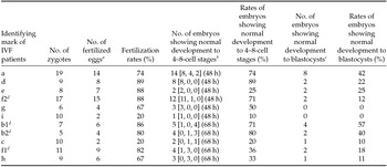
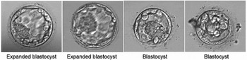
Table 1 shows the early development of human embryos produced by conventional IVF treatments using ejaculated spermatozoa and oocytes from nine infertile couples. Rates of embryos showing normal development to blastocysts (see typical examples of blastocysts in Fig. 1) were greatly varied among these treatments with the wide range from 0 to 57%, and relatively high rates were obtained in the patients a and b (b1 and b2) (42, 57 and 40%, respectively). In patients d, f2 and g, rates of embryos showing normal development to 4–8 cell stages during the first 48-h period (89, 71 and 50%, respectively) were similar to the rate obtained in patient a (74%). However, rates of embryos showing normal development to blastocysts (22, 12 and 0%, respectively) were relatively lower, compared with patient a (42%). In patients e, f1 and h, rates of embryos showing normal development to 4–8-cell stages during the first 48-h or 68-h period after insemination (25, 36 and 33%, respectively) were relatively lower than in patients a and b (b1 and b2) (74, 71 and 80%, respectively). In patients c and i, moreover, fertilization rates were likely lower (20 and 20%, respectively) than any other patients (67–89%). These results suggest that low ability of embryos (in vitro fertilized eggs) to form blastocysts in patients c–i may be due to non-fertilization, unsuccessful fertilization, or defective development of early embryos either between 1-cell and 4-cell stages or between 4-cell and blastocyst stages.
Early development of human embryos produced by the conventional IVF treatments in this study

a Successful fertilization was determined by the presence of two pronuclei visible at 19 h after insemination.
b Total embryos which were developed to 4–8 cell stages at 48 or 68 h after insemination: 4 cell stage, 5–7 cell stage, 8 cell stage (observation of embryos at the period after insemination).
c Total embryos which were developed to blastocysts until 140 h after insemination.
d The conventional IVF treatments were made twice for patient b (b1 and b2, the first and second treatments, respectively) and patient f (f1 and f2, the first and second treatments, respectively).
Typical examples of blastocysts obtained in this study.

Results of immunodetection of SPACA1 proteins
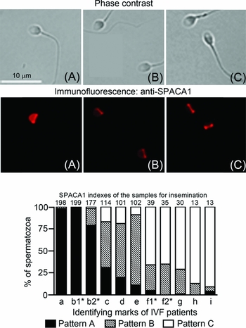
Indirect immunofluorescence of spermatozoa with the anti-SPACA1 antibody revealed three detection patterns of antigens in the principal and equatorial segments of the acrosome (Fig. 2 upper panel). Specifically, the acrosomal equatorial segment was strongly stained in almost all of the spermatozoa. However, immunofluorescence levels in the acrosomal principal piece were largely varied among spermatozoa and could be classified into three categories: (A) strong, (B) intermediate or faint, and (C) almost no immunofluorescence. Hao et al. (Reference Hao, Wolkowicz, Shetty, Klotz, Bolling, Sen, Westbrook, Coonrod, Flickinger and Herr2002) also observed two kinds of immunodetection patterns of SPACA1 proteins in human ejaculated spermatozoa, although there was not description on the individual differences in the percentages of detection patterns in their report. In this study, thus, we immunostained the balance of the sperm samples (surplus spermatozoa) prepared for the insemination to oocytes and compared the obtained results of the detection patterns of the SPACA1 proteins among 11 samples from nine patients. As shown in Fig. 2 lower panel, there were large differences among the patients. To simplify the results, we calculated the SPACA1 indexes according to the following numerical formula:
 $$\begin{eqnarray*}
{\rm SPACA1\; indexes}\; &=& \;{\rm [(\% \; of\; A\hbox{-}pattern\; sperm)}\; \times \;{\rm 2]}\\
&&{\rm + }\;{\rm [(\%\; of\; B\hbox{-}pattern\; sperm)}\; \times \;{\rm 1]}
\end{eqnarray*}$$
Immunodetection patterns of sperm acrosome-associated 1 (SPACA1) proteins in human sperm samples prepared for insemination to oocytes. In the upper panel, photographs A, B and C indicate typical representatives of different detection patterns of SPACA1 proteins in the acrosomal region. In all spermatozoa, strong immunofluorescence was observed in the equatorial segment. However, immunofluorescence levels in the acrosomal principal piece were largely varied among spermatozoa and could be classified into three categories; (A) strong, (B) intermediate or faint, and (C) almost no immunofluorescence. A graph of the lower panel shows individual differences in the results of SPACA1 detection patterns among patients a–i. The SPACA1 indexes were calculated according to the following numerical formula {[(% of A-pattern sperm) x 2] + [(% of B-pattern sperm) × 1]}. *The conventional IVF treatments were made twice for patient b (b1 and b2, the first and second treatments, respectively) and patient f (f1 and f2, the first and second treatments, respectively). For each preparation, 100 spermatozoa were observed with a differential interference microscope equipped with epifluorescence.

The obtained SPACA1 indexes could be divided into three classes; high class [higher than 150 points; patients a, b (b1 and b2)], intermediate class (between 75 and 150 points; patients c, d and e) and low class [lower than 75; patients f (f1 and f2), g, h and i]. In the samples classified into high class, almost all spermatozoa exhibited pattern A. By contrast, intermediate class and low class samples contained mainly B-pattern and C-pattern spermatozoa, respectively. In addition, the samples of seven healthy fertile volunteers (Fig. S1) were classified into the high [volunteers j (SPACA1 index: 151 points) and o (189 points)] and intermediate classes [volunteers k (122 points), l (104 points), m (115 points), n (144 points) and p (127 points)].
Relationship between outcomes of conventional IVF and results of the immunodetection of SPACA1 proteins
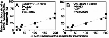
In patients a, b (b1 and b2), d, e, f (f1 and f2), g and h with relatively high fertilization rates (67–89%), high [40–57%; patients a and b (b1 and b2)], intermediate (22–25%; patients d and e) and low rates [0–18%; patients f (f1 and f2), g and h] of embryos showing normal development to blastocysts (Table 1) were obtained in the IVF treatments with the sperm samples which were classified into high [177–199 points; patients a and b (b1 and b2)], intermediate (101–102 points; patients d and e) and low classes [13–39 points; patients f (f1 and f2), g and h] of SPACA1 indexes (Fig. 2), respectively. Thus, we examined the relationship between outcomes of conventional IVF and results of the immunodetection of SPACA1 proteins. As shown in Fig. 3 A for all patients (patients a–i), a significant positive correlation was observed between ‘rates of embryos showing normal development to blastocysts’ and ‘SPACA1 index’ (r = 0.829, P = 0.00162). Moreover, when the results of patients c and i (lower fertilization, Table 1) were excluded from the statistical analyses, the correlation coefficient was enhanced to 0.983 (P = 0.000002; Fig. 3 B). In addition, no significant correlation was obtained between ‘fertilization rates’ and ‘SPACA1 indexes’ (Fig. S2) and between ‘rates of embryos showing normal development to blastocysts’ and ‘rates of morphologically abnormal spermatozoa’ (Fig. S3). These results suggest possible linkage of distribution of SPACA1 proteins in the acrosomal principal segment with blastocyst-forming ability of embryos.
(A) Relationship between SPACA1 indexes and rates of embryos showing normal development to blastocysts in conventional IVF treatments. (B) Data is as in panel (A) except that patients c and i with lower fertilization rates (see Table 1) were excluded from panel B.

It is considered that a spermatozoon which fertilizes an oocyte in the ampulla oviduct (in vivo fertilization) is required to overcome the putative hardship produced by the uterus and then to accomplish timely the capacitation-associated changes in the oviduct (Holt & Fazeli, Reference Holt and Fazeli2010, Reference Holt and Fazeli2015; Aitken & Nixon, Reference Aitken and Nixon2013; Kawano et al., Reference Kawano, Araki, Yoshida, Hibino, Ohnami, Makino, Kanai, Hasuwa, Yoshida, Miyado and Umezawa2014). This indicates that the embryo (in vivo fertilized egg), which is derived from normal oocyte and highly selected spermatozoon (probably with best quality), has the high ability to undergo early development to blastocysts. In IVF treatments, however, spermatozoa are able to skip the hardship produced by the uterus and various restrictions which are necessary for the fertilization with the oocyte in vivo. This suggests that embryos (in vitro fertilized eggs) may be derived from morphologically normal oocytes and spermatozoa with various qualities (namely, non-selected spermatozoa) in IVF treatments. Thus, it is possible that different qualities of the spermatozoa could have larger impacts on the ability of embryos (in vitro fertilized eggs) to form blastocysts.
In this study, results of development of the embryos (in vitro fertilized eggs) to blastocysts were largely varied among patients and significantly correlated with the SPACA1 indexes of the sperm samples (Fig. 3), though fertilization rates were not associated with these indexes (Fig. S2). Before IVF treatments, the spermatozoa with high motility were collected by the swim-up method (Table S3) but had large variations in the SPACA1 indexes among the samples (Fig. 2). Thus, the obtained embryos (in vitro fertilized eggs) were derived from morphologically normal oocytes and spermatozoa with various SPACA1 conditions. If the SPACA1 conditions are supposed to indicate sperm qualities, it is reasonable that results of development of embryos (in vitro fertilized eggs) to blastocysts were associated with the SPACA1 indexes of the sperm samples. However, further experiments are necessary to disclose what detailed conditions of the spermatozoa are indicated by the SPACA1 indexes. As mentioned above, low ability of embryos (in vitro fertilized eggs) to form blastocysts in patients c–i of this study may be due mainly to non-fertilization, unsuccessful fertilization, or defective development of the embryos either between 1-cell and 4-cell stages or between 4-cell and blastocyst stages (Table 1). In mammalian spermatozoa, indispensable molecules for fertilization and egg activation have already been identified and include acrosomal serine proteases (Kawano et al., Reference Kawano, Kang, Yamashita, Koga, Yamazaki, Hata, Miyado and Baba2010), IZUMO1 (Okabe, Reference Okabe2013) and phospholipase C-zeta (Swann & Lai, Reference Swann and Lai2013). Moreover, the activation of paternal genome from the spermatozoon (embryonic genome activation, EGA) occurs in human embryos mainly between the 4- and 8-cell stages and transcripts from paternal genome are present and functional in early embryo between 4–8-cell and blastocyst stages (Niakan et al., Reference Niakan, Han, Pedersen, Simon and Pera2012). In our future research, we should examine the relationship between SPACA1 indexes and sperm indispensable molecules for fertilization, egg activation and EGA.
In conclusion, this study provides initial data to promote large-scaled clinical investigation to demonstrate that the SPACA1 indexes are valid as molecular biomarkers which can predict effectiveness of conventional IVF of infertile couples.
Acknowledgements
We thank all the patients and volunteers who participated in this study for their kind cooperation. We are very grateful to the staff of the Department of Obstetrics and Gynecology, Shiga University of Medical Science, Japan for their technical assistance. This work was supported by a grant from the Japan Society for the Promotion of Science, no. 25450394.
Disclosures
The authors have nothing to disclose.
Authors contributions
K.-K.: study conception and design, collection of samples, acquisition of data, analyses and interpretation of data, and preparation and revision of the manuscript. H.-H.: co-supervision of the research group, study conception and design, analyses and interpretation of data, and preparation and revision of the manuscript. F.-K.: collection of samples, acquisition of data, and interpretation of data. T.-M.: co-supervision of the research group, interpretation of data, and preparation of the manuscript.
Supplementary Material
To view supplementary material for this article, please visit http://dx.doi.org/10.1017/S0967199415000623.