Introduction
Neuroma of the facial nerve (NFN) is a rare benign tumour involving the cell sheath of Schwann. It accounts for less than 1% of all intrapetrous tumours. Reference Cornelius, Sauvaget, Tran Ba Huy and George1 It can affect any portion of the nerve from the cerebellopontine angle to the parotid ending with a predilection for labyrinthine (geniculate ganglion) and tympanic portion. Reference Pulec2 Although NFN were mostly managed by surgical resection, the timing of surgery is controversial due to the inevitability of post-operative House–Brackman grade III facial palsy. Reference Pathapati, Barla, Dayal, Gati and Lakota3–Reference Lee and Lee5 It has been demonstrated that fractionated stereotactic radiotherapy (FSRT) has been effective for acoustic neuromas of the cerebellopontine angle or internal auditory canal with minimal morbidity. Reference Xu, Pan, Alonso, Dekker and Bambakidis6 Our purpose is to systematically review the literature about FSRT on the treatment of NFN.
Clinical case
Our patient was a 70-year-old-woman who progressively presented for 1 year with an asymmetry of the smile, left palpebral closure defect and hearing loss. The clinical examination revealed left peripheral facial paralysis grade V of House–Brackmann. Otoscopy found a normal-looking eardrum without retrotympanic mass, and vestibular examination was normal. The neurological examination did not show any dysfunction of others cranial pairs. The general examination does not reveal any signs of neurofibromatosis. Tonal audiometry found a slight conductive hearing loss of 25 db with a Rinne of 15 db. Schirmer’s test showed preservation of lacrimal secretion on the side of the tumour.
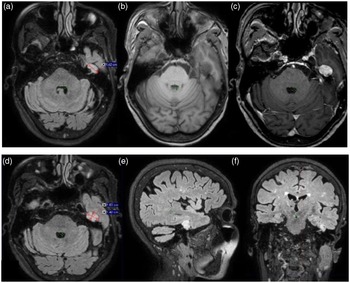
The magnetic resonance imaging (MRI) scan revealed an intrapetrous mass, measuring 10 x 17 mm, centred on the tract of VII nerve and the left geniculate ganglion. There was a homogenous well-circumscribed enhancement after gadolinium injection. It was hypointense on T1-weighted image (WI), hyperintense on T2WI with a high signal on diffusion-weighted image (DWI) and high apparent coefficient diffusion (ADC) values (Figure 1).
(a) Axial FLAIR images demonstrating an oval-shaped lesion centred on the tract of VII nerve and the left geniculate ganglion. (b) Axial T1-weighted images showing the lesion with low signal intensity. (c) Axial post-contrast showing the well-defined lesion with intense and homogenous enhancement. (d) Axial, (e) sagittal and (f) coronal FLAIR images showing the tract of the left facial nerve and the geniculate ganglion in close contact with the ipsilateral oval-shaped hyperintense lesion.

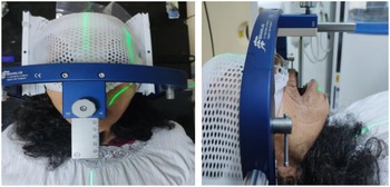
The surgery was not indicated in team board due to the high risk of a permanent facial paralysis. We opted for FSRT. We used stereotactic BrainLab mask with an endo-buccal wedge for positioning (Figure 2). An axial acquisition was acquired with a slice thickness of 1mm.
CT simulation: stereotactic BrainLab mask with an endobuccal wedge.

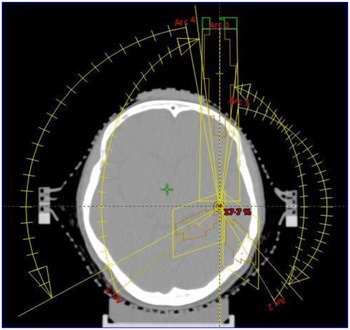
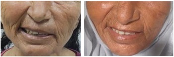
A neuronavigation MRI, for delineation, was performed 10 days prior the computed tomography (CT) simulation. An MRI image fusion between CT was well conducted. Gross tumour volume corresponded to the visible lesion on T1WI, taking into account the 3D heavily T2-weighted sequence (FIESTA) for a better assessment of the remaining cranial nerves. The planning was performed using the TPS Eclipse (TPS, V13) with an additional jaw (X,Y) maximum field size 4 cm x3 cm. The patient received a total dose of 18 Gy every other day with 6 Gy per fraction on the 80% isodose line. Five non-coplanar arcs were performed in the dynamic conformal arc (DCA) technic selected in our procedure (Figure 3). The plan was calculated using the AAA v13.7 algorithm. The treatment was delivered by an accelerator (Clinac iX, Varian Medical Systems, Palo Alto, CA, USA). FSRT was well tolerated, with neither immediate nor delayed toxicities. Six-month MRI follow-up revealed a slight decrease in tumour size. At 1-year follow-up, the patient began noticing an improvement in her left-sided facial palsy and in her left eyelid. There was no change in the tumour size on MRI. At 18 months, we noticed an improvement of her facial weakness (House–Brackmann grade V to IV) (Figure 4) with stable tumour size.
Five non-coplanar arcs (130°, 110°) in the DCA technic.

Left peripheral facial palsy grade V versus IV of House–Brackmann, respectively, before and after treatment.

Discussion
The NFN is extremely rare and accounts for 0·15% to 0·8% of all intracranial tumours. Reference Pathapati, Barla, Dayal, Gati and Lakota3 Multisegment involvement is also common. Reference Chen, Tseng, Hung and Chen4 The extension into the cerebellopontine angle constitutes a diagnostic challenge due to the difficulty to distinguish the lesion from vestibular schwannoma. Reference Lee and Lee5 The bilateral form was never reported, and an association with a neurofibromatosis is rare. Reference Xu, Pan, Alonso, Dekker and Bambakidis6,Reference McRackan, Wilkinson, Brackmann and Slattery7 It can occur at any age with an average age in the fourth decade with a sex ratio of 1· Reference Quesnel and Santos8 The symptoms are usually progressive with an average delay of a 3-year diagnosis and depend essentially on the site of the tumour. It can clinically mimic acoustic neuromas when arising from cerebellopontine angle or internal auditory canal and are often not distinguished until operative intervention. Reference McClelland, Dusenbery, Higgins and Hall9
The CT is the modality of choice for assessing bony changes in the tympanic and mastoid segments, typically showing an enlarged fallopian canal compared with the contralateral side. Reference McRackan, Wilkinson and Rivas10 MRI is the investigation of choice for the diagnosis and characterisation of facial schwannomas. Reference Quesnel and Santos8 It has a high sensitivity for detecting small lesions and high specificity for differentiating a facial and vestibular schwannoma. Reference Pathapati, Barla, Dayal, Gati and Lakota3
The management of non-vestibular schwannomas is relatively limited. Management strategies include radiological monitoring, microsurgical resection, microsurgery combined with radiosurgery or upfront radiosurgery. The lack of large series and heterogeneous data makes it difficult to suggest a definitive treatment strategy. Although treatment is mostly surgical, the timing of intervention is still controversial. Some authors claimed that it is beneficial to operate as early as possible except in cases of poor general status or advanced age. Reference McClelland, Dusenbery, Higgins and Hall9–Reference Chung, Ahn, Kim, Nam, Kim and Lee11 However, others use electromyography to decide on the optimal timing for intervention when the denervation is up than 50%. Reference Abada, Belamkaddem and Moujahid12 Wilkinson et al. indicate resection and grafting in case of an enlarging tumour with facial function House-Brackmann (H–B) IV, compressive symptoms or in the case of stereotactic radiation failure. Reference Wilkinson, Hoa and Slattery13 Abstention with close supervision is only recommended in case of neuromas with normal facial function or minimal paresis H–B less than III. Reference Cho, Choi, Lim and Cho14
Radiation is an emerging treatment modality for facial schwannomas. Its aim is to avoid further tumour growth and to preserve residual facial function. Reference Quesnel and Santos8 Understanding of the mechanisms of radiation on facial nerve schwannomas is limited by the low incidence of these tumours. Thus, given the similarity in location between small acoustic neuromas and cerebellopontine angle facial nerve neuromas of similar size, and in second part, because of the accumulation of longitudinal data for radiation of vestibular schwannomas in which the facial nerve is in the radiation field, FSRT has been established to be a treatment option. Reference Quesnel and Santos8
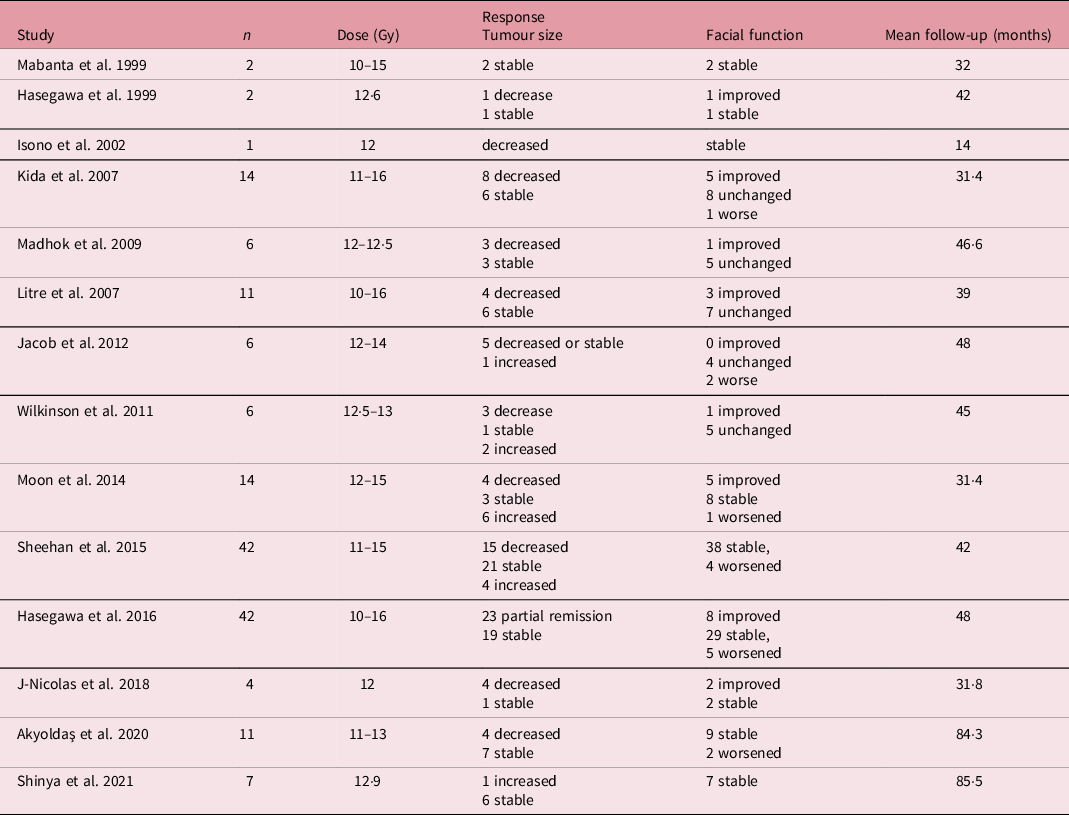
The risks of FSRT include tumour regrowth, deterioration of facial function, hearing loss and possible malignant degeneration. Reference Lee and Lee5,Reference Wilkinson, Hoa and Slattery13 There have been a few case studies reporting the outcomes of FSRT for FNS, and there was no comparing study between radiosurgery and FSRT. Reference Shi, Jain and Kim15 The comparison between both modalities was derived primarily from the existing literature with regard to the treatment of acoustic neuromas, which demonstrated FSRT to have less permanent trigeminal nerve morbidity than stereotactic radiosurgery with comparable efficacy. Reference Söderlund Diaz and Hallqvist16 Hillman et al. studied two patients with facial nerve tumours receiving FSRT of 25 Gy in five fractions, and the facial nerve function was significantly improved for both of them without any tumour growth. Reference Hillman, Chen and Fuhrer17 In a study of patients with non-acoustic intracranial schwannomas undergoing FSRT, 4 patients having NFN were treated with a dose of 50 Gy delivered in 25 fractions. We found that 50% of patients had subjective improvement in facial symptoms, and 50% noted a regression in tumour size. Reference Nishioka, Abo and Aoyama18 Showalter et al. studied five patients with FNS treated with 50·4 Gy in 1·8- to 2-Gy fractions and reported 100% tumour control rates. Reference Showalter, Werner-Wasik, Curran, Friedman, Xu and Andrews19 Shi et al. followed eight patients treated for FNS with FSRT to a median dose of 50·4 Gy in 1·8 or 2·0 Gy fractions. Reference Shi, Jain and Kim15 Six patients had improvement in clinical symptoms, one patient had stable clinical findings and one patient had worsened House–Brackman grade due to cystic degeneration. In Choi et al. study, six patients were treated with FSRT at a median marginal dose of 18 Gy (range, 15–33 Gy) in one to three sessions. After a median follow-up of 29 months, tumour control was achieved in 41 of the 42 lesions. Eighteen of 42 lesions (43%) decreased in size; 23 tumours (55%) remained stable. Reference Choi, Soltys and Gibbs20 In the same way, Hong et al. reported an excellent tumour control of 100% for three patients treated with FSRT at 21 to 25 Gy in three to five fractions with good tolerance. The progression-free survival was 71 months clinically and 41 months radiologically.21 Many studies have also noted stabilisation or improvement in facial function after SRS (Table 1).
Review of literature for radiosurgery of facial nerve schwannomas

A recent meta-analysis aimed to compare the results of SRS with surgical treatment. Reference Hong, Kagawa, Sato and Nomura21 However, most series included heterogeneous groups consisting of patients who had a primary surgical decompression and those who received SRS as the initial treatment. Therefore, it is difficult to make a direct comparison. FSRT seems to be efficient in the treatment of FNS with good tolerance. The choice of FSRT over SRS was derived from a radiobiological standpoint in which there appears to be an intuitive value to fractionation by limiting damage to normal structures while potentially allowing for repair between fractions.
Conclusion
This article reports the first experience in our institute in the treatment of FNS with FSRT. Tumour control at 18 months was efficient with improvement in pre-treatment symptoms without morbidity. More data and longer follow-up are needed to better evaluate the tumour control and the quality of life.
Acknowledgements
Competing interests
None.