Case report
A 50-year-old woman presented to us with several years' history of nasal obstruction associated with rhinorrhoea and hyposmia. She had initially been treated at another institution, with topical nasal steroids alone for a presumed diagnosis of rhinosinusitis. When this failed, a septoplasty and bilateral inferior turbinoplasties had been performed.
Shortly after surgery, the patient's symptoms had become worse. Clinically, adhesions had been noted between the inferior turbinate, the nasal septum and high up in the nasal vault. Due to the unusual extent of adhesion formation, a biopsy had been performed. Surprisingly, this had shown the presence of amyloid. A full investigation for a systemic cause had been negative.
The patient had remained symptomatic with slowly worsening symptoms, until she was referred to our practice several years later.
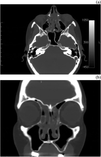
At this stage, the patient's major symptoms were nasal obstruction, hyposmia and post-nasal drip. Endoscopic assessment was limited by what appeared to be significant adhesions between the septum and the lateral nasal wall, and thickened mucosa overlying the middle turbinate with total obliteration of the space medial to the middle turbinate. Interestingly, computed tomography (CT) of the paranasal sinuses showed disease restricted to the nasal cavity and ethmoids, with sparing of the remainder of the sinuses (Figure 1).
(a) Axial and (b) coronal computed tomography scans of the paranasal sinuses. Mucosal thickening is noted along the roof of the nose. The maxillary sinuses are clear. The disease appears, both symptomatically and radiologically, to be restricted to the nasal cavity alone.

Revision surgery was offered, with a guarded prognosis. The aim was to provide symptomatic relief of the nasal obstruction, together with potential improvement in the other symptoms.
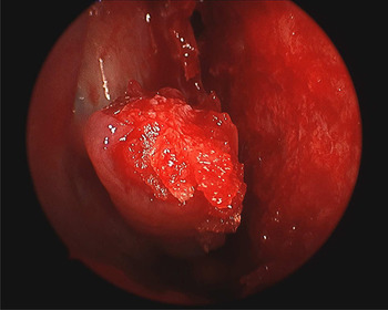
Surgery was performed with image guidance to facilitate clearance of disease high up in the nasal vault, along the skull base and adjacent to the cribriform plate. The intra-operative findings were much worse than those seen during pre-operative endoscopic assessment. There was additional submucosal involvement of the entire nasal cavity and ethmoid sinuses (Figure 2), but the maxillary sinuses were not involved. The affected tissue was meticulously dissected. Due to the extensive involvement of the middle turbinates, they were partially resected. The frontal and maxillary sinuses were not addressed as they appeared clinically normal. Silastic® splints (0.13 mm thick) were used to line the nasal cavity in an attempt to reduce the formation of adhesions.
Intra-operative endoscopic view showing submucosal infiltration of the nasal and ethmoid mucosa.

Formal histopathological analysis confirmed the presence of amyloid deposits.
Our usual protocol of oral antibiotics and saline douching was followed post-operatively. The Silastic splints were removed six weeks after surgery. Small adhesions were noted at that time, and divided under local anaesthetic in the consulting room. There was a significant improvement in the patient's symptoms of nasal obstruction, and her sense of smell started to return. However, even at that time there was evidence of local recurrence high up in the septum, adjacent to the olfactory groove.
Literature review
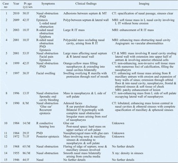
A rigorous search of the literature located only 15 previous reported cases (the earliest in 1946) which constituted true cases of isolated primary sinonasal amyloidosis (Tables I and II). Five other cases purported to be idiopathic primary sinonasal amyloidosis, but were found to be associated with conditions such as multiple myeloma, or to affect multiple sites within the body.
Previously reported isolated sinonasal amyloidosis: presentation and assessment

No = number; pt = patient; y = years; F = female; PND = post-nasal drip; MT = middle turbinate; CT = computed tomography; MRI = magnetic resonance imaging; L = left; IT = inferior turbinate; R = right; M = male
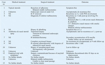
Previously reported isolated sinonasal amyloidosis: treatment and outcomes

*Drug used for myeloma. No = number; GA = general anaesthetic; R = right; IT = inferior turbinate; y = years; PNS = postnasal space; mth = months; post-op = post-operative; CT = computed tomography
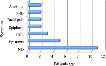
Idiopathic primary sinonasal amyloidosis affects women much more frequently than men (11/15) and afflicts all age groups, with a mean presenting age of 34 years (range, 8 to 86 years). Nasal obstruction is the most common presenting symptom, being reported in 78 per cent (Figure 3). Facial pain, when present, is a worrying feature and may be associated with significant bony erosion.
Presenting symptoms of isolated sinonasal amyloidosis in the present study. PND = post-nasal drip; CHL = conductive hearing loss; NO = nasal obstruction; n = number of patients

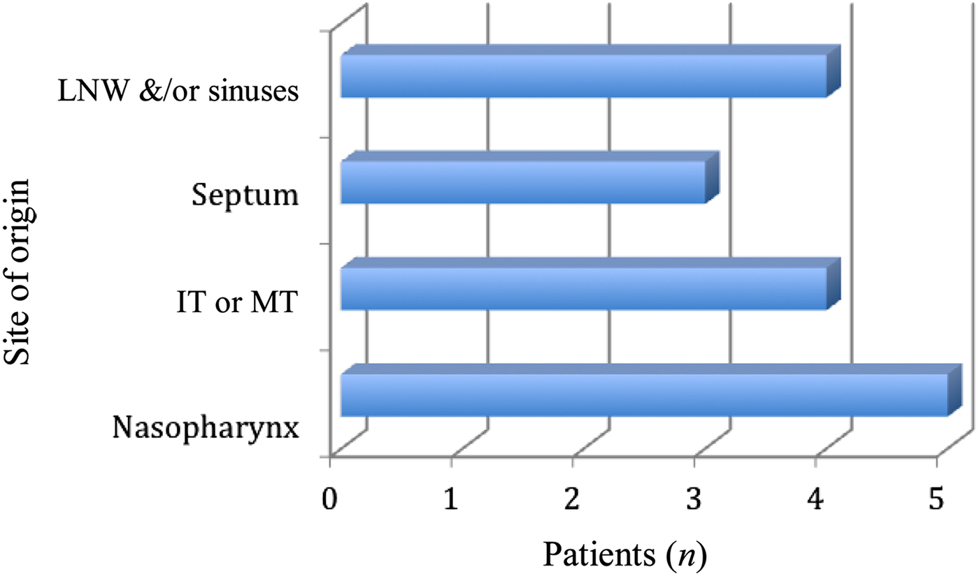
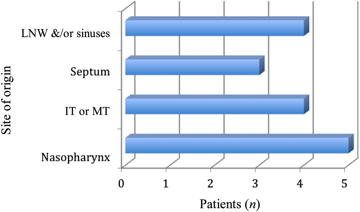
The most common sites of origin are the conchae and nasopharynx (Figure 4).
Sites of origin of isolated sinonasal amyloidosis in the present study. LNW = lateral nasal wall; IT = inferior turbinate; MT = middle turbinate
Medical therapy has not been successful in ameliorating symptoms. Of the 15 reported cases, 11 were offered surgery, one was unfit for surgery, and it is unreported whether or not surgical intervention occurred in the remaining two cases. Where reported, surgery was associated with symptomatic improvement in 8/11 (72 per cent) cases. Five of the 11 surgically managed patients were asymptomatic and recurrence-free post-operatively (45 per cent), whereas three of the 11 were symptomatically improved but experienced recurrences.
Radiological findings are not pathognomonic. Enhancement with contrast might be noted on both CT and magnetic resonance imaging (MRI). Bony erosion is seen with advanced lesions.
Discussion
Amyloid is a pathological proteinaceous substance deposited between cells in various tissues and organs of the body, in a variety of clinical settings.1
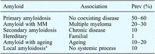
Amyloidosis is the accumulation of extracellular deposits of fibrous, beta-pleated sheet protein in one or more body parts, resulting in organ failure. A detailed classification has been presented in Table III.
Classification of amyloidosis

*Isolated organ deposits. Prev = prevalence; MM = multiple myeloma
Amyloidosis most commonly affects individuals between 50 and 70 years of age, with a male-female predominance of between 3:1 and 3:2.Reference McAlpine and Fuller2 The larynx is the most common site of involvement in the head and neck region. Nasal involvement is rare.Reference Carrol3
Isolated sinonasal amyloidosis is extremely rare, and is a localised, idiopathic, primary process involving amyloid light chain Prasad et al. Reference Prasad, Somayaji, Aroor and Abdulla5 reviewed the literature and reported 23 previously reported cases of isolated sinonasal amyloidosis; they also described one further case. We too have reviewed the literature, and have identified only 15 previously described cases of true idiopathic isolated sinonasal amyloidosis.
The diagnosis and management of this disorder presents a clinical challenge to the practising otorhinolaryngologist. Isolated sinonasal amyloid presents with common rhinological symptoms that mimic chronic rhinosinusitis, i.e. nasal obstruction, nasal discharge, epistaxis, glue ear and post-nasal drip. In children, the symptoms are easily attributed to enlarged adenoids.
On examination, the mass is seen as a yellowish, polypoidal lesion mimicking inflammatory polyps. Endoscopy might alternately reveal the presence of what seem to be simple adhesions.
Interestingly, CT and MRI scans of the paranasal sinuses might not show any unusual features, and in isolated sinonasal amyloidosis might indeed be entirely normal. The imaging characteristics of sinonasal amyloidosis have not been identified. Chin et al. Reference Chin, Fatterpeckar, Kao, Chen and Som6 have reported that on MRI amyloid has a signal intensity similar to that of skeletal muscle on T1- and T2-weighted sequences. Amyloid enhances minimally after contrast administration, but peripheral enhancement might be present. On CT imaging, ‘fluffy’ bone changes might be noted adjacent to the amyloid deposit, although they are not a consistent radiological feature. Bony erosion has been noted in advanced cases, and in one case the entire maxillary antrum and orbital floor was eroded.
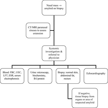
Amyloidosis is confirmed by tissue biopsy. In the unoperated nose, the presence of ‘adhesions’ should raise suspicion of an underlying infiltrative process. In the post-surgical nose, early and unusually extensive ‘adhesion’ formation should be biopsied to exclude the rare possibility of amyloidosis or other benign or malignant processes.Reference Panda, Saravanan, Purushotamam, Gurunathan and Mahesha7 Systemic causes of amyloidosis, such as multiple myeloma, tuberculosis and rheumatic diseases, must be excluded.Reference Simpson, Strong, Skinner and Cohen8Figure 5 presents a clinical algorithm for the diagnosis and management of idiopathic isolated sinonasal amyloidosis.
Suggested algorithm for assessment of a suspected amyloidosis case. CT = computed tomography; MRI = magnetic resonance imaging; FBC = full blood count; UEC = urea electrolytes creatinine; LFT = liver function test; ESR = erythrocyte sedimentation rate; B-J = Bence–Jones

• Isolated primary sinonasal amyloidosis is extremely rare
• The disease mimics common otolaryngological problems
• When restricted to the sinonasal tract, consider surgery for symptomatic relief
• Surgery causes no exacerbation, and relieves symptoms in most patients
There is no satisfactory treatment for systemic amyloidosis. For localised amyloidosis, however, symptomatic removal is appropriate. Although recurrence rates are as high as 50 per cent, conservative medical treatment such as intranasal corticosteroids has not proved useful. Surgery at least provides symptomatic improvement in the majority of patients.
More aggressive medical treatment such as chemotherapy has not been successful. Melphalan, an alkylating chemotherapeutic agent used for multiple myeloma, has been trialled in one case.Reference Johner, Widen and Sahgal9 This drug has significant side effects, and was discontinued after 45 days with no improvement in symptoms.
There is no evidence in the literature to support the use of radiotherapy for this benign condition. For isolated sinonasal amyloidosis, occurring as it does in a critical area of the skull with proximity to the orbit, optic nerve and pituitary gland, radiotherapy is best avoided.
Although there is a concern that surgically induced trauma to the paranasal mucosa could incite reactive amyloid deposition, this seems not to be the case. In addition, the judicious use of Silastic splints to prevent apposition of raw mucosal surfaces might limit the degree of adhesion formation and/or further amyloid deposition.
Conclusion
Sinonasal amyloid is a rare entity that presents with common symptoms and signs. The treating clinician must have a high index of clinical suspicion in order to reach the diagnosis. In the absence of curative treatment, surgery is aimed at symptom alleviation. Surgery improves symptoms in the majority of patients. Recurrence rates are high, but surgery does not seem to worsen the condition.