I. INTRODUCTION
Galvanizing has been one of the most practical commercial methods for prevention of steel corrosion by providing a galvanic protection to the steel substrate and a barrier protection as well. To improve further the performance of galvanized steel (or galvanized iron: GI), a small amount of alloying elements such as aluminum (Al) and magnesium (Mg) is added to the zinc coating. Reference Dutta, Halder and Singh1 The commercial grades of galvanized steel alloyed with Al are Galfan (Zn + 5 wt% Al) and Galvalume (Zn + 55 wt% Al + 1.6 wt% Si). Reference Schuerz, Fleischanderl, Luckeneder, Preis, Haunschmied, Mori and Kneissl2 They exhibit higher corrosion resistance than the conventional GI steel because of the formation of aluminum-based thin oxide film (Al2O3) known as the protective passive film on the coating surface. Reference Shih, Hsu, Sun and Chung3–Reference Palma, Puente and Morcillo6 Addition of Mg to Zn coating also improves greatly the corrosion resistance of galvanized steel. Reference Hosking, Ström, Shipway and Rudd7–Reference Vlot, Bleeker, Maalman and van Perlstein12 Successful examples of the coating development are Super Zinc, Super Dyma, and Dymazinc by Nippon Steel Corporation (Tokyo, Japan) and ZAM (Zn-Al-Mg alloy coated steel product) by Nisshin Steel Corporation (Tokyo, Japan). Since 1960s, many attempts have been made to develop Zn–Mg alloy coating with superior corrosion resistance. The effect of Mg in Zn–Mg alloy coatings on the corrosion resistance of galvanized steel has been proposed by numerous researchers. Reference Hosking, Ström, Shipway and Rudd7–Reference Vlot, Bleeker, Maalman and van Perlstein12 Nevertheless, the exact mechanism has not been fully clarified.
The Zn–Mg alloy coated steels are commercially produced by the traditional hot dip coating process. Although this process is one of the most reliable methods, it has major disadvantages such as the formation of dross in the molten Zn bath, formation of intermetallic compound layers between the coating layer and steel substrate, and generation of polluted wastes. Moreover, due to the burning phenomenon of Mg in the molten bath, no more than 3 wt% Mg can be added successfully in the commercial operation. To overcome such industrial and environmental problems, the dry coating method has been considered as an alternative commercial option. Reference Kazutoshi, Hiroshi, Masao, Hidetoshi, Koki, Junji, Shoji, Shingo and Hirohiko13 Out of many dry coating methods, the physical vapor deposition (PVD) technique by evaporation is considered as a promising one for commercial application of high volume production.
The Zn–Mg alloy coated steel produced by PVD method has a superior corrosion resistance in chloride-containing environment. Reference Volovitch, Allely and Ogle11,Reference Larché, Prosek, Nazarov and Thierry14 Particularly, Volovitch et al. Reference Volovitch, Allely and Ogle11 have reported that the high corrosion resistance of the coated steel is closely associated with the stability of the simonkolleite [Zn5(OH)8Cl2] known as a protective corrosion product. In spite of the superior corrosion resistance in chloride-containing environment, its major drawback is the surface darkening of the Zn–Mg alloy coating layer when exposed to high humidity environment of over 60% relative humidity at a temperature over 50 °C. Reference Kwak, Kim, Jung, Nam, Lee, Hong, Kim, Jung, Eom, Choi, Kim, Park, Lee and Nam15,Reference Yasushi, Masanori, Hiroshi, Tadaaki, Kazushi and Yasumi16 The surface of the Zn–Mg alloy coated steel produced by PVD process easily turns to dark within a short time when its surface is exposed to an extremely thin layer of water film in high humid and temperature environment. The present authors have suspected that Mg contained in the Zn–Mg alloy coated layer plays an important role for the surface darkening phenomena, and a Mg-based corrosion product could be formed on the coating surface by precipitation. Although a great deal of attention has been focused on the electrochemical corrosion behaviors of the Zn–Mg alloy coated steel in wet–dry environment containing chloride, no attention has been paid to the surface color degradation phenomenon appeared mostly in the humid environment at high temperature. In fact, during shipment and storage of the Zn–Mg alloy coated steel sheets, its surface can be easily turned to dark if it is exposed to the high humid environment at high temperature. Since the surface darkening phenomenon may lead to degrading of the coating quality regardless of its superior corrosion resistance, it is very critical industrial problem for the Zn–Mg alloy coating. To develop a proper mitigation method for this surface darkening problem, it is necessary to clearly understand the nature and formation mechanism of the surface darkening phenomenon.
This study is mainly focused on understanding the nature and formation mechanism of the dark rust formed on the Zn–Mg alloy coated steel produced by a PVD method. Various analytical techniques including transmission electron microscopy (TEM), field-emission scanning electron microscopy (FE-SEM), Auger electron spectroscopy (AES), glow discharge spectroscopy (GDS), x-ray photoelectron spectroscopy (XPS), and Fourier transformation infrared (FT-IR) spectroscopy are utilized for the analysis on the surface film.
II. EXPERIMENTAL PROCEDURES
A. Specimen preparation and microstructure examination
The Zn–Mg alloy coated steel was obtained by a high speed PVD using evaporation method. The nominal composition of the alloy coating was 90 wt% Zn-10 wt% Mg. Firstly, the cold rolled steel was cleaned by sputter etching method before deposition. The coating thickness on the specimens was in the range of 1–2 μm. The microstructure and morphology of specimens were analyzed by the FE-SEM and energy dispersive spectroscopy (EDS). The specimen was ultrasonically cleaned in ethanol, dried in air, and coated with platinum before observation of the surface morphology by FE-SEM. To determine the composition of coating layer as a function of depth, GDS was used. In addition, the TEM was used for the cross-sectional observation.
B. Corrosion test
Two different kinds of corrosion tests were used to understand the nature of the dark rust, and they were the immersion test and exposure test to a high humidity and temperature environment. The immersion test was performed by immersing the specimens in boiling deionized (DI) water at 100 °C for 5 min. Before immersion, the specimens were ultrasonically rinsed in ethanol and dried in air. Instead of DI water, tap water, 0.2 M NaHCO3 solution and 0.2 M K2CO3 solution containing 0.1 M ZnCl2 were also used as the other immersion test solution to evaluate the effect of bicarbonate ion on the prevention of surface darkening. The reason why the tap water was used is that the tap water contains bi-carbonate ion (HCO3 −). According to the ion chromatography analysis, the amount of HCO3 − in the tap water is around 81.3 mg/L. By the addition of 0.1 M ZnCl2 to the 0.2 M K2CO3 solution, Zn2+ ion used for the precipitation of Zn-based protective film on the coating surface can be supplemented.
After the test, the surface was analyzed by FE-SEM, EDS, GDS, XPS, and scanning vibrating electrode technique (SVET).
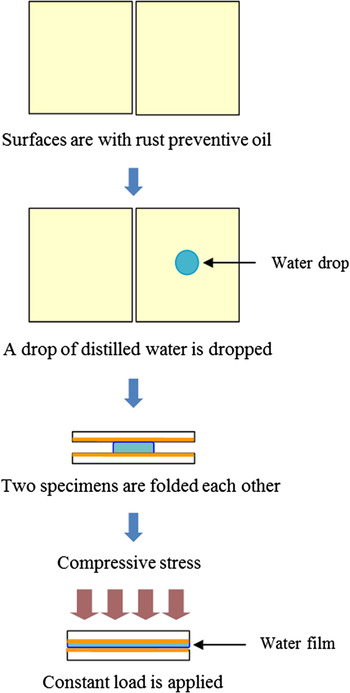
A high temperature–humidity exposure test was performed to simulate the corrosion by water films condensing on the surfaces of narrow gaps of the galvanized steel roll. The two specimens were ultrasonically rinsed in ethanol and dried in air. After two drops of DI water were dropped on the surface of one steel specimen, the other specimen was folded on this specimen to make them in face-to-face contact. Then, a constant load (compressive stress) was applied to press these two specimens together in such a way that the small amount of water in the gap between these two specimens cannot be evaporated easily. Figure 1 illustrates the exact description of the experimental procedure. The temperature and humidity levels were kept at 50 °C and at 95%, respectively, for 3 days. After the test, the surface was analyzed by digital camera and AES.
High temperature–humidity exposure test procedure.

III. RESULTS AND DISCUSSION
A. Microstructure analysis
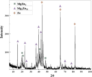
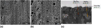
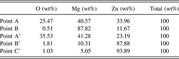
Figure 2 presents X-ray diffraction (XRD) patterns of the Zn–Mg alloy coated surface of as-received specimen. Detected phases are two intermetallic compounds of MgZn2 and Mg2Zn11. Figure 3(c) exhibits the cross sectional image of the coating layer analyzed by TEM, and Table I lists the result of the EDS quantitative analysis at each point marked on Fig. 3. From these analyses, it is found that the light and dark images in Fig. 3(c) are identified as MgZn2 phase and Mg2Zn11 phase, respectively. Besides these two main intermetallic phases, however, presence of metallic Mg phase was also identified by the backscattered electron images (BEI) of the Zn–Mg coating surface and EDS quantitative analysis examined by FE-SEM. Figures 3(a) and 3(b) and Table I show BEI of the coating surface and the result of quantitative EDS analysis, respectively. Considering that the black-colored region in BEI indicates the presence of the low atomic weight element and that the atomic percent of Mg is much higher than that of Zn at the point marked “A” on the Fig. 3(a), it is estimated that metallic Mg phases are also present along with MgZn2 and Mg2Zn11 intermetallic compounds. This may be due to the fact that the Zn–Mg alloy coating was developed by a nonequilibrium process in the PVD method and the metallic Mg phases could be formed abnormally on the coating layer.
XRD spectra of Zn–Mg alloy coated surface.

(a) Surface morphology of Zn–Mg alloy coated steel examined by FE-SEM in the BEI mode; (b) magnified view of (a); (c) cross sectional view of Zn–Mg alloy coating layer examined by TEM.

EDS quantitative analysis at each point marked on Fig. 3.

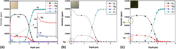
From Fig. 3(c), the outermost coating layer is covered with a thin film of Mg-based oxide (MgO) and it can also be identified by the GDS analysis as shown in Fig. 4(a). Larché et al. Reference Larché, Prosek, Nazarov and Thierry14 have reported that the presence of the MgO film on the outermost surface plays a positive role in the corrosion resistance of Zn–Mg alloys since the film is stable enough to ensure good barrier properties to ion and electron transfer comparing to other oxide films such as Zn-based oxide (ZnO). Considering the resistance of MgO (1012–1013 Ω m at 573 K) is almost 1010–1011 order higher than that of ZnO (94 Ω m at 433 K), Reference Samsonov17 it is expected that the cathodic reaction of oxygen reduction can be effectively suppressed on the MgO so that the corrosion resistance can be improved under the condition that the oxides have the same physical size such as the thickness.
GDS analysis on (a) as-received Zn–Mg alloy coated specimen, and (b) and (c) coating surface after immersion test in 0.2 M NaHCO3 solution and DI water, respectively.

B. Surface morphology after corrosion test
1. Surface analysis after immersion test
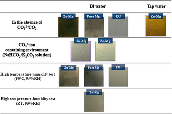
Figure 5 presents the surface morphology after various immersion tests examined by digital camera. Contrary to the steel with electro-galvanized steel (EG; pure Zn coating), Zn–Mg alloy coated steel specimens exposed to boiling DI water turned to dark in 5 min. In the case of the steel with pure Mg coating, the surface was also changed to dark brown within 10 min. However, in this case, the extent of surface color change is not as severe as that of Zn–Mg alloy coated steel and the time required to change the surface color was longer. The two different Zn–Mg alloy coated steel specimens shown at the second row of Fig. 5 show the result of the immersion test using solutions containing the carbonate ion (CO3 2−). The left one is the coating surface after immersion test in 0.2 M NaHCO3 solution and the right one is the surface after immersion test in 0.2 M K2CO3 solution. Contrary to the results of immersion test in DI water, no discernible change in the surface color was observed. The reason for this phenomenon will be discussed in detail later in this section.
Surface morphology of Zn–Mg alloy coated, pure Mg coated and EG steels after various immersion and high-temperature humidity tests.

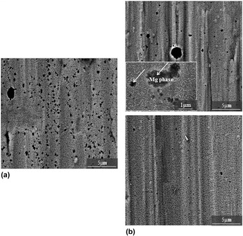
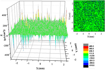
To determine the nature of the dark surface shown in Zn–Mg alloy coated steel, the cross-sectional observation was carried out by TEM and the results are shown in Fig. 6(d). The coating layer was covered with a thin corrosion product with thickness of around 100 nm. It is estimated that the thin layer formed on the coating surface is closely associated with the surface darkening phenomenon. Figures 6(a) and 6(b) exhibit the surface morphology of the coating before and after the immersion test in DI water by utilizing FE-SEM. For the specimen after the immersion test, it is evident that the needle-like corrosion products are formed on the outermost surface. In addition, as clearly seen in Fig. 6(c), the corrosion products are presented in the dark region examined by digital camera. Therefore, it can be strongly suspected that the nature of dark rust is closely associated with the corrosion product on the outermost surface. Some researches have indicated that the black staining phenomenon of zinc coated steel is due mainly to the oxygen deficient phenomenon (ZnO1−x ) occurring in zinc oxide. Reference Yanping, Zuyu and Yanming18 It can lead to the change of band gap energy related to the optical property. This kind of phenomenon has also been identified after the immersion test in this research. In the case of the EG, surface has some local staining like dot. However, as it is seen clearly in Fig. 5, dominant dark color was not observed. Therefore, although the oxygen deficient phenomenon in ZnO can degrade the surface to a certain extent, it seems that it is not directly associated with the dark rust formation discussed here, and the dark rust formation cannot be fully explained by the mechanism of oxygen deficiency in ZnO. To understand the nature of the corrosion product formed on the coating surface, GDS analysis was additionally carried out. According to the analysis shown in Figs. 4(b) and 4(c), there is a clear increase in Mg peak on the outermost surface in case of the specimen immersed in DI water for 5 min contrary to the case of the specimen immersed in 0.2 M NaHCO3 solution. The effect of solution containing carbonate ion on the suppression of surface darkening is mainly discussed in Sec. III. B. 2. The results of various analyses on the surface of the steel with pure Mg coating and Zn–Mg alloy coated steel after immersion test suggest that Mg plays a crucial role in the formation of dark rust formed on the coating surface. It is known that when two dissimilar metals are coupled electrochemically or galvanically, the active metal is preferentially corroded while the noble one is protected from corrosion. Reference Jones19 Since Mg is more electroactive than Zn and also more electroactive than MgZn2 and Mg2Zn11, Mg has a tendency to dissolve preferentially when Mg is electrochemically coupled with MgZn2 and Mg2Zn11 in the Zn–Mg coating. In the Zn–Mg alloy coating, since the area of Mg phase acting as an anode is much smaller than that of MgZn2 and Mg2Zn11 phases acting as cathode, the dissolution of Mg can be accelerated drastically. Figure 7 shows the surface morphology examined by FE-SEM in the BEI mode before and after the immersion test for 40 s. Although large Mg phases appearing as black-colored region in Fig. 7 still remain, the amount of fine size of Mg phases is clearly reduced, suggesting that the metallic Mg phases are dissolved at a short period of immersion. Furthermore, the remaining large Mg phases after the immersion test in 40 s are precisely observed and the results are shown in magnified view of Fig. 7(b). It appears that the needle-like corrosion products were scarcely precipitated only on the remaining Mg phases. It implies that the Mg phases act as anode and the high anodic dissolution could occur preferentially at those sites. To confirm this phenomenon, ion-chromatography (ICP) analysis was performed on the dissolved Zn and Mg ion after immersion test in DI water. The amount of dissolved Mg ion was 0.05 ppm, whereas that of dissolved Zn ion was less than 0.01 ppm. This ICP analysis result is consistent with the discussion above. It is believed that this is the reason why the extent of surface staining of Zn–Mg alloy coated steel is more severe than that of pure Mg coated steel. Although an electrochemical potential map analyzed by SVET, which is found in Fig. 8, cannot give the potential level at each phase in the microstructure, it can be surely indicated that there are a lot of local anodes and cathodes in the coating surface.
FE-SEM images showing the surface morphology of Zn–Mg alloy coated steel (a) before, and (b) and (c) after immersion test in DI water for 5 min and 25 s, respectively; (d) TEM image showing the cross section of the coated steel after immersion test in DI water for 5 min.

Surface morphology of the coating surface examined by FE-SEM in the BEI mode (a) before and (b) after the immersion test for 40 s; (c) magnified view of (b).

Potential map of Zn–Mg alloy coated steel after immersion test in DI water, analyzed by SVET.

In summary, the dark rust is closely associated with the precipitation of the Mg-based corrosion product in a form of Mg(OH)2. Although it is known that the color of pure Mg(OH)2 is white, its color can be changed to gray or other dark colors depending on the kind and level of impurity. Considering the fact that the optical properties are very sensitive to variations in metal particle size and/or shape, Reference Wallinder, Augustsson and Leygraf20 it could be also possible that accelerated dissolution of Mg from the coating surface makes the surface rougher, leading to the scattering of visible ray and therefore it could contribute additionally to the surface color change to dark.
Lindstrom et al. Reference Lindstrom, Johansson, Thompson, Skeldom and Svensson21 have also reported that Mg surface is covered with dark Mg(OH)2 when Mg is exposed to corrosive wet environment, especially in the absence of CO2. In addition, it is known that the solubility of Mg(OH)2 in aqueous water at 100 °C is around 4 mg/L which is only one third of that in the water at room temperature (25 °C). Reference Ambrose, Gnanam and Ramasamy22 This suggests that as the temperature of the aqueous water increases, the solubility of Mg(OH)2 in the water is decreased and therefore easy precipitation of Mg(OH)2 on the coating surface can be facilitated. Considering these facts, the dark rust formation of Zn–Mg alloy coated steel in aqueous solution at high temperature could be well explained by the preferential dissolution of Mg from the coating layer and easy precipitation of Mg(OH)2 on the coating surface. The preferential dissolution of Mg and subsequent precipitation of Mg(OH)2 are basically attributed to the pre-existing metallic Mg on the coating layer. To avoid the formation of abnormal Mg phase on the coating layer during the PVD process, the conditions of the coating process should be optimized or efficient surface treatment should be developed. However, detailed discussion on the prevention methods are beyond the scope of this paper and the present study is focused sorely on the mechanistic study of the dark rust formation.
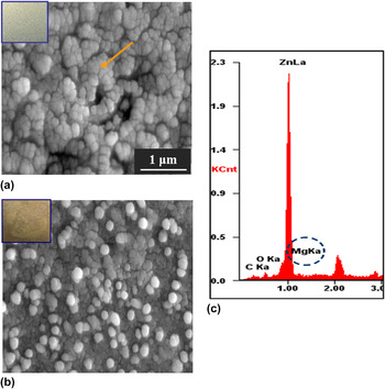
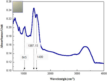
Contrary to the result of immersion test in DI water, dark rust is not observed from Zn–Mg alloy coated steel specimens when tested by immersion in a solution containing bi-carbonate ion (HCO3 −) and Zn2+ ion. The extent of surface color change to dark was less severe when immersed in tap water containing a small amount of carbonate ion than when immersed in DI water. Secondly, little change of surface color was observed after the test using solutions containing the carbonate ion such as NaHCO3 and K2CO3. After immersion test in the solutions containing NaHCO3 and K2CO3, the specimen surface did not show any discernible change in the color and pattern by observation with naked eye. As shown in Fig. 4(b), Mg peak was not detected on the outermost surface in case of the specimen immersed in 0.2 M NaHCO3 solution, which is attributed to the precipitation of Zn-based thin film on the surface. The morphology Zn-based film is presented in Fig. 9. Consequently, the surface darkening phenomenon can be avoided. Considering the fact that pH of the solution containing 0.2 M NaHCO3 is around 8.3, the thin film of Zn-based corrosion product is assumed to be hydrozincite [Zn5(OH)6(CO3)2] since the hydrozincite becomes stable between pH 7 and 8.5 of the bicarbonate buffer solution. Reference Sontheimer and Kuch23 The surface analysis by FT-IR, which is found in Fig. 10, clearly supports this assumption. The peaks at 1499, 1387.15, and 845 cm−1 are attributed to hydrozincite. Reference Prosek, Nazarov, Bexell, Thierry and Serak10 According to Chung et al., Reference Chung, Lin, Chang and Shih24 the presence of hydrozincite as a passive film is widely accepted as being advantageous for the longevity and protectively, which is primarily attributed to a superior insolubility comparing to oxidation products such as simple oxides and chloride species. Figure 9(b) showing FE-SEM image obtained from the carbonate treated specimen after immersion test by using DI water for 30 min indicates that the hydrozincite particles which can act as a protection barrier against the formation of dark rust gradually disappear by dissolution in boiling DI water, and therefore the coating layer containing Mg is exposed to the DI water. As a result, the dark rust is gradually formed on the coating surface. A number of test results obtained by a variety of analysis equipment such as FE-SEM, EDX, GDS, and FT-IR supports the fact that the outermost layer of carbonate treated specimen is composed of hydrozincite and there is no evidence that Mg rich layer such as MgCO3 is present on the coating surface. This could be due mainly to the fact that the Gibbs free energy of formation for hydrozincite (−3160 kJ/mol at room temperature to 100 °C) is much lower than that for MgCO3 (−1100 kJ/mol at room temperature to 100 °C), suggesting that the hydrozincite could be preferentially formed on the coating surface. Nevertheless, it would not be possible to entirely exclude the fact that some Mg rich layers are also present on the coating surface.
Surface morphology of the coating surface (a) before and (b) after immersion test in 0.2 M NaHCO3 solution, (c) EDS analysis on the coating surface.

FT-IR spectra of the coating surface after immersion test in 0.2 M NaHCO3 solution.

2. Surface analysis after high temperature–humidity test
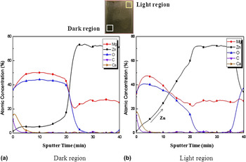
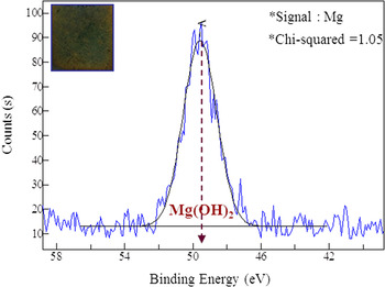
At the third and fourth row of Fig. 5, the surface morphology examined by digital camera after high temperature–humidity exposure test are presented. Since the condition of high temperature–humidity exposure test is different from that of immersion test, the mechanism of dark rust formation is not the exactly same as that of immersion test. Contrary to immersion test, an extremely small amount of water exists on the coating surface during this test. This suggests that the precipitation rate of metallic ions, specifically Mg ions, increases since their solubility limit exceeds rapidly because of a small volume of solution in the thin water film condensed on the surface. Therefore, MgO/hydroxide can be formed rapidly by precipitation from the water. Reference Liu, Zanna, Ardelean, Frateur, Schmutz, Song, Atrens and Marcus25 As seen clearly, the extent of surface change was the most severe in the case of Zn–Mg alloy coated steel due to the combined effects of galvanic action between Mg and two intermetallic phases of MgZn2 and Mg2Zn11, and fast precipitation of Mg ions. Although a similar pattern was observed in the case of the pure Mg, the extent of surface change was not as severe as that of Zn–Mg alloy coated steel. In the case of the EG, there was no discernable change on the coating surface. This result shows that it is pretty much the same as the result of immersion test. Considering these results of Zn–Mg alloy coated steel and pure Mg shown in Fig. 5, it seems that the dark surface is caused by precipitation of Mg(OH)2. Figure 11 can surely support this assumption by AES analysis. As seen in Fig. 11, there are locally darker part and lighter part from the same test specimen. The AES analysis from the dark region indicates that the region is composed of Mg oxide/hydroxide. Very small amount of Zn is present on the outermost surface. On the other hand, in the case of the comparatively light region, Mg oxide/hydroxide is not sufficiently present to make the surface completely dark; Zn peak continuously increases as a function of depth. Little change in the surface color is observed due to the lack of Mg oxide/hydroxide. To clarify the nature of dark rust formed on the surface, XPS analysis is additionally conducted and Fig. 12 also supports that outermost layer is composed of Mg(OH)2. In addition, to determine the effect of temperature on the surface color degradation phenomena, an exposure test was carried out at room temperature (25 °C). As it is clearly seen at the bottom of Fig. 5, little change can be observed. It seems that relatively high temperature is required to form the dark rust on the coating surface, suggesting that the anodic dissolution of Mg is getting faster at high temperature. As a result, the precipitation rate of Mg in the form of Mg(OH)2 becomes faster, too. Furthermore, considering the fact that the solubility of Mg(OH)2 in aqueous water decreases as the temperature of the water increases, the dark rust formation due to the precipitation of Mg(OH)2 on the coating surface will be inevitable when the Zn–Mg alloy coated steel is exposed to a limited amount of aqueous environment at elevated temperature.
Surface morphology of Zn–Mg alloy coated steel after high temperature–humidity exposure test and AES analysis at (a) dark region and at (b) light region.

XPS analysis on the coating surface after immersion test in DI water for 5 min.

IV. CONCLUSIONS
This research is focused on the understanding of the nature of dark rust, and its formation mechanism on the surface of Zn–Mg alloy coated steel for which a PVD method is used. The results are briefly summarized as follows:
-
(1) The coating layer of the Zn–Mg alloy coated steel is composed of metallic Mg phase and two intermetallic compounds of MgZn2 and Mg2Zn11.
-
(2) The formation of dark rust is observed from both Zn–Mg alloy coated steel and pure Mg coated steel after the immersion test in DI water at high temperature. Preferential anodic dissolution occurs at the Mg phase in the coating surface of the Zn–Mg alloy coated steel, and it is estimated that the formation of Mg(OH)2 on the outermost surface of the coating layer is main responsible for the dark rust. In addition, it could be also possible that the preferential anodic dissolution of Mg from the coating surface leads to the rougher surface. The scattering of visible ray due to the rougher surface can contribute additionally to the surface color change to dark.
-
(3) The darkening effect is more serious when the Zn–Mg alloy coated steel is exposed to an extremely small amount of condensed water on the steel surface. It is due to the preferential dissolution of metallic Mg phases by the galvanic action with intermetallic phases of MgZn2 and Mg2Zn11 mainly present in the Zn–Mg coating layer, and easy precipitation of Mg2+ ion in a form of Mg(OH)2 in a limited volume of the condensed water film on the steel surface.