Introduction
The Ti6Al4V alloy has low density, high corrosion resistance, wear resistance, and fatigue strength, which makes it the most commonly used implant material in hip and knee replacement surgeries [Reference Patel, Hamlekhan, Royhman, Butt, Yuan, Shokuhfar, Sukotjo, Mathew, Jursich and Takoudis1]. Despite its widespread use in orthopedics, Ti6Al4V based implants have a long-term failure problem with an average lifetime of only 15–20 years, which is a pressing issue for orthopedic patients [Reference Saini2, Reference Ramiah, Ashmore, Whitley and Banniste3]. On the one hand, the geriatric population in the world, who are at a higher risk to develop orthopedic complications, is constantly growing, and on the other hand, the age spectrum of patients receiving orthopedic implants is broadening. The average age of people receiving hip replacement surgery is getting younger overtime [Reference Sansone4], and nowadays nearly half of the patients receiving orthopedic implants are younger than 65 years old. Thus, a significant portion of the population receiving a hip and knee replacement requires a high-risk revision surgery, while younger and more active patients may need multiple revision surgeries [Reference Li and Webster5, Reference Bayliss, Culliford, Monk, Glyn-Jones, Prieto-Alhambra, Judge and Price6]. Clearly, currently used Ti6Al4V based implants do not satisfy the needs of the patients.
For the last couple of decades, scientists investigated alternate means to reduce the incidence of orthopedic implant failures to prolong their average lifetime. The underperformance of Ti6Al4V based orthopedic implants can in part be attributed to the presence of vanadium element in its chemistry, which was associated with increased expressions of pro-inflammatory factors, and eventually lead to osteolysis in the juxtaposed bone tissue [Reference Li, Jiang, Peng, Li, Yang, Liu and Wang7], as well as to the incomplete osseointegration (integration of prosthesis to juxtaposed bone tissue) upon its implantation into bone tissue due to the bioinert nature of this alloy [Reference Webster and Ejiofor8]. When a Ti6Al4V alloy is implanted into a bone defect, instead of promoting bone cell adhesion, it is typically encapsulated by a fibrous tissue, isolating it from the juxtaposed bone, and thus providing limited osseointegration [Reference Xie and Luan9]. Hence, there is a need to engineer new implants with specifically designed surfaces to induce better biological outcomes for orthopedic applications. One such approach is to modify Ti6Al4V surfaces to enhance its bioactivity and promote osseointegration [Reference Webster10]. In the literature, various surface modification methods were investigated for Ti6Al4V alloy, i.e., hydrothermal treatment, sol–gel coatings, sandblasting, physical and chemical vapor deposition, and etc. [Reference Duraccio, Mussano and Faga11] Among these methods, anodization is an attractive approach due to its simplicity, low cost, and ability to control physical and chemical properties of the oxide film fabricated on Ti6Al4V surfaces. Anodization is an electrochemical technique where Ti6Al4V is used as the anode and typically an inert material (i.e., platinum or graphite) is used as the cathode. The electrodes are connected to a DC power source and anodization process undergoes inside an electrolyte. During anodization, micron and nanofeatured surface topography of Ti6Al4V alloy can be altered to mimic that of bone surface, which is documented to promote bone cell functions on Ti6Al4V surfaces, enhance bioactivity, and osseointegration [Reference Webster and Ejiofor8]. In addition, anodization can also be used to obtain desired color coding [Reference Webster and Ejiofor8] and enhance corrosion resistance of Ti6Al4V samples [Reference Patel, Hamlekhan, Royhman, Butt, Yuan, Shokuhfar, Sukotjo, Mathew, Jursich and Takoudis1].
In the literature, anodization of Ti6Al4V is typically investigated using fluorinated electrolytes to fabricate nanofeatured surfaces. For instance, Macak et al. anodized Ti6Al4V using an electrolyte mixture of 1 M (NH4)2SO4 + 0.5 wt% NH4F and the results demonstrated growth of self-organized porous oxide structures on Ti6Al4V surfaces [Reference Macak, Tsuchiya, Taveira, Ghicov and Schmuki12]. Park et al. anodized Ti6Al4V fixtures inside a 1 wt% NH4F + glycerol + water electrolyte and reported the formation of well-arranged and self-organized nanotubular titanium oxide layer on the Ti6Al4V fixtures [Reference Stępień, Handzlik and Fitzner13]. Cumulatively, the use of fluorinated electrolytes led to the formation of nanotubular structures on Ti6Al4V, which improved bone cell adhesion, proliferation, and cellular functions in vitro compared to conventional Ti6Al4V [Reference Webster and Ejiofor8]. However, the fragile nature of the nanotubular structures and weak delamination strength of the oxide based nanotubular coatings prevented translation of anodized nanotubular Ti6Al4V from bench to bedside. Furthermore, the use of fluorinated electrolytes left behind fluoride based residues on anodized Ti6Al4V surfaces, which brought the risks of fluoride release from the anodized surfaces to potentially react with calcium mineral of bone and red blood cells and potentially lead to long-term failure of anodized implants. Thus, alternative anodization strategies to fabricate bioactive oxide films without the use of fluorinated electrolytes on Ti6Al4V are required.
Few researchers reported possibility of using chloride-containing electrolytes in combination with perchloric, oxalic, and formic acid to anodize metallic surfaces and demonstrated chloride ions to play a similar role as fluoride during anodization [Reference Richter, Panaitescu, Willey and Menon14, Reference Park, Oh and Bae15]. It is important to note that chloride ions provide a less hazardous alternative to the highly toxic fluorinated electrolytes in industrial applications where the potential hazards can be corrosion, environmental influence, skin/lung exposure, and etc. [Reference Cheong, Yam, Ng, Hassan, Ng and Low16] Research showed that it was possible to obtain nanotubular structures having 20 nm diameter in various chloride-containing electrolytes, i.e., 0.4 M NH4Cl + 0.5 M formic acid, 0.4 M NH4Cl + 0.05 M sulfuric acid, and 0.4 M NH4Cl + 0.5 M formic acid electrolytes [Reference Cheong, Yam, Ng, Hassan, Ng and Low16, Reference Ishibashi, Yamaguchi, Kimura and Niwano17]. Aside from chloride-containing electrolytes, various acid and salt solutions were also investigated as potential electrolytes. For instance, Cui et al. anodized Ti6Al4V in 1.0 M H2SO4 solution at 150 V and 0.5 M Na2SO4 solution at 100 and 130 V, followed by immersion in simulated body fluid for 7 days. The results showed deposition of calcium phosphate minerals on Ti6Al4V surfaces [Reference Cui, Kim, Kawashita, Wang, Kokubo and Nakamura18]. In another study, Narayanan et al. reported anodization of Ti6Al4V using a 0.3 M phosphoric acid containing electrolyte where a titanium oxide layer was obtained independent of anodization duration [Reference Narayanan and Seshadri19]. In these studies, the lack of biomimetic topography formation on anodized Ti6Al4V surfaces, along with weak mechanical adhesion of the oxide films onto the metallic substrates remain to be addressed to propose the anodization procedures for orthopedic applications.
This paper addressed the aforementioned issues by introducing micropit structures that were fabricated on Ti6Al4V alloy surfaces using a fluoride-free electrolyte (aqueous NH4Cl solution) for anodization. Surface chemical composition, wettability, crystallinity, and oxide layer delamination were investigated. Calcium phosphate deposition onto the anodized micropit samples was also assessed using simulated body fluid up to 30 days. This study provided a detailed fabrication route to form micropit surface topography on Ti6Al4V alloy surfaces and showed preliminary data supporting its potential use for orthopedic applications.
Results and discussion
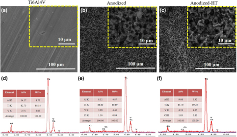
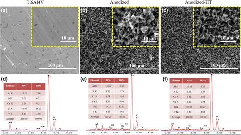
The surface morphologies of anodized Ti6Al4V samples using 0.2, 0.4, and 0.6 M NH4Cl electrolytes under 10, 20, and 30 V up to 3 min are provided in the Supplementary material figures (Figs. S1–S3). For some of the investigated anodization conditions, micron-phase surface topographies appeared on Ti6Al4V samples (i.e., anodization using 0.6 M NH4Cl electrolyte under 10 V for 3 min). Among all the investigated anodization conditions, the use of 0.4 M NH4Cl and application of 30 V potential for 2 min was chosen due to the lack of crack formation on the oxide surfaces and the repeatability of obtaining micropit surface morphologies on Ti6Al4V surfaces. The surface of nonanodized Ti6Al4V alloy, micropit structures formed on anodized Ti6Al4V (named Anodized), and heat treated anodized Ti6Al4V samples (named Anodized-HT) could be observed in Fig. 1. In this research, we observed formation of random micron-sized features (micropits) upon anodization. The micropits had approximately 2–3 μm inner diameters [Fig. 1(b)] and it was clear that annealing process at 500 °C for 2 h preserved micropit structures on anodized Ti6Al4V samples [Fig. 1(c)]. The oxide layer formed upon anodization of Ti6Al4V samples had an approximate thickness of 14.6 µm (Fig. S4). Surface investigations showed that micropits were not present throughout the whole surface. They had nonuniform distribution across the Ti6Al4V surface and located only inside larger pit formations. Furthermore, no morphological difference between alpha and beta phases was present, which could be correlated with similar etching rates of alpha and beta phases inside the electrolyte, as well as limited electrical field induced migration of large chloride ions inside Ti6Al4V matrix. Energy dispersive spectroscopy (EDS) analysis displayed the chemical composition of the Ti6Al4V samples before and after anodization [Figs. 1(d) and 1(e)], as well as after heat treatment [Fig. 1(f)]. The EDS spectra confirmed presence of Ti, Al, and V elements for all of the samples and Cl was present for the Anodized and Anodized-HT samples. Though further research is required, we believe that chloride ion induced pitting, where pits nucleate at microscopic scales at localized active sites, coupled with rapid breakdown anodization, which is the local breakdown of the oxide film by chloride ions and formation of irregular patterns on the surface, was responsible for the formation of surface features in this study.
(a–c) SEM images and (d–f) EDS spectra of (a) and (d) Ti6Al4V, (b) and (e) Anodized, (c) and (f) Anodized-HT samples. Insets show higher magnification images of samples.

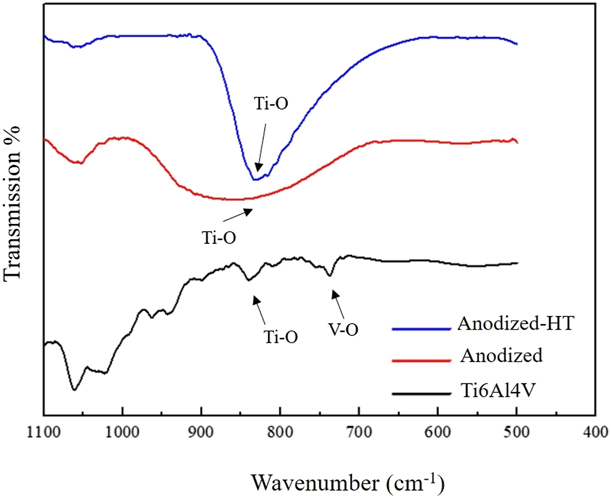
The influence of anodization and the follow-up heat treatment process on the surface chemical structure of Ti6Al4V samples was investigated by Fourier transform infrared (FTIR) spectroscopy and the results were displayed in Fig. 2. Stretching vibrations consistent with Ti–O bonding were detected around 850 cm−1 for all the samples [Reference Popa20]. It was also observed that Anodized and Anodized-HT samples expressed more intense Ti–O bands compared to Ti6Al4V (nonanodized) samples, which could be attributed to the growth of oxide layer during anodization and the follow-up heat treatment. For the Ti6Al4V samples, V–O stretching vibrations were detected around 720 cm−1; however, in-line with previous researchers [Reference Patel21], no vanadium or aluminum peaks appeared for the Anodized and the Anodized-HT samples.
FTIR spectra of Ti6Al4V (black line), Anodized (red line), and Anodized-HT (blue line) samples.

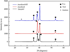
To investigate the crystal structure of the samples, XRD scans were taken and the results were presented in Fig. 3. The XRD spectra showed characteristic α and β peaks belonging to Ti6Al4V for all samples. Ti-α peaks appeared at 35.4°, 38.5°, 40.5°, and 53.3° (JCPDS file #44-1294), which belong to the (100), (002), (101), and (102) planes, respectively, and Ti-β peak appeared at 39.5° (JCPDS file #44-1288), which belongs to the (110) plane. XRD characterization did not show any crystalline phase for the oxide based micropit surfaces, indicating an amorphous crystal structure which lacked ordered planes to appear on XRD spectra. For the case of Anodized-HT samples, the diffraction peaks were ascribed to the anatase phase at 25.37° and 38.67° (JCPDS #01-0562), which were correlated with (101) and (112) planes, respectively. It is important to note that formation of crystalline titanium oxide is favored in orthopedic applications since presence of anatase was correlated with greater biocompatibility compared to amorphous titanium oxide [Reference López-Huerta, Cervantes, González, Hernández-Torres, García-González, Vega, Herrera-May and Soto22, Reference Cervantes, López-Huerta, Vega, Hernández-Torres, García-González, Salceda, Herrera-May and Soto23].
XRD spectra of Ti6Al4V (black line), Anodized (red line), and Anodized-HT (blue line) samples.

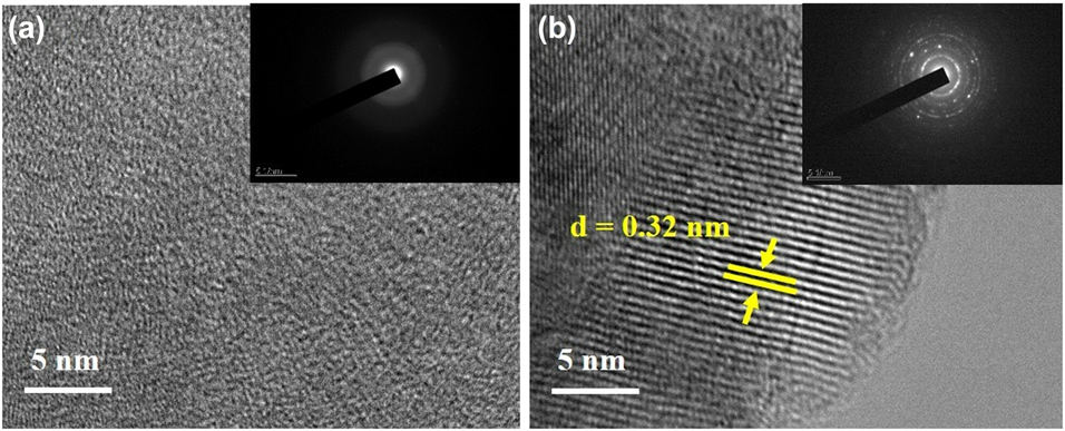
High-resolution transmission electron microscopy (HR-TEM) images for the Anodized and Anodized-HT samples were displayed in Figs. 4(a) and 4(b), respectively. TEM characterizations confirmed the amorphous nature of the anodized oxide layer on Ti6Al4V samples [Fig. 4(a)] and the corresponding selected area electron diffraction (SAED) pattern further supported this finding. For the case of Anodized-HT samples [Fig. 4(b)], the HR-TEM image and the corresponding SAED pattern were in-line with the XRD results and demonstrated anatase phase upon heat treatment of the Anodized sample at 500 °C. The lattice fringes obtained from the HR-TEM image exhibited a d-spacing of 0.32 nm which corresponded to (101) plane of anatase [Reference Wang, Chen, Zhang, Huang, Li and Yu24]. Thus, these results further confirmed transformation of the amorphous oxide layer of Anodized samples to anatase after the heat treatment process.
HR-TEM images of (a) Anodized and (b) Anodized-HT samples. Insets show SAED patterns obtained from these samples.

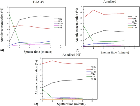
To investigate surface chemical composition of the samples, XPS depth profile analysis was computed [Figs. 5(a)–5(c)]. Among all the elements present on the surface of Ti6Al4V samples [Fig. 5(a)], carbon (indicated with green line) had the highest concentration and it decreased upon anodization and heat treatment procedures. Carbon was a contaminant on Ti6Al4V surfaces and it could potentially originate due to the organic solvents used to clean the specimen surfaces. During anodization, the electrolyte etched the Ti6Al4V surfaces and led to a decrease in the adsorbed carbon content on specimen surfaces [Fig. 5(b)]. When anodized Ti6Al4V samples were heat treated, carbon contamination on the surfaces decomposed and its concentration further decreased [Fig. 5(c)]. Upon sputtering the sample surfaces, carbon content exhibited a drastic decrease independent of the sample type, which suggested surface adsorption to be the main mechanism for the presence of carbon contamination. As opposed to carbon, oxygen content (indicated with the red lines) followed an opposite trend where its concentration increased after anodization compared to Ti6Al4V samples. This was an expected result since the purpose of anodization in this study was to grow an oxide layer on Ti6Al4V surfaces. When the anodized surfaces were sputtered, there were no significant changes in the oxygen concentrations since the thickness of the micropits were well above micron range. The titanium concentrations of the samples (indicated with black lines) were low on all specimens, yet its concentration increased significantly upon sputtering, which was probably due to the removal of carbon contamination on the surfaces. Once the top surfaces were sputtered, it was observed that Ti6Al4V sample had the highest titanium concentration, followed by Anodized-HT and Anodized samples. Aluminum and vanadium concentration of Ti6Al4V surface followed a similar trend where they were the highest on Ti6Al4V samples, followed by Anodized and Anodized-HT samples. Cumulatively speaking, the surface compositions of Ti6Al4V, Anodized, and Anodized-HT samples were different from each other and these differences would certainly influence biological properties of the samples and alter their bioactivity.
XPS depth profiles of (a) Ti6Al4V, (b) Anodized, and (c) Anodized-HT samples. Samples were sputtered with Ar+ five times each for 2 min.

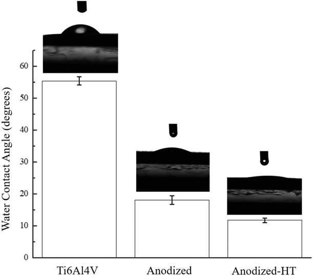
Sessile drop water contact angles for Ti6Al4V, Anodized, and Anodized-HT were reported in Fig. 6. It was observed that water contact angles for Anodized and Anodized-HT samples were significantly lower than Ti6Al4V samples, indicating hydrophilic nature of these surfaces. The water contact angle for the Anodized samples was 20° and the Anodized-HT samples was 12°, both of which were significantly lower compared to Ti6Al4V, indicating hydrophilic nature of the anodized specimens. The increase in hydrophilicity could be explained with changes in chemical composition and topography of the Anodized and Anodized-HT sample surfaces compared to Ti6Al4V. As evidenced in the XPS results, the surface chemical composition of the Ti6Al4V, Anodized, and Anodized-HT samples differed from each other. Anodization removed the carbon contamination from the alloy surfaces and introduced oxide based rough surface features. In addition, surface roughness in the micro- and nanoscales also changed upon anodization of Ti6Al4V, which has been documented to influence hydrophobicity of materials [Reference Patel, Hamlekhan, Royhman, Butt, Yuan, Shokuhfar and Takoudis25].
Sessile drop water contact angles of Ti6Al4V, Anodized, and Anodized-HT samples. Values are reported as mean ± EM; N = 5 and n = 4.

Microscratch testing of anodized Ti6Al4V samples showed that the critical load to induce delamination of the oxide layer from the anodized samples is 0.7 N. It was speculated that the use of fluorinated electrolytes during anodization was one of the reasons for the weak adhesion of anodized titanium oxide layer onto the metal substrate, where electrical field induced migration and accumulation of fluoride ions at the bottom of nanotubular features was the reason for weak adhesion of the oxide layer [Reference Macak, Tsuchiya, Ghicov, Yasuda, Hahn, Bauher and Schumiki26]. Since fluoride ions did not exist for the utilized electrolyte in this study, this failure mechanism was not active, and thus a stronger adhesion of the oxide layer onto the Ti6Al4V substrate was obtained. Though more research is required on the mechanism of oxide film delamination from the underlying substrate, to the best of our knowledge, this is the first published data quantifying the delamination force of anodic oxide layer grown using a chloride based electrolyte on Ti6Al4V substrate.
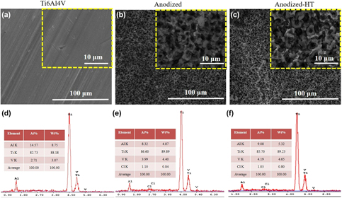
Figures S5 and 7 show SEM images of the samples after 15 and 30 days exposure to 1 × simulated body fluid (SBF), respectively. By comparing Figs. 1 and 7, it could be observed that surface morphology of Anodized and Anodized-HT samples changed upon interacting them with 1×SBF for 30 days; however, no visible change was observed for Ti6Al4V samples. EDS spectra detected the presence of Ca and P elements on all the samples and the concentrations of these elements were much higher on Anodized and Anodized-HT samples compared to Ti6Al4V. The bioinert nature of Ti6Al4V could be attributed to the lack of significant CaP mineral formation on these surfaces. Upon anodization, hydrophilicity of the samples increased, along with changes in surface chemical composition. Since both of these parameters influence bioactivity of materials, increased CaP mineral formation was observed on Anodized samples compared to Ti6Al4V. For the case of Anodized-HT, sample surface transformed to crystalline anatase phase. Since crystalline titanium oxide phases were documented to enhance bioactivity, it is possible that the heat treatment process also contributed to the formation of CaP crystals on Anodized-HT samples [Reference Patel21].
(a–c) SEM images and (d–f) EDS spectra of (a) and (d) Ti6Al4V, (b) and (e) Anodized, (c) and (f) Anodized-HT samples immersed in 1×SBF for 30 days. Insets show higher magnification images of samples.

It is important to note that Anodized and Anodized-HT samples fabricated using the optimized parameters (0.4 M NH4Cl electrolyte under 30 V potential for 2 min) had desired surface properties for orthopedic applications. Specifically, Anodized and Anodized-HT samples had a higher surface roughness, increased surface area, and hydrophilicity compared to nonanodized Ti6Al4V samples. In addition, anodized samples favored higher levels of mineralization upon soaking inside 1×SBF. Thus, the aforementioned changes in the surface properties upon anodization could potentially enhance in vitro bone cell adhesion, proliferation, and functions on orthopedic implant surfaces. The present research obtained preliminary results for anodization of Ti6Al4V to fabricate surface micropits and showed its potential to enhance calcium phosphate mineralization; however, further studies are necessary to investigate mechanical, chemical, and biological properties of anodized micropit surfaces to assess its suitability for orthopedic applications.
Conclusion
This research presented anodization as a potential method to fabricate micropit structures on Ti6Al4V alloy. Specifically, anodization using aqueous solutions of different chloride based electrolytes, including NH4Cl, up to 30 V potential were investigated. Results showed successful fabrication of micropit surfaces on Ti6Al4V alloy using 0.4 M NH4Cl electrolyte under 30 V potential for 2 min. The critical load to induce delamination for anodized micropit samples was 0.7 N. The results also suggested that fabrication of micropit surfaces via fluoride-free anodization could be an effective method to enhance calcium phosphate mineral formation on Ti6Al4V alloys. Though further research including chemical and biological properties is required, anodized micropit structures on Ti6Al4V can potentially enhance osseointegration of the implants to the juxtaposed bone tissue and prolong average lifetime of orthopedic implants.
Experimental details
Sample preparation
Ti6Al4V rod having 14 mm diameter was cut into 1 mm thick pieces using a precision cutter (IsoMet Low Speed Precision Cutter, Buehler, Esslingen, Germany). The obtained pieces were sonicated with acetone, 70% ethanol, and ultrapure water each for 20 min, respectively. The cleaned Ti6Al4V samples were anodized using an electrochemical set. For anodization experiment, a platinum mesh (Thermo Fisher Scientific, Lancashire, UK) was used as cathode and the cleaned Ti6Al4V samples were used as anode. The platinum mesh and Ti6Al4V samples were connected to a direct current (DC) power supply (Genesys 300 V/5, TDK Lambda, Achern, Germany) using a copper wire and 0.4 M NH4Cl was used as the electrolyte. Samples were anodized for 2 min using 30 V potential under magnetic stirring at room temperature. The distance between anode and cathode was 4 cm. Once anodization was complete, samples were rinsed with ultrapure water, dried at room temperature, and heat treated at 500 °C for 2 h in an open-air environment to induce phase transformation of the anodized oxide layer. In this study, nonanodized samples, anodized samples, and heat treated samples were denoted as Ti6Al4V, Anodized, and Anodized-HT, respectively.
Surface morphology
Samples were characterized using scanning electron microscope (SEM; FEI, Nova Nano 430, Brno, Czech Republic) to observe their surface morphology. 20-kV accelerating voltage was used and images were captured using secondary electrons. The thickness of the oxide layer was measured by obtaining micrographs from cross-sections of the anodized Ti6Al4V samples cut across one of its pits.
Surface chemistry
Molecular bonds on Ti6Al4V surface were characterized by Fourier transform infrared reflection spectroscopy (FTIR; Perkin Elmer 400, Perkin Elmer, Waltham, Massachusetts) using the attenuated total reflection (ATR) configuration. The spectra were collected in the 500–1100 cm−1 range with a resolution of 4.00 cm−1 and the background signal was subtracted from the obtained reflectance. For each sample, the average of two spectra from five different fields was reported. To analyze the chemistry of the top surface, X-ray photoelectron spectroscopy (XPS; PHI 5000 Versaprobe, Physical Electronics, Chanhassen, Minnesota) spectra were collected. An Al Kα X-ray source was used with an approximate vacuum pressure of 4.5–10.9 Pa during measurements. High resolution scans of the Ti 2p, O 1s, Al 2p, V 2p, Cl 2p, and C 1s were recorded from a spot diameter of 100 μm. To obtain depth profile analysis and analyze the variation of elements across different depths, sample surfaces were sputtering with Ar1+ for 4 times each for 2 min.
Crystallinity
Crystallinity of the samples was analyzed with X-ray diffractometer (XRD; D/Max-2200, Rigaku, Tokyo, Japan). Using Cu-K radiation (λ = 1.54 Å) and operated at 30 mA and 40 kV, spectra were collected between 20° and 60° diffraction angles (2θ) at a scanning rate of 0.5°/min. To image and confirm crystallinity of the surface oxide layer, transmission electron microscopy (TEM; JEM 2100F, JEOL, Tokyo, Japan) was used. For these experiments, oxide based micropit structures were carefully removed from the underlying Ti6Al4V alloy, dispersed in ethanol, centrifuged for 1 h, and then transferred to copper grids for TEM analysis. Samples were characterized in high-resolution (HR) and SAED modes at 200 kV accelerating voltage. SAED patterns were collected from a circular area approximately 0.15 μm in diameter.
Mechanical property characterization
Anodized specimens were tested for its mechanical properties using a MicroScratch Tester (Anton Paar MST3, CSM Instruments, Buchs, Switzerland). Samples were fixed on the instrument and a shear force was applied on the sample surface with an indenter. A Berkovich type indenter having a 5-μm diameter tip was used to scratch a 0.4-mm long region of the sample with a loading rate of 0.2 N/min. Experiments were repeated for five times.
Water contact angle
Sessile drop water contact angles were measured using a drop shape analysis system (EasyDrop; KRÜSS GmbH, Hamburg, Germany). The contact angles of 3 µL double distilled water droplets were measured 10 s after drops were introduced onto Ti6Al4V, Anodized, and Anodized-HT surfaces. Measurements were repeated for five times.
Preparation of SBF
Ti6Al4V, Anodized, and Anodized-HT samples were soaked in 1×SBF to assess calcium phosphate crystal formation on their surfaces. To prepare SBF NaCl (8.035 g/L), NaHCO3 (0.355 g/L), KCl (0.225 g/L), K2HPO4·3H2O (0.231 g/L), MgCl2·6H2O (0.311 g/L), 1 M HCl (39 mL), CaCl2 (0.292 g/L), and Na2SO4 (0.072 g/L) were dissolved in distilled water, respectively. Afterward, the solution was buffered with Tris (6.118 g/L) and 1 M HCl as per established protocols [Reference Kokubo and Takadama27]. Once SBF was prepared, Ti6Al4V, Anodized, and Anodized-HT samples were immersed in SBF at 37 °C up to 30 days. The presence of calcium and phosphate minerals on the samples was determined by energy dispersive spectroscopy (EDS) using a detector (AMETEK-EDAX, Mahwah, New Jersey).
Supplementary material
To view supplementary material for this article, please visit https://doi.org/10.1557/jmr.2019.56.
Acknowledgments
This work was financially supported by METU Research Funds (Grant No. BAP-08-11-2017-019) and The Scientific and Technological Research Council of Turkey (Grant No. 117M187). The author would like to thank Çağlar Karaoğlu, Doğuhan Sarıtürk, Iman Kamfa, İlker Aydın, İpek Koçak, and Yağmur Göçtü for their help in optimizations of anodization parameters.