

High-voltage electrical injuries (HVEIs) are a problem in the society because of their high morbidity and mortality rate.Reference Saracoglu, Kuzucuoglu and Yakupoglu 1 , Reference Prindeze, Jo and Paul 2 In addition, most victims are young and have just started jobs at the workplace. The high post-HVEI amputation rate has resulted in serious socioeconomic problems because patients are often incapable of returning to the workplace and their families are also affected. When electric current enters the human body via the skin, it runs along the organs that have smaller resistance, and when it reaches a certain level, the tissue cells on the current conduction path cause depolarization and cell membrane electric cracking. Further, the heat generated by electric current because of the body’s resistance can result in the coagulation and carbonization of tissue proteins, destroying the integrity of tissue cells. HVEIs often lead to serious physical injuries of deep limb tissues, especially the damage and necrosis of muscles, in which the physical appearance of the limb is normal, or only limb swelling occurs, but the deep skeletal muscles often exhibit sandwich-like, jumping, and segmental injuries. According to the features of HVEI, the clinical diagnosis and treatment of such patients require repeated surgical explorations to understand the extent and scope of organ damage. Previously, methods such as intraoperative naked eye observation, striated-muscle electrical stimulation, and the intraoperative frozen section method have been used to identify muscle necrosis. Hunt and HeckReference Hunt and Heck 3 introduced nitroblue tetrazolium (NBT) as a means of identifying the post-HVEI muscle activities. In addition, tissue oxygen consumption can also be used to identify the degree of degeneration and necrosis in muscles and tissues after HVEI. However, similar to the intraoperative frozen section method, the detection of biological oxygen consumption is limited by sampling sites; hence, it proves difficult to fully evaluate the extent of HVEI, and such studies are still at the stage of animal experimentation.Reference Jia-ke, Li-gen and Quan-wen 4 HVEI normally occurs at the exposed parts of limbs, from which the current could easily pass through the neurovascular tissues with lower resistance; therefore, damage of deep muscles and blood vessels may not be obvious to the patient. Therefore, high voltage -induced progressive injuries of muscles and blood vessels, as well as the resistance from the resultant limb swelling and inflammations, are independent risk factors for amputation.Reference Hsueh, Chen and Pan 5 It is difficult for many surgeons to find the proper amputation level in limbs and maintain the motions. A thorough analysis of limb vigor and vascular injury after an HVEI is very important for guiding subsequent treatment and prognosis. Recently, multi-slice, spiral computed tomography-angiography (CTA) has been used clinically and provides a safe, non-invasive, reliable, and novel alternative for preoperative evaluation and postoperative follow-up in patients with arterial stenosis and occlusion.Reference Chin and Rubin 6 Nevertheless, CTA visualizes intimal changes, such as hydropic degeneration, thickening, and shedding, with a resolution that is not significantly better than that of ultrasound examination.Reference Li, Chai and Sheng 7 , Reference Vedung, Arturson and Wadin 8 The use of 256-slice spiral computed tomography (CT) perfusion scanning could aid in the determination of muscular activity and be used to select the optimal phase for displaying the vessels in order to assess vascular injury and quantitatively and qualitatively investigate limb vigor.
METHODS
Clinical Data
Patients with HVEI who underwent a multi-slice spiral CT perfusion examination in the Second Affiliated Hospital of Kunming Medical University, China were included in this study. Inclusion criteria included: (1) injury from voltage between 380 and 100 kV, (2) time since exposure to the high voltage being within 7 days, and (3) provision of written informed consent by the patients. Multi-slice spiral CT perfusion examination of patients who were electrically shocked by voltages of <380 V or >100 kV was not performed because of very-mild or too-severe injury, respectively. There were 3 radiologists who reviewed the scans. The patients were blinded to the outcome except for the perfusion dose of iopromide and the ray dose. This study was conducted in accordance with the declaration of Helsinki. This study was conducted with approval from the Ethics Committee of the Second Affiliated Hospital of Kunming Medical University. Written informed consent was obtained from all participants.
CT Examination
CT perfusion scanning of HVEI limbs was performed using a 256-slice spiral CT scanner (Brilliance iCT; Philips Healthcare, Eindhoven, Holland). The scanning range was 20-30 cm. A bolus of 60-80 ml of the contrast medium iopromide (370 mg/ml, 1.5 ml/kg body weight) followed by 45 ml of physiological saline was intravenously injected at a flow rate of 4-5 ml/8-10 seconds (upper limb for 8 seconds and lower limb for 10 seconds) into the median cubital vein (or external jugular vein for some special patients) on the healthy side using a double-tube high-pressure syringe. Then the Jog-mode scanning was performed. Non-ionic contrast medium, a widely used group of global contrast agents, was utilized. Less-serious side effects included nausea, flushing of the skin, and swelling of the eyelid. Serious side effects included shock, tachycardia, and pulmonary edema. The following scanning parameters were used: the detector collimation was 128×0.625 mm, total z-axis coverage was 200-250 mm, tube voltage was 120 kV, tube current was 220 mAs, layer thickness was 2.5 mm, table speed was 183 mm/second, pitch was 0.915, gantry rotation speed was 0.4 second/rotation, matrix was 512×512, and field of view was 500×500 mm. The spiral scanning was repeated 10-12 times at 6-7-second intervals. A total of 2500-3000 images were acquired. During the scan period, the area outside the regions of interest was protected with a lead apron to minimize the radiation dose that the patients were exposed to patients.
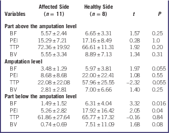
Vascular stenosis or occlusions were determined through the CTA images, and the perfusion results were statistically analyzed using SAS9.1 software. Data were expressed as mean±SD and are listed in Table 1.
Comparisons of BF, PEI, TTP, and BV Between Affected and Healthy Sides (
 $$\overline{\mib x} $$
±s)

Abbreviations: BF, blood flow (ml/minute/100 g); PEI, peak enhancement intensity (HU); TTP, time to peak (seconds); BV, blood volume (ml/100 g).
Perfusion parameters used for limb scanning on the affected and healthy sides included blood flow (BF), peak enhancement intensity (PEI), time to peak (TTP), and blood volume (BV), and the results were expressed as mean±SD. Considering the non-normal distribution of data and relatively small sample size, BF, PEI, TTP, and BV of different regions on the affected and healthy sides were compared. Wilcoxon’s rank sum test with a 0.05 level of significance was used. P<0.05 was considered statistically significant.
Parameter comparison (BF, PEI, TTP, and BV) was performed between the amputation level on the affected side (amputation level-affected) and the corresponding level on the healthy side (corresponding level-healthy), between amputation level-affected and the part above the amputation level on the affected side (part above the amputation level-affected), and between amputation level-affected and the part below the amputation level on the affected side (part below the amputation level-affected). Data regarding the affected side were collected from 11 patients and data regarding the healthy side from 8 patients. Fisher’s discriminant analysis, Bayes’ discriminant analysis, and distance discriminant analysis were used. The κ test was used to assess the consistency of interpretation, with a κ value of >0.8 indicating very good agreement, and values of 0.6-0.8, 0.4-0.6, and <0.4 showing good agreement, moderate agreement, and poor agreement, respectively.
To ensure the stability of the equation, the correlation coefficients among four variables (BF, PEI, TTP, and BV) were calculated. There were strong linear correlations between BV and BF (r=0.87, P<0.0001), between BV and PEI (r=0.65, P=0.0026), and between BF and PEI (r=0.51, P=0.0241). To avoid collineation, variables were selected using gradual discriminant analysis, with an inclusion criterion of P=0.10 and an exclusion criterion of P=0.15.
Quantitative data were shown as mean±SD, and SAS software (SAS Institute Inc., version 9.1) was used to carry out statistical analysis. The perfusion parameters, like BF, PEI, TTP, and BV, were all compared between the affect limb and the corresponding limb using a paired t-test on the normally distributed data, and using the rank test of paired comparison for data in non-normal distribution. P<0.05 means the difference was statistically significant.
Data Analysis and Evaluation
All images were input into Philips EBW workstation for general analysis of functional CT. Time-density curves (TDC) and parameter images of each pixel were acquired. The affected and healthy limbs were analyzed using multiplanar reconstruction, maximum intensity projection, volume rendering techniques, and an advanced vessel analysis software.
RESULTS
A total of 53 patients with HVEI who underwent a multi-slice spiral CT perfusion examination were included in the study. The CT scan would be taken within 3-5 days after the injury, and the amputation would be performed within the following 2 days. Among 48 patients who underwent examination, 16 underwent amputations (all male patients, at an average age of 22±14.01 (range, 5-57) years), 5 were electrically injured by 8000-35000 V, 8 had unilateral upper limb injury, 4 had unilateral lower-limb injury, 2 had bilateral limb injury, and 10 underwent decompression. The time from exposure to high voltage to scanning of the patients was less than a week.
CTA
CT inspection was performed on 53 patients among whom 48 were subjected to multi-slice CT perfusion imaging, and clear vascular CTA images were obtained. CTA revealed 14 cases of vascular stenosis, interruption, or occlusion, 28 cases without obvious vascular abnormality, and 45 cases with muscle injury.
Comparisons of perfusion parameters used for limb scanning on the affected and healthy sides are shown in Table 1. From Table 1, BF and PEI values in the part below the amputation level on the affected side were significantly lower than those in the corresponding level on the healthy side (P<0.05). TTP and BV values in the part below the amputation level on the affected side were slightly, but not significantly, lower than those in the corresponding level on the healthy side (P>0.05). BF, PEI, and BV values in the part above the amputation level on the affected side were slightly, but not significantly, lower than those in the corresponding level on the healthy side (P>0.05). TTP value in the part above the amputation level on the affected side was slightly, but not significantly, greater than that in the corresponding level on the healthy side (P>0.05); BF, PEI, BV, and TTP values at the amputation level on the affected side were slightly, but not significantly, lower than those at the corresponding level on the affected side (P>0.05). Taken together, BF and PEI values in the part below the amputation level on the affected side were significantly lower than those in the corresponding level on the healthy side.
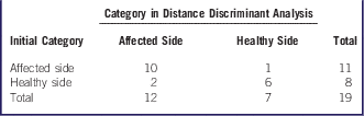
Discriminant models of BF, PEI, TTP, and BV values used for limb scanning on the affected and healthy sides at the amputation level were constructed. BF and TTP values were selected for distance discriminant analysis (Table 2).
Distance Discriminant Analysis Results

The misjudgment rate was 14.29% (1/7), accuracy rate was 82.95% (17/19), sensitivity was 83.3% (10/12), specificity was 85.7% (6/7), positive predictive value was 90.9% (10/11), negative predictive value was 75% (6/8), and the κ value was 0.67 (>0.6), indicative of good consistency. BF and TTP values helped to predict the amputation level on the affected side and the corresponding level on the healthy side.
The BF, PEI, TTP, and BV values were compared between the part above the amputation level on the affected side and the amputation level on the affected side. The comparison results are shown in Table 3.
Comparisons of BF, PEI, TTP, and BV Values Between the Part Above the Amputation Level on the Affected Side and Amputation Level on the Affected Side

Abbreviations: BF, blood flow; PEI, peak enhancement intensity; TTP, time to peak; BV, blood volume.
The BF value in the part above the amputation level on the affected side was significantly greater than that at the amputation level on the affected side (t=2.99, P=0.042). The PEI, TTP, and BV values in the part above the amputation level on the affected side were slightly, but not significantly, greater than at the amputation level on the affected side (P>0.05).
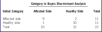
Discriminative models of BF, PEI, TTP, and BV values in the part above the amputation level on the affected side and at the amputation level on the affected side were constructed for Bayes discriminant analysis (Table 4).
Discriminative Models of BF, PEI, TTP, and BV Values in the Part Above the Amputation Level on the Affected Side and at the Amputation Level on the Affected Side Were Constructed for Bayes Discriminant Analysis

Misjudgment rate was 9.09% (1/11), accuracy rate was 86.36% (19/22), sensitivity was 90.0% (9/10), specificity was 83.33% (10/12), positive predictive value was 81.81% (9/11), negative predictive value was 90.9% (10/11), and the κ value was 0.73 (>0.6), indicative of good consistency. BF, PEI, TTP, and BV values help to predict the amputation level on the affected side and the part above the amputation level on the affected side.
Perfusion parameters in the part below the amputation level on the affected side and at the amputation level on the affected side are shown in Table 5.
BF, PEI, TTP, and BV Values in the Part Below the Amputation Level on the Affected Side and at the Amputation Level on the Affected Side (
$$\overline{\mib x} $$
±s, n=11)

Abbreviations: BF, blood flow; PEI, peak enhancement intensity; TTP, time to peak; BV, blood volume.
The BF, PEI, and BV values in the part below the amputation level on the affected side were significantly lower than those in the amputation level on the affected side (P<0.05). The TTP value in the part below the amputation level on the affected side was slightly, but not significantly, greater than at the amputation level on the affected side. Therefore, the BF, PEI, and BV values in the part below the amputation level on the affected side were significantly lower than those at the amputation level on the affected side.
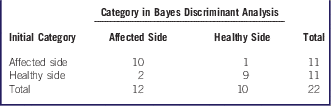
Discriminant models of BF, PEI, TTP, and BV values in the part below the amputation level on the affected side and at the amputation level on the affected side were constructed for Bayes discriminant analysis (Table 6).
Discriminant Models of BF, PEI, TTP, and BV Values in the Part Below the Amputation Level, Like Table 4

Misjudgment rate was 10.00% (1/10), accuracy rate was 86.36% (19/22), sensitivity was 83.33% (10/12), specificity was 90% (9/10), positive predictive value was 90.9% (10/11), negative predictive value was 81.81% (9/11), and the κ value was 0.73 (>0.6), indicative of good consistency.
Case 1
A 5-year-old boy was admitted because of HVEI (6% third-degree burn) to the head, face, right hand, and two feet. The skin on the wrist and back of the right hand was burned black and covered by eschar. After admission, amputation was suggested by the physicians from the burn department. Through preoperative CT perfusion examination, the right ulnar and radial arteries were shown to be normal, the soft tissue in the right forearm was seen to be swollen, but muscle tissue in the perfusion region was not necrotic. In the exploratory operation, neither vascular embolism nor deep-tissue necrosis was observed. Therefore, debridement and skip grafting were performed on the right upper extremity without amputation, thus the right upper extremity was salvaged.
Case 2
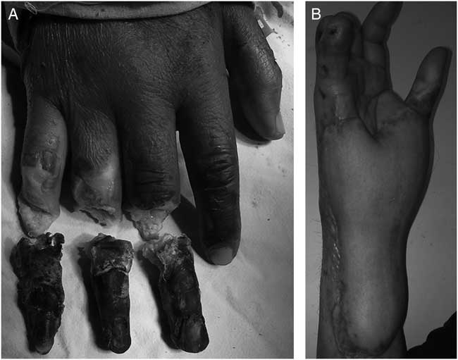
A 45-year-old male was admitted because of HVEI (4% fourth-degree burn) to the head, both upper extremities, and the right foot. The right wrist was covered by ring-like eschar. The skin of the last segment of middle, ring, and little fingers, and the whole thumb were burned black and had become necrotic. The thenar and hypothenar muscles of the right palm were covered by eschar. Amputation was suggested by the physicians from the burn department and CT perfusion examination was performed before surgery. In the CTA images, the whole-length right ulnar artery was uneven in thickness, with a rough tubal wall and stenotic wrist lumens, and the right radial artery was interrupted 5 cm above the wrist joint. The superficial palmar arch, deep palmar arch, and interdigital artery were displayed, but not very clearly. The interosseous artery showed good blood circulation. On day 4 after HVEI, during debridement on the right forearm, we found that the muscle of the thenar and hypothenar were dark red with necrotic flexor digitorum superficialis muscle tendon, exposed ulnar and radial arteries, and median nerve, thrombosis and necrosis in ulnar and radial arteriovenous. Through preoperative 256-slice CT perfusion examination, the blood circulation in the right hand was observed to be fair, and the interosseous artery was supplied by collateral circulation. To salvage the right hand, the right forearm was covered with acellular pigskin and blood circulation was observed. After surgery, the blood circulation in the right hand was satisfactory, and on day 11 after HVEI, an abdominal flap was grafted onto the right forearm, and the middle, ring, and little fingers of the right hand were amputated (Figure 1A). At 3 weeks after surgery, the abdominal flap pedicle was separated. The right hand exhibited a satisfactory appearance (Figure 1B) and retained its basic functions, and the patient could live independently.
(A) The Amputated Middle, Ring, and Little Fingers of the right hand. (B) At 3 Weeks After Surgery, the Right Hand Shows Satisfactory Appearance.

DISCUSSION
HVEI
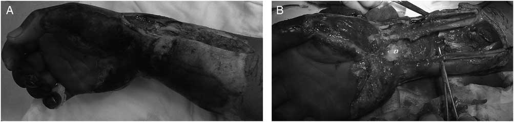
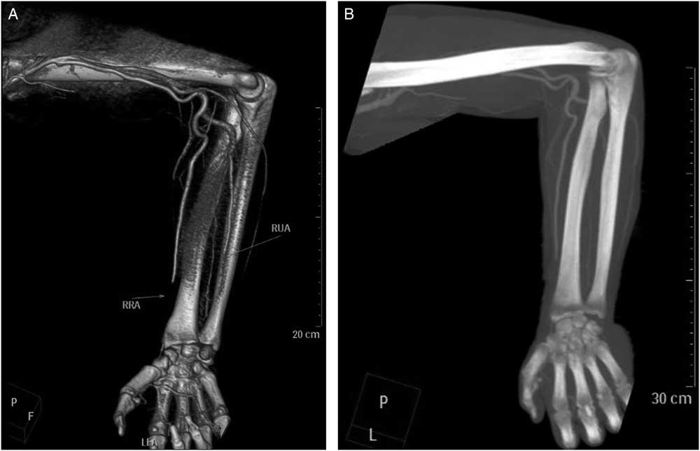
HVEI refers to the injuries caused by exposure to high-voltage electricity (≥1000V) far beyond human tolerance level (≥1000 V), including injuries to the heart, brain, skin, or muscle.Reference Dim, Amanari and Nottidge 9 After contact, the “entrance” and “exit” regions that the current passes through in the body would be burned most, as would be limb folds (such as axillary, elbow, etc.). The evolution of HVEI-caused tissue damage can be divided into two stages: instant injury when the body contacts the current followed by progressive injury occurring several hours later.Reference Chaudhry, Shaikh and Ahmad 10 Some scholars have found that when vessels are injured, the middle vascular layer contraction, intimal elastic membrane elongation, and vasculitis would appear, but no thrombosis would occur instantly.Reference Lee, Canady and Hammer 11 The patients enrolled in this study did not experience instant embolism, but over a longer time period, 21 patients developed muscular necrosis, consistent with findings from other studies, and this might be because the vessels themselves underwent pathological changes, which might delay the blockage of blood vessels, leading to muscular necrobiosis.Reference Baxter 12 - Reference Muehlberger, Krettek and Voqt 14 The current passing through blood vessels might easily lead to vessel wall injury and blood vessel obstruction; therefore, clinicians pay more attention to retaining HVEI patients’ limbs. Although efforts have been made to prevent electrical injury, according to the reports of foreign burn centers, the incidence rate is still gradually increasing.Reference Arnoldo, Purdue and Kowalske 15 The HVEI area is large (Figure 2A and B), and the injuries at the “entrance” and “exit” of the current are often covered by a wide range of necrotic tissues; therefore, deep tissues and muscles may develop occult necrosis. According to the characteristics of HVEI, clinical diagnosis and treatment for such patients normally require repeated surgical explorations to understand the extent and scope of tissue injury. Previously, intraoperative naked-eye observation, electrical stimulation of the striated muscle, and the intraoperative frozen-section method were routinely used to identify muscular necrosis. Hunt and HeckReference Hunt and Heck 3 applied NBT to identify HVEI muscular activities. The amputation rate in foreign reports is 65%.Reference Ferreiro, Meléndez and Regalado 16 According to our hospital’s statistics, among the 367 HVEI cases in the last 3 years, the amputation rate was 29.8%. Amputation is performed if: the limbs’ blood circulation is completely cut off, leading to development of necrosis in the limbs; tissues such as blood vessels, nerves, muscles, and bones are so severely injured that repair or reconstruction cannot be performed; or if serious infections occur, particularly those of an anaerobic nature. However, amputation inevitably causes disability; hence, a cautious attitude must be maintained. With rapid developments of multi-slice spiral CTA, because of its high temporal and spatial resolution, as well as its powerful post-processing capabilities, multi-slice CTA has made CTA-imaging techniques show their unique advantages in clearly displaying small arteries (Figure 3).
(A) Case 2 Preoperation; (B) During Debridement of the Right Hand, the Ulnar and Radial Arteries were Embolic.

(A) Volume-Rendering and (B) Maximum-Intensity Projection Images: The Superficial Palmar Arch, Deep Palmar Arch, and Interdigital Artery are Pale, but the Interosseous Artery has Good Blood Circulation. The Right Ulnar Artery (RUA) and Right Radial Artery (RRA) are not Occlusive, and Superficial and Deep Palmar Arches are Visible.

CT Perfusion Imaging
Routine CT scanning includes dual or 3-stage scanning when performing routine, enhanced, or specifically requested scanning; however, electrical injuries are more complex, showing such characteristics as multi-segments and jumping, so grasping their phases using routine scanning is challenging. In order to compensate for this limitation and to get the best CTA image, perfusion scanning should be performed. Perfusion scanning is a new functional CT inspection method, and its theoretical concepts were proposed by AxelReference Axel 17 in 1980, and then designed and completed by Berninger et al and Norman et al.Reference Norman, Axel and Berninger 18 , Reference Berninger, Axel and Norman 19 The theoretical bases of CT perfusion imaging are the dilution principles of nuclear medicine radiotracers and the laws of central volume: BF=BV/mean transit time.Reference Eastwood, Lev and Azhari 20 , Reference Ota, Takase and Rikimaru 21 After intravenous bolus injection of contrast agents, peer-level dynamic scanning is performed on selected levels, which could generate the TDC of each pixel in this level, with time as the abscissa and the increased CT values after the contrast agent is injected as the ordinate. On the basis of this curve, various perfusion indexes could then be calculated, followed by gradient color assignments to form the perfusion images; therefore, the blood perfusion characteristics of organs and lesions could be understood. Perfusion imaging is one method using imaging methods to visualize the in vivo perfusion processes and to perform quantitative or semi-quantitative analysis. In this study, Philips 256-slice iCT (Amsterdam, Netherlands) was used for perfusion scanning, which could capture a 256-layer image when rotating in a circle, within 0.27-0.4 seconds, with a slice thickness of 1-2 mm, spatial resolution of 24 lp/cm, as well as a 1024×1024 reconstructed matrix resolution (the highest reported currently). Further, its resolution and scanning speed are 3-4 times the previous ones, thus greatly reducing the time constraints, improving the spatial and temporal resolution, and maximally guaranteeing real-time and authentic image reconstruction. The BF value in the part above the amputation level on the affected side was significantly higher than that at the amputation level on the affected side. We used JOG-scanning technology to get CT perfusion imaging of more organs instead of conventional CT perfusion imaging, which would only get partial organ imaging limited by the detector width (z-axis-covered range:10-40mm), including single-slice helical CT to 64-slice helical CT. The Jog technology is the wide-range perfusion scanning technology developed by Philips, and it can perform repeated helical scanning over a wider range, thus allowing the perfusion data to be acquired over this range. Considering the impact of sampling times on TDC, the z-axis coverage could be up to 200-280 mm; hence, it could provide the basis for a wide range of limb perfusion imaging. The BF, PEI, TTP, and BV values are of practical significance in the determination of whether limb salvage is required. Thus, we can determine the amputation site in new patients based on the discriminant models of BF, PEI, TTP, and BV values, which provide valuable evidence for selection of the amputation site in the clinic.
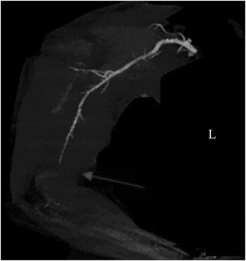
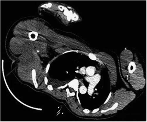
Clinical values of intelligent 256-slice CT perfusion imaging for limbs with HVEI; with the progress in new multi-slice CT technologies, it is possible to achieve the isotropism of images and increased spatial resolution to clearly display vascular lesions (Figure 4). Multi-slice spiral CT perfusion imaging (MSCTP) is one technology being used in recent years, which could reflect the blood supply conditions of the target organ at the parenchymal level, thus providing the functional status of tissues and organs. Perfusion imaging has been widely used to check organs such as the liver, brain, etc.; it could better show microscopic perfusion conditions of living tissues. Electricity has been widely used in production and daily life, and incidences related to it have increased year by year. MSCTP is the most widely used method for studying the organ perfusion status; after muscles are injured, they appear as low-density shadows in routine and enhanced CT scanning images (Figure 5), but in perfusion scanning, high perfusion prompts hyperemia and edema of the related regions, low perfusion prompts ischemia of the corresponding regions, and non-perfusion prompts necrosis of the corresponding regions. Tissue injury due to high voltage normally appears as “sandwich”-like necrosis of muscles because of different tissue resistances; the deep muscle tissue might develop necrosis under the intact skin surface area, thus clinical observation alone would be insufficient for the accurate determination of the extent of deep-tissue injuries. The application MSCTP could be to obtain the perfusion information of limb muscles, thus helping to determine the vitality of muscle tissues. Perfusion technology is presently only being applied to study HVEI using animal models.Reference Zhang, Wei and Shang 22 There was an early increasing of the TDC curve in normal skeletal muscles and became stable later on. When limb ischemia and necrosis occur, the early increasing of the TDC curve becomes slow, and no significant increasing peak can be seen; in perfusion images, muscle injuries appear as perfusion delay at the lesions, and when the muscles are completely necrotic no blood perfusion is seen. The 256-slice CT scanning technique could provide qualitative and quantitative image data for assessing the activities of muscle tissues and flap transplantation, and it has such advantages as enabling reasonable and timely treatment, reducing morbidity and complications, faster scanning speed, one-step and repeated scanning, as well as its use for preoperative and postoperative assessment, etc. Whether it could exhibit clinical importance for determining amputation or amputation range needs further investigation because of the small number of patients and a limited time period used in this study. Moreover, future studies should be performed to understand limb salvage after HVEI using magnetic resonance imaging.
Distal Brachial Artery is Interrupted Like a Mouse Tail.

Low-Density Shadow of Injured Triceps Brachii and Latissimus Dorsi Muscle.

Acknowledgments
Thanks for the help of all colleagues in the burn center of the Second Affiliated Hospital of Kunming Medical University, especially guidance and suggestions by Professor Fu Jinfeng and Professor Zeng Ming.
Author Contributions
P. Ma: Research concept, analysis of data, writing the paper, and final approval of the article. Y. Sun: Study design, drafting the article, revising it critically for important intellectual content. X. Zhang: Analysis of data, writing and revision of the article. H. Liu: Collection and assembly of data, analysis of data, and interpretation. C. Liu: Collection of data and final approval of the article. J. Zhao: Collection and assembly of data.
Ethical Statement
This study was conducted in accordance with the declaration of Helsinki. This study was conducted with approval from the Ethics Committee of the Second Affiliated Hospital of Kunming Medical University. Written informed consent was obtained from all participants.
Conflict of Interest
All authors have no conflict of interest regarding this paper.