INTRODUCTION
We do not simply inhabit our bodies, we literally use them to think with
(Seitz, 2000)In our everyday lives, we constantly interact with the environment through our bodies. The way we interact influences and shapes our understanding of both the external world and ourselves. The interaction between our body and the external stimuli has attracted increasing attention from both basic science and clinical communities.
One of the most intriguing and studied phenomena in this regard is the concept of embodied cognition (Barsalou, Reference Barsalou1999, Reference Barsalou2008; Gibbs, Reference Gibbs2006; Wilson, Reference Wilson2002). In contrast to the symbolic view of cognitivism that considers amodal symbols as the basis of mental activities, the embodied cognition view considers body and action at the core of cognition (Barsalou, Reference Barsalou1999). According to this integrated mind–body view, mental activities result from the interaction of multiple brain systems, which are linked to action, perception, and introspection. As such, perception, cognition, and action are closely related. The majority of the accounts of embodied cognition underlined the key role of simulation in cognition (e.g., Decety & Grèzes, Reference Decety and Grèzes2006). As defined by Barsalous, “Simulation is the reenactment of perceptual, motor, and introspective states acquired during experience with the world, body, and mind (Barsalous, Reference Barsalou2008, p. 618).” For example, visual mental simulation is the result of predicted sensory feedback formulated by a forward model on the basis of motor inputs, which are in turn the consequence of the body representation (Grush, Reference Grush2004; Schubotz, Reference Schubotz2007).
On these grounds, what happens when the peripheral motor feedback system does not function properly? Is mental imagery, and in particular motor simulation, influenced by alterations of the peripheral motor inputs and the body representation?
These questions are the starting point of the present work, and we sought to answer them by exploring the mental rotation (MR) performance in patients with myotonic dystrophy type 1 (DM1), an autosomal dominant inherited neuromuscular disorder. As such, DM1 patients may represent an ideal model for exploring the extent to which peripheral motor alterations can impact on cognitive functions as MR.
Mental Rotation: Cognitive and Neural Mechanisms
In MR tasks, individuals are required to compare pairs of object stimuli presented at different degrees of rotation and to judge whether these objects are identical or if one is the mirror image of the other. The classic finding is that the time to respond depends on the angular disparity between the objects: higher disparity leads to longer reaction times (Shepard & Metzler, Reference Shepard and Metzler1971). This pattern has been interpreted as reflecting the fact that people perform such tasks by “mentally rotating” an object as it would occur if the object were physically rotated (Thayer, Johnson, Corballis, & Hamm, Reference Thayer, Johnson, Corballis and Hamm2001).
Several researchers tried to test this idea by comparing performance in MR and physical rotation (Gardony, Taylor, & Brunyé, Reference Gardony, Taylor and Brunyé2014; Wiedenbauer & Jansen-Osmann, Reference Wiedenbauer and Jansen-Osmann2008; Wiedenbauer, Schmid, & Jansen-Osmann, Reference Wiedenbauer, Schmid and Jansen-Osmann2007; Wohlschläger & Wohlschläger, Reference Wohlschläger and Wohlschläger1998). In particular, Wohlschläger and Wohlschläger (Reference Wohlschläger and Wohlschläger1998) showed similar effects of angular disparity in response times (RTs) between MR task and a physical rotation task. Other studies have shown a positive effect of physical rotation training in a subsequent MR task (Wiedenbauer & Jansen-Osmann, Reference Wiedenbauer and Jansen-Osmann2008; Wiedenbauer et al., Reference Wiedenbauer, Schmid and Jansen-Osmann2007). More recently, Gardony et al. (Reference Gardony, Taylor and Brunyé2014) asked participants to rotate virtual 3D Shepard and Metzler objects mentally and physically, and observed similar patterns of response for MR and physical rotation, suggesting shared processes. The authors, however, stated that it is still premature to definitively ascertain that MR strictly mirrors the continuous physical rotation. Also, neuroimaging research provides still mixed results for the motor–MR link (see below).
A debate in the literature is also related to which extent the type of stimulus elicits different MR strategies, and consequently, a different involvement of the motor system. A variety of distinct types of stimuli have been indeed employed, such as 3D abstract objects (Cohen et al. Reference Cohen, Kosslyn, Breiter, DiGirolamo, Thompson, Anderson, Bookheimer, Rosen and Belliveau1996; Shepard & Metzler, Reference Shepard and Metzler1971), letters and numbers (Corballis & Sergent, Reference Corballis and Sergent1989), and hands, legs, and whole-body figures (Parsons, Reference Parsons1994).
Two main views can be recognized. According to some authors, although some stimuli – such as body parts – elicit motor simulation to greater extent, motor processes are intrinsically included in all visuospatial mental transformations, thus regardless of the type of stimulus to rotate (Amick, Schendan, Ganis, & Cronin-Golomb, Reference Amick, Schendan, Ganis and Cronin-Golomb2006; Paivio, Reference Paivio1986; Shepard, Reference Shepard1987). On the other hand, other studies showed that visuospatial and motor processes are cooperative but dissociated processes in MR (e.g., Dominey, Decety, Broussolle, Chazot, & Jeannerod, Reference Dominey, Decety, Broussolle, Chazot and Jeannerod1995; Jeannerod & Decety, Reference Jeannerod and Decety1995; Stevens, Reference Stevens2005). In particular, abstract objects were shown to trigger a visuospatial strategy centered on the object, whereas body parts are more prone to promote a motor simulation centered on the body (Kosslyn, Ganis, & Thompson, Reference Kosslyn, Ganis and Thompson2001; Munzert, Lorey, & Zentgraf, Reference Munzert, Lorey and Zentgraf2009; Sirigu & Duhamel, Reference Sirigu and Duhamel2001).
From the embodied cognition perspective, recent research investigated whether stimuli that trigger motor simulation, such as body parts, could facilitate MR performance by promoting the use of sensorimotor embodied strategies. Interestingly, Amorim et al. (Reference Amorim, Isableu and Jarraya2006) “anthropomorphized” abstract 3D Shepard and Metzler’s shapes by adding a head to the shapes of cubes and found that, as compared with the classical abstract stimuli, embodied stimuli yielded faster responses. Iachini, Ruggiero, Bartolo, Rapuano and Ruotolo (Reference Iachini, Ruggiero, Bartolo, Rapuano and Ruotolo2019) explored changes in MR during life span using different kinds of stimuli (hands, human/animal faces, and abstract stimuli). The authors found a general facilitation for concrete stimuli, but, interestingly, they observed that during childhood a specific facilitation occurred for body-related stimuli rather than simply for concrete ones.
Furthermore, during MR of body parts, the biomechanical constraints of the body affect the performance and mental imagery of body parts. For example, laterality judgments of hand stimuli take longer when individuals have palms in physically awkward postures than in canonical positions (Parsons, Reference Parsons1994).
MR is severely impaired in several movement disorders, including Parkinson’s disease and dystonia, and the extent to which the performance is affected seems to strictly depend on the stimulus type, and thus the strategy used. In particular, patients with Parkinson’s disease, having reduced motor capacity, showed more impairments in the MR performance with body parts than with 3D objects or letters (Amick et al., Reference Amick, Schendan, Ganis and Cronin-Golomb2006; Dominey et al., Reference Dominey, Decety, Broussolle, Chazot and Jeannerod1995).
Patients with dystonia, a neurological syndrome characterized by sustained muscular contractions that lead to abnormal postures and repetitive movements, showed deficits in MR of body parts (Fiorio, Tinazzi, & Aglioti, Reference Fiorio, Tinazzi and Aglioti2006; Katschnig et al., Reference Katschnig, Edwards, Schwingenschuh, Aguirregomozcorta, Kägi, Rothwell and Bhatia2010). In dystonia, motor disturbances may involve a single-body region, such as the hand in the writer’s cramp syndrome, which represents the most common form of focal hand dystonia (Hallett, Reference Hallett1998). Interestingly, the study by Fiorio et al. (Reference Fiorio, Tinazzi and Aglioti2006) showed that writer’s cramp patients had difficulties in mentally rotating hands but not feet, suggesting that the impairment was caused by the anatomical constraints of the real hand, and thus could not be generalized to the other (unaffected) body parts.
On the other hand, having good motor abilities (e.g., balance ability, coordinative ability) has been associated with a better MR performance, in children and older adults (Jansen & Heil, Reference Jansen and Heil2010; Jansen & Kaltner, Reference Jansen and Kaltner2014), and this relationship has been especially observed in MR performance with body-related stimuli (Jansen & Kaltner, Reference Jansen and Kaltner2014).
Regarding the neural bases of MR (Cona & Scarpazza, Reference Cona and Scarpazza2019; Zacks, Reference Zacks2008), two meta-analyses showed that cortical regions that are consistently activated during MR include posterior parietal and visual cortex, premotor cortex, and supplementary motor areas (see also Bode, Koeneke, & Jäncke, Reference Bode, Koeneke and Jäncke2007; Cona, Marino, & Semenza, Reference Cona, Marino and Semenza2017; Cona, Panozzo, & Semenza, Reference Cona, Panozzo and Semenza2017). In particular, previous studies showed a modulation in pre-SMA involvement as a function of the angle of rotation: the higher the angle of rotation, the greater the pre-supplementary motor area contribution (Cona & Semenza, Reference Cona and Semenza2017; Cona, Marino et al., Reference Cona, Marino and Semenza2017; Milivojevic, Hamm, & Corballis, Reference Milivojevic, Hamm and Corballis2009; Richter et al., Reference Richter, Somorjai, Summers, Jarmasz, Menon, Gati, Georgopoulos, Tegeler, Ugurbil and Kim2000). Interestingly, the meta-analysis by Cona and Scarpazza (Reference Cona and Scarpazza2019) found that, as compared with other visuospatial functions, MR tasks are selectively supported by activations in the left inferior occipital gyrus and a region in the right postcentral sulcus, between superior parietal lobule and the somatosensory cortex. These cortical regions are deputed to the integration of information that comes from various modalities (visual and/or somatosensory), a process critical in MR. The processes of mental imagery – a function inherently involved in MR tasks – imply indeed the mental reproduction of physical objects’ properties, which are not limited to visual modality (Barsalou, Reference Barsalou1999; Kosslyn et al., Reference Kosslyn, Ganis and Thompson2001). MR thus relies upon the integration of multimodal information. Furthermore, in view of the tight link between MR and physical rotation, which in turn leads to the idea of shared mechanisms between mental simulation and motor execution, it has been suggested that the primary motor cortex (M1) might support MR. Nevertheless, the existing studies provided mixing results: some studies found the M1 involvement (Pelgrims, Michaux, Olivier, & Andres, Reference Pelgrims, Michaux, Olivier and Andres2011; Tomasino, Borroni, Isaja, & Ida Rumiati, Reference Tomasino, Borroni, Isaja and Ida Rumiati2005), whereas others did not show any causal contribution of M1 to MR (Cona, Marino et al., Reference Cona, Marino and Semenza2017; Sauner, Bestmann, Siebner, & Rothwell, Reference Sauner, Bestmann, Siebner and Rothwell2006).
Myotonic Dystrophy Type 1
DM1 is one of the most common form of inherited neuromuscular disorders in humans (Harper, van Engelen, Eymard, Rogers, & Wilcox, Reference Harper, van Engelen, Eymard, Rogers and Wilcox2002). It is an autosomal dominant disease and results from a triplet cytosine thymine guanidine (CTG) repeat expansion on chromosome 19, which is excessively amplified (Day & Ranum, Reference Day and Ranum2005). The number of CTG repeats can vary from 50 to more than 4000 in these patients and is correlated with disease severity and negatively correlated with the age at onset (Ashizawa et al., Reference Ashizawa, Dubel, Dunne, Dunne, Pizzuti, Caskey, Perryman, Epstein and Hejtmancik1992).
Classic DM1 is characterized by CTG repeat expansion from 100 to 1000, slow progression of disorders’ severity, wasting and muscle weakness, and myotonia. Despite the heterogeneity of this condition, indeed, the most frequent signs are dominated by muscular impairment, including hypotonia and muscle weakness, and myotonia (i.e., difficulties and delayed relaxation, thus prolonged contraction, of the skeletal muscles after voluntary contraction or electrical stimulation). In DM1, myotonia and muscle weakness involve predominantly distal limb muscles. Cataract and cardiac conduction abnormalities are often observed (Ricker et al., Reference Ricker, Koch, Lehmann-Horn, Pongratz, Otto, Heine and Moxley1994). Individuals affected may become physically disabled over several decades.
Other abnormalities include cognitive dysfunction and sleep breathing disorders. In DM1, intellectual impairments have been observed in 24%–75% of patients and may range from selective cognitive deficits – such as dysfunctions in attention, visuospatial/constructional abilities, and verbal memory – to personality and social problems, up to severe mental decline (Modoni et al., Reference Modoni, Silvestri, Pomponi, Mangiola, Tonali and Marra2004; Serra et al., Reference Serra, Cercignani, Bruschini, Cipolotti, Mancini, Silvestri, Petrucci, Bucci, Antonini, Licchelli and Spanò2016). There are, however, very few studies that have explored cognitive alterations associated with DM1, and so far no study has ever investigated possible impairment in MR performance.
MRI studies showed white matter abnormalities and brain atrophy in DM1, in particular white matter lesions are located within anterior temporal lobes (Kornblum et al., Reference Kornblum, Reul, Kress, Grothe, Amanatidis, Klockgether and Schröder2004). Gray matter reductions have been also described in several cortical regions and recently observed also in thalami and hippocampi using voxel-based morphometry (Antonini et al., Reference Antonini, Mainero, Romano, Giubilei, Ceschin, Gragnani, Morino, Fiorelli, Soscia, Di Pasquale and Caramia2004; Weber et al., Reference Weber, Roebling, Kassubek, Hoffmann, Rosenbohm, Wolf, Steinbach, Jurkat-Rott, Walter, Reske and Lehmann-Horn2010).
Aims and Hypotheses
This study was aimed at exploring the role of peripheral motor feedback in mentally rotating different kinds of stimuli and at evaluating, for the first time, possible MR impairments in DM1 patients.
In line with the embodied cognition perspective, we expected that DM1 patients exhibited an impairment in MR performance and that such impairment would be more pronounced for MR of body parts (e.g., hand stimuli) than abstract objects. The rationale behind this hypothesis is the following: the peculiar signs in DM1 patients involve myotonia and muscular weakness of distal limbs, which cause abnormality in posture and movements. This condition may lead to alterations in the use of peripheral motor feedback and in turn to modifications of the body representation and the processes it mediates. If MR of body parts stimulates – and is prominently supported by – the activation of motor representations for motor simulation, DM1 patients’ abnormalities would be reflected in a decline in MR performance, especially in tasks involving body parts.
MATERIALS AND METHODS
Patients and the Healthy Controls
All analyses were performed in 24 DM1 patients (11 females; mean age: 43.7 years ± 11.35 standard deviation, SD; educational level: 12.7 years ± 3.20 SD) and in 24 age- and education-matched control subjects (14 females; mean age: 43.5 years ± 13.01 SD; educational level: 12.7 years ± 3.31 SD). For DM1 patients, diagnoses were confirmed by genetic testing in all cases. CTG repeat expansion sizes were determined in all DM1 patients (mean = 485 SD = 322; range 74–1100 repeats). None of the DM1 patients and control subjects had (other) neuromuscular or central nervous system disorders, or past medical history. The Muscular Impairment Rating Scale (MIRS) was used to assess disease severity (Mathieu, Boivin, Meunier, Gaudreault, & Begin, Reference Mathieu, Boivin, Meunier, Gaudreault and Begin2001). The MIRS mean value was 3.75 ± 0.73 SD. Informed written consent was obtained from all participants. The study was conducted in accordance with the ethical standards of the Declaration of Helsinki and following the guidelines of the Myotonic Dystrophy Association.
Neuropsychological Testing
Cognitive functioning and mood were assessed in both patients and healthy controls using a comprehensive neuropsychological test battery that involved: The Mini-Mental State Examination (MMSE) to evaluate global cognitive function, the Beck Depression Inventory (BDI) for depression, the Trail Making Test (TMT) – part A and B (for complex attention), the Forward Digit Span test and Backward Digit Span test (memory function); the Rey Complex Figure Test copy and recall (visuospatial perception and constructive dexterities, and memory); the Semantic and Phonemic Fluency (language and executive functions); the Stroop task (attention and inhibition); the Rey Auditory Verbal Learning Test (RAVLT; memory); the Raven matrices (intelligence and reasoning abilities).
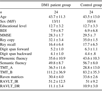
The scores in neuropsychological performance in both MD1 patients and healthy participants are presented in Table 1. Despite the fact that patients scored worse than the control subjects on almost all tests, their global cognitive level remains within the normal performance range (e.g., MMSE = 28.3 ± 1.7 SD).
Sociodemographic information and neuropsychological scores in patients and controls (mean ± standard deviation)

BDI = Beck Depression Inventory; MMSE = Mini-Mental State Examination; TMT = Trail Making Test; RAVLT = Rey Auditory Verbal Learning Test (IR = Immediate Recall; DR = Delayed Recall).
MR Paradigm
Two computerized MR tasks were administered, involving pictures of objects and hands, and were the same as used in the study by Cona, Marino et al., Reference Cona, Marino and Semenza2017; Cona, Panozzo et al., Reference Cona, Panozzo and Semenza2017. The experiment was run using the E-Prime software system. The two types of MR tasks involved the presentation of pictures of 3D objects and hands, and were administered in a counterbalanced order across participants. The pictures of abstract objects and hands had a size of 26.2° × 13.7° in visual angle if viewed from a 60 cm distance. In both tasks, pairs of stimuli were simultaneously presented and could appear at four angles of orientation (0°, 50°, 100°, and 150°). All the stimuli were rotated around the vertical axis. Each MR task comprised 56 pairs of stimuli: 7 “same” and 7 “mirror” pairs of different stimuli, presented with four possible MR angular disparities (0°, 50°, 100°, and 150° of rotation). Participants were asked to judge whether the stimulus presented on the right was the same or the mirror version of the stimulus presented on the left by saying “sì” (yes; same stimuli) or “no” (no; mirror stimuli). Verbal responses and times of decision responses were measured using E-Prime software system: “yes” and “no” responses were associated with two specific keys on the PC keyboard. As soon as the participant gave the response, the neuropsychologist stopped the chronometer by pressing the corresponding key on the keyboard so that the decision time and the type of response were recorded by E-Prime system. The sequence and timing of events in a single trial was as follows: a fixation cross was presented on the screen for 500 ms, followed by the target stimuli (objects or hands), which remained on the screen as long as the participants gave a response. Then, a blank screen was presented for 2500 ms.
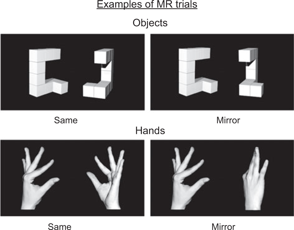
The object stimuli were similar to 3D Shepard and Metzler object figures obtained from the dataset by Ganis and Kievit (Reference Ganis and Kievit2015). The objects were white on a black screen and had a natural-looking shading effect. The hand stimuli were 3D pictures of upright hands, constructed with a digital camera and picture-editing software (Adobe Photoshop C24 Version 11.0). The hands were white on a black background. Figure 1 illustrates some examples of trials comprising hand stimuli and object stimuli, in both same and mirror conditions.
Examples of experimental trials of mental rotation (MR) tasks. The panels display pairs of stimuli (abstract objects and hands) at 150° of rotation in “same” conditions and in “mirror” conditions (where the right stimulus is the mirror version of the stimulus on the left).

As recommended by Ganis, Keenan, Kosslyn and Pascual-Leone (Reference Ganis, Keenan, Kosslyn and Pascual-Leone2000), the stimulus on the left part of the screen was always a left hand whereas the stimulus on the right part of the screen was a left hand in 50% of the pairs and a right hand in the other 50%, in order to avoid visuomotor interference.
The variables taken into account were: group (patients, control), stimulus type (hands, objects) × match condition (same, mirror) × angle (0°, 50°, 100°, and 150°).
Mean proportion of accuracy and mean RTs only for correct responses were collected for each level as dependent variables.
RESULTS
MR Performance
For both the accuracy and the RTs, only the effects that are relevant for the purposes of the work will be presented. Such effects will be displayed also in Figures 2 and 3 (but see Tables 2 and 3, for an overview of all the significant and nonsignificant effects on accuracy and RTs, respectively).
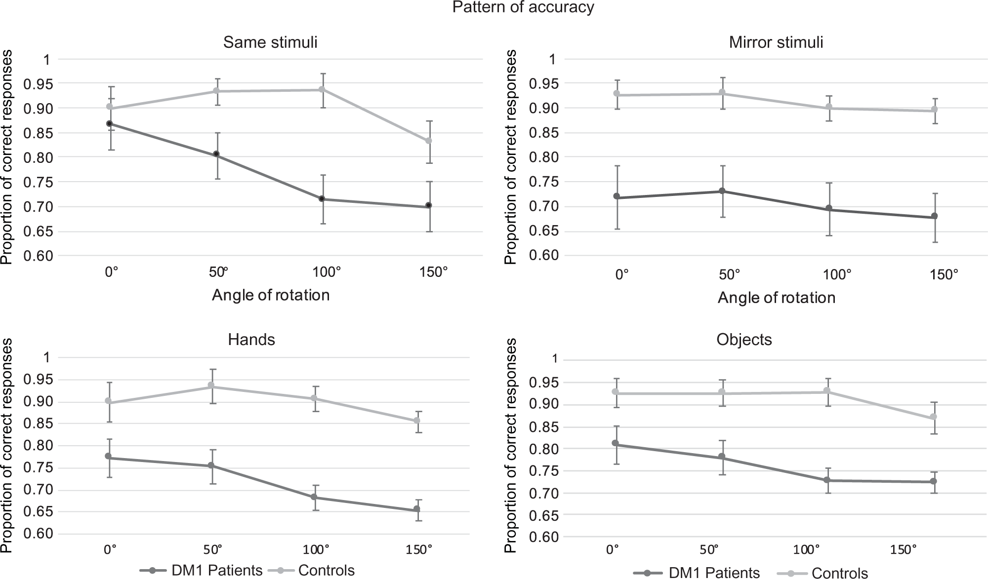
Accuracy performance in Hand and Object MR tasks, in DM1 patients and healthy controls. The mean proportion of correct responses as a function of the degree of rotation (0°, 50°, 100°, and 150°) are shown separately for same and mirror stimuli (upper panel) and for hands and object stimuli (lower panel), in DM1 patients and healthy controls. Error bars represent standard errors.

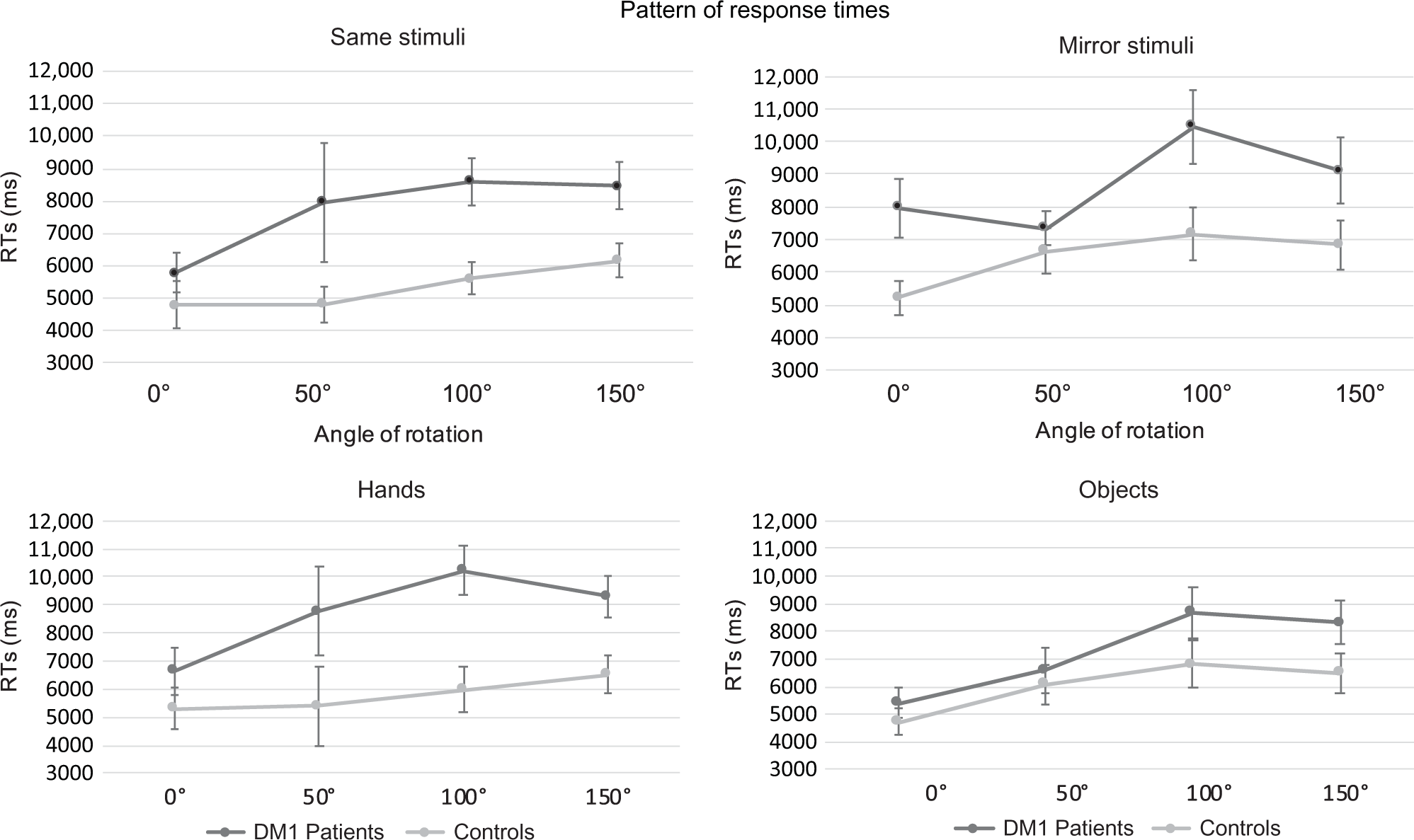
Response times in Hand and Object MR tasks, in DM1 patients and healthy controls. The mean response times as a function of the degree of rotation (0°, 50°, 100°, and 150°) are shown separately for same and mirror stimuli (upper panel) and for hands and object stimuli (lower panel), in DM1 patients and healthy controls. Error bars represent standard errors.

Overview of the main effects and interactions in the accuracy of MR tasks

The factors included in the analysis were: Stimuli (objects vs. hands), Group (patients vs. controls), Match (same vs. mirror), and Angle (0°, 50°, 100°, and 150°).
Significant effects with p < .05 are indicated with an asterisk.
Overview of the main effects and interactions in the response times of MR tasks

The factors included in the analysis were: Stimuli (objects vs. hands), Group (patients vs. controls), Match (same vs. mirror), Angle (0°, 50°, 100°, 150°).
Significant effects with p < .05 are indicated with an asterisk.
Marginally significant effects are indicated with the § symbol.
The analysis of MR performance accuracy showed a main effect of Group [F(1,46) = 23.77; p < .001;  $\eta _p^2$ = .34], with patients having a lower level of performance as compared to controls. Importantly, the significant Group × Angle interaction [F(3,138) = 3.27; p < .05; $\eta _p^2$ = .06] suggests that patients had impaired MR performance only when there was an angular disparity between the stimuli (Figure 2). Bonferroni-corrected post hoc comparisons showed indeed that no significant differences were observed for the 0° trials, whereas a significantly worse performance was observed in patients for 50°, 100°, and 150° trials (all ps < .001).
In order to better test the null effect at 0°, thus to quantify and compare the relative likelihood of the data under the alternative (H1) and the null (H0) hypothesis, we analyzed the Bayes factor (BF10) of the contrasts of interest (Morey & Rouder, Reference Morey and Rouder2015; Rouder, Morey, Speckman, & Province, Reference Rouder, Morey, Speckman and Province2012). A BF10 value ≥ 3 indicates robust evidence for the alternative over the null hypothesis (Jeffreys, Reference Jeffreys1946), whereas a BF10 value < 1/3 indicates evidence for the null hypothesis. Values of BF10 ranging between 1/3 and 3 indicate that the evidence is insensitive at distinguishing between the alternative and the null hypotheses (Dienes, Reference Dienes2014). We run the analyses using JASP 0.10 software (available on https://jasp-stats.org/) using the default setting: r scale fixed effects = .5, r scale random effects = 1, r scale covariates = .35, and samples = auto. A Bayesian repeated-measures ANOVA was carried out on proportion of accuracy in 0° trials, with match (same, mirror), stimuli (hands, objects), and group (patients, control) as variables.
The model that received the strongest support against the null hypothesis was the interaction model between Stimuli × Match × Group by the BF of 8.89, which was preferred to the model with only Group (BF = 4.77) as well as to the other remaining models (all BFs < 3). To explore the interaction model, we performed Bayesian independent t tests, which revealed that the MR performance was not significantly different between patients and controls when the stimuli were same (BFhands = .34; BFobjects = .30), whereas a significant worse performance in patients as compared to controls was observed for mirror stimuli (BFhands = 3.93; BFobjects = 25.87). This corroborates previous evidence suggesting that individuals rely upon different strategies when the stimuli match or not (Cona, Panozzo et al., Reference Cona, Panozzo and Semenza2017; Gardony et al., Reference Gardony, Taylor and Brunyé2014; Hochberg & Gellman, Reference Hochberg and Gellman1977).
The analysis of RTs was performed only for correct responses (see Figure 3, Table 3). As four patients obtained 0% of accuracy in some of the conditions, they were not included in the RT analysis.
The analysis revealed a significant main effect of Group [F(1,42) = 4.86; p < .05; $\eta _p^2$ = .10] and a significant Group × Stimuli configuration interaction [F(1,42) = 4.61; p < .05; $\eta _p^2$ = .09]. Bonferroni-corrected post hoc comparisons showed that, as compared to the control group, the patient group took longer to judge the rotation of stimuli for hands (p < .05), whereas no significant differences were found with objects stimuli.
A three-way Group × Match × Angle interaction was also significant [F(3,126) = 2.54; p = .05; $\eta _p^2$ = .06]. Bonferroni-corrected post hoc comparisons showed a significant slower performance in patient group in the “same” condition when the stimuli had 100° and 150° of rotation (p < .05), but not for 0° and 50°; whereas for the “mirror” condition, a significant slower performance was observed in patients only for the 0° and 100° of rotation (p < .05).
As for the analysis of accuracy, we performed a Bayesian repeated-measures ANOVA on RTs in 0° trials. The models that included Group as factor did not reach the BF of 3 (BFGroup = 1.45; BFGroup × Simulus = 1.19; BFGroup × March = 1.02) suggesting the absence of a substantial difference between patients and controls in 0° trials.
Relationships between MR Performance and Neuropsychological Scores
In order to investigate possible influences of the level of various cognitive abilities in MR, we analyzed the correlations between performance scores on neuropsychological tests and accuracy on MR tasks, in both DM1 patients and healthy controls. When analyzing the performance on MR with abstract objects, we found that significant correlations with performance in Stroop and Raven tests, both in patients (r = −.51; p = .01 and r = .55; p = .005, respectively) and controls (r = −.61; p = .002 and r = .51, p = .01, respectively). Control group showed significant correlations between object MR performance and the scores both in the forward digit span test (r = .55, p = .005) and the backward digit span test (r = .72; p < .001), whereas such correlations were not significant in the patient group (r = .36 and r = .28, respectively). By contrast, only patients showed a significant correlation with the scores in the Rey Complex Figure Test Copy (r = .62; p = .001; r in controls = .33; p > .05).
In the hand MR performance as well, the correlation with the scores in the Rey Complex Figure Test Copy was significant only for the patient group (r = .46; p = .024) whereas no significant relation was observed for controls (r = .13; p > .05), reflecting probably a more prominent contribution of motor and constructive impairment in hand MR performance in patients. Significant correlations between performance in the Rey Complex Figure Test Recall (visuospatial memory) and hand MR performance were found in both patients (r = .56; p = .004) and controls (r = .48; p = .02).
Finally, in patients only, significant correlations were shown between hand MR performance and scores in both the Raven test (r = .60; p = .002) and the Rey Auditory Verbal Learning Test (r = .45; p = .005).
DISCUSSION
The present study investigates, for the first time, possible alterations in MR in patients with DM1. As such, the findings of this study are relevant in two ways: to gain new insights into the neurocognitive mechanisms of MR and to better explore the cognitive profile of DM1 patients.
To address these issues, we used kinds of stimuli that varied for the extent to which they would emphasize the use of motor simulation (body parts) versus visuospatial imagery (abstract objects).
The results of the pattern of accuracy showed no differences between DM1 patients and controls in trials with 0° degree of rotation between the two stimuli when they were identical, thus indicating that DM1 patients had no deficits in perceptual discrimination. On the other hand, the significant reduced level of accuracy observed in DM1 patients for all rotated stimuli (50°, 100°, and 150°) demonstrates the presence of deficits in MR operations.
Two main sources might be proposed to account for the reduced MR performance in DM1 patients: visuospatial difficulties and constructional dysfunctions due to alterations in motor peripheral inputs. Indeed, patients only showed a significant correlation between MR performance – in both object and hand MR tasks – and the scores in the Rey Complex Figure Test Copy, which evaluates constructional abilities and visuomotor integration skills, behavioral monitoring and performing mental manipulations. Interestingly, previous studies found that patients had mainly visuospatial construction but not spatial orientation abnormalities (Johnson et al. Reference Johnson, Abresch, Carter, Kilmer, Fowler, Sigford and Wanlass1995; Sistiaga et al., Reference Sistiaga, Urreta, Jodar, Cobo, Emparanza, Otaegui, Poza, Merino, Imaz, Marti-Masso and López De Munain2010; Winblad, Jensen, Månsson, Samuelsson, & Lindberg, Reference Winblad, Lindberg and Hansen2006). For example, the study by Zalonis et al. (Reference Zalonis, Bonakis, Christidi, Vagiakis, Papageorgiou, Kalfakis, Manta and Vassilopoulos2010) revealed that, among the various functions, the most affected function (together with fluid intelligence) was the constructional visuomotor skill assessed by the Rey Complex Figure Test Copy. Furthermore, the work by Romeo et al. (Reference Romeo, Pegoraro, Ferrati, Squarzanti, Sorarù, Palmieri, Zucchetta, Antunovic, Bonifazi, Novelli and Angelini2010) showed that such two abilities were the only functions able to distinguish between DM1 and DM2 patients, with DM1 patients being more affected. This study also identified frontal and parieto-occipital alterations in DM1 patients, demonstrating that such alterations strongly correlated with poorer performance in tasks evaluating constructional visuomotor tests. Since these areas are activated also during MR tasks, DM1 patients’ impaired performance could be also accounted for by the dysfunction of these areas (Cona, Marino et al., Reference Cona, Marino and Semenza2017; Cona, Panozzo et al., Reference Cona, Panozzo and Semenza2017; Ganis et al., Reference Ganis, Keenan, Kosslyn and Pascual-Leone2000; Kosslyn et al., Reference Kosslyn, Ganis and Thompson2001; Zacks, Reference Zacks2008). In line with Romeo et al.’s study, we found significant correlations between the scores in Raven test (fluid intelligence) and performance in MR tasks with object stimuli (for both groups) and hand stimuli (only for DM1 patients).
The pattern of RTs revealed that DM1 patients were slower than controls in performing MR task, but selectively with hand stimuli. The selective impairment for rotating hand stimuli can be attributed mainly to DM1 patients’ abnormalities in motor representation and manipulation, likely due to modifications of body representation. This might be caused by the alterations in the use of peripheral motor inputs, as DM1 disorder involves myotonia and muscles weakness of distal limbs. Indeed, according to the classic neuropsychological observations, an online representation of body posture, labeled “body schema”, is shaped from multimodal sensory inputs (including somatosensory, proprioceptive, visual, and vestibular inputs) and interacts with motor system to support the guidance of movements (Cole & Paillard, Reference Cole, Paillard, Bermudez, Marcel and Eilan1995; Schwoebel et al., Reference Schwoebel, Friedman, Duda and Coslett2001).
Neuropsychological evidence provided by Sirigu et al. (Reference Sirigu, Duhamel, Cohen, Pillon, Dubois and Agid1996) suggests that not only actual but also mentally simulated movements depend on representations included in body schema. The findings of the present study are in line with previous evidence in other disorders involving movement alterations: impairments in rotating body parts were shown in patients with dystonia (Fiorio et al., Reference Fiorio, Tinazzi and Aglioti2006; Katschnig et al., Reference Katschnig, Edwards, Schwingenschuh, Aguirregomozcorta, Kägi, Rothwell and Bhatia2010), Parkinson’s disease (Amick et al., Reference Amick, Schendan, Ganis and Cronin-Golomb2006; Dominey et al., Reference Dominey, Decety, Broussolle, Chazot and Jeannerod1995), and even in patients with conversion paralysis, a psychiatric condition that induces a loss of voluntary motor function without an organic cause (de Lange, Roelofs, & Toni, Reference de Lange, Roelofs and Toni2007; de Lange, Toni, & Roelofs, Reference de Lange, Toni and Roelofs2010). Similar results were found in patients with complex regional pain syndrome, who presented with a specific impairment in mentally rotating the affected hand (Moseley, Reference Moseley2004; Schwoebel et al., Reference Schwoebel, Friedman, Duda and Coslett2001). This is, however, the first time that the processes underlying MR were investigated in patients with DM1. In this regard, these findings deepened our understanding on the role of motor system in the service of MR and can be discussed in the light of embodied cognition (Wilson, Reference Wilson2002). In a broader sense, embodied cognition is conceptualized as the fact that cognitive processes and mental representations are rooted in the body and in its interaction with the external world, and that body representations serve to act on cognitive processes (Wilson, Reference Wilson2002). Our findings are coherent with the view of embodied cognition, indicating a tight relation between body and cognitive processes underlying MR. Our study indeed showed that MR is influenced by body and body representations, and demonstrated that, when these are altered and impaired, MR is affected in turn. Muscle weakness, myotonia, and posture abnormalities – characteristic signs of DM1 – would have an impact on monitoring and manipulating patients’ body schema: The altered body representation would lead indeed to less accurate and reliable use of such monitoring/manipulating processes, which have a key role in MR.
Interestingly, in a recent work, Goldinger, Papesh, Barnhart, Hansen and Hout (Reference Goldinger, Papesh, Barnhart, Hansen and Hout2016) shared skeptical thoughts about the tendency to generalize the concept of embodied cognition to every cognitive phenomenon or activity, but they highlighted and emphasized the MR as an exception, stating the importance of motor system in carrying out MR operations.
Finally, considering together the results on accuracy and RTs, we found that the specific MR impairment for hand stimuli in DM1 patients was observed only in the pattern of RTs. By contrast, the accuracy pattern did not show a significant difference in the patients’ impairment between hands and objects. Yet, when looking at the graphs (Figure 2), at the mean proportion of accuracy and at SD of means for hands (Patients: M = .72, SD = .18; Controls: M = .90, SD = .07) and objects (M = .76, SD = .16; Controls: M = .91, SD = .11), it appears that patients’ performance was lower with hands than objects. It might be thus possible that a greater impairment for hands did exist but was masked in the pattern of accuracy because of other concurrent factors, such as the relatively small sample size and/or the high variability in the level of performance, especially among patients.
The discrepancy in the pattern of findings between accuracy and RTs might also be due to the fact that different processes and abilities come into play in MR performance, as also evidenced from the analysis of correlations with neuropsychological tests. Importantly, a possible explanation of this (apparent) discrepancy comes from the studies that explored the relationships between mental and physical rotation. The study by Gardony et al. (Reference Gardony, Taylor and Brunyé2014) showed indeed a similar pattern of RTs for MR and physical rotation, suggesting shared processes underlying them. Accordingly, it is possible that the alterations in the use of peripheral inputs to support the MR of hand stimuli are mainly expressed in the RTs as they would mirror the pattern of the actual rotation of hands. In such a way, RTs would reveal the impact of altered motor representations on MR processes to a greater extent than accuracy pattern would do.
An issue that deserves a future exploration is, however, the specific contribution of both central and peripheral motor alterations in producing MR deficits in DM1 patients. Furthermore, the interplay between motor system and cognition in MR paradigms needs to be taken into account not only in basic research but also in clinical domains as it could offer important insights in driving rehabilitation programs (De Vries & Mulder, Reference De Vries and Mulder2007, for a review). Since the rationale behind the classical use of motor imagery in clinical setting lies in the assumption that motor imagery and motor execution share the same neural circuits (Jackson, Lafleur, Malouin, Richards, & Doyon, Reference Jackson, Lafleur, Malouin, Richards and Doyon2001; Johnson-Frey, Reference Johnson-Frey2004), this rationale might be extended to other functions, such as MR. In this way, motor imagery would represent a crucial process of mental practice to support and enhance the performance in other – more cognitive – domains.
ACKNOWLEDGMENTS
This research received no specific grant from any funding agency, commercial or not-for-profit sectors. The present work was carried out within the scope of the research program Dipartimenti di Eccellenza (art.1, commi 314-337 legge 232/2016), which was supported by a grant from MIUR to the Department of General Psychology, University of Padua.
CONFLICT OF INTEREST
The authors declare no conflict of interest.