Introduction
Toxoplasma gondii is a ubiquitous obligate intracellular parasite that can infect any nucleated host cell of warm-blooded vertebrates, including humans (Jones and Dubey, Reference Jones and Dubey2012). Its transmission mainly occurs by the ingestion of undercooked meat containing tissue cysts, or the consumption of water or food contaminated with oocysts (Elsheikha, Reference Elsheikha2008). This opportunistic pathogen infects around one-third of the worldwide population, with varying prevalence rates in different countries (Hill and Dubey, Reference Hill and Dubey2002). In general, most infected individuals present a chronic infection and cysts are formed in the brain, skeletal muscle, heart and other organs with no evident symptoms or only mild ones (Jensen et al., Reference Jensen, Camejo, Melo, Cordeiro, Julien, Grotenbreg, Frickel, Ploegh, Young and Saeij2015). However, in immunocompromised patients, such as those with AIDS or those undergoing immunocompromising treatments (transplantation and cancer chemotherapies), Toxoplasma infections lead to severe clinical symptoms and even death (Montoya and Remington, Reference Montoya and Remington2008). Furthermore, congenital toxoplasmosis is one of the main causes of miscarriages, stillbirth and fetal abnormalities (Robbins et al., Reference Robbins, Zeldovich, Poukchanski, Boothroyd and Bakardjiev2012). Despite its detrimental impact on public health, there are still no effective methods for the prevention of toxoplasmosis (Dubey, Reference Dubey2008; Boothroyd, Reference Boothroyd2009).
Like all members of the phylum Apicomplexa, T. gondii possesses an apical complex consisting of three secretory organelles: microneme, rhoptry and dense granules. Notably, rhoptry secretes effector proteins (ROPs), which play a significant role in host cell invasion by forming and modifying parasitophorous vacuoles (Bradley and Sibley, Reference Bradley and Sibley2007; Boothroyd and Dubremetz, Reference Boothroyd and Dubremetz2008). Among secreted ROPs, ROP18, a Ser/Thr protein kinase, has been identified as a key factor responsible for the virulence of T. gondii by interacting with host proteins and phosphorylating other ROP proteins (Saeij et al., Reference Saeij, Boyle, Coller, Taylor, Sibley, Brooke-Powell, Ajioka and Boothroyd2006; Taylor et al., Reference Taylor, Barragan, Su, Fux, Fentress, Tang, Beatty, Hajj, Jerome, Behnke, White, Wootton and Sibley2006; Sinai, Reference Sinai2007). In rodent hosts, ROP18 has been found to phosphorylate immunity-related GTPases (IRGs), such as Irga6, Irgb6 and Irgb10, and prevent their accumulation at parasitophorous vacuole membranes (PVMs), as a result, the clearance of susceptible and moderately virulent strains is impaired (Fentress et al., Reference Fentress, Behnke, Dunay, Mashayekhi, Rommereim, Fox, Bzik, Taylor, Turk, Lichti, Townsend, Qiu, Hui, Beatty and Sibley2010; Steinfeldt et al., Reference Steinfeldt, Konen-Waisman, Tong, Pawlowski, Lamkemeyer, Sibley, Hunn and Howard2010). Moreover, ROP18 also phosphorylates activating transcription factor 6 β (ATF6β) and p65 guanylate binding protein 1 (GBP1) in humans (Virreira Winter et al., Reference Virreira Winter, Niedelman, Jensen, Rosowski, Julien, Spooner, Caradonna, Burleigh, Saeij, Ploegh and Frickel2011; Yamamoto et al., Reference Yamamoto, Ma, Mueller, Kamiyama, Saiga, Kubo, Kimura, Okamoto, Okuyama, Kayama, Nagamune, Takashima, Matsuura, Soldati-Favre and Takeda2011; Yamamoto and Takeda, Reference Yamamoto and Takeda2012). These phosphorylation events increase the resistance of the PVM against host immune defences and enhance the acute virulence of the parasites. Furthermore, research on factors interacting with ROP18 in host cells indicates that ROP18 also affects other cellular processes, such as protein degradation (Yang et al., Reference Yang, Hou, Hao, Rho, Wan, Luan, Gao, Yao, Pan, Xie, Qian, Liao, Zhu and Zhou2017), host cell apoptosis (Xia et al., Reference Xia, Kong, Zhou, Wu, Yao, He, He and Peng2018) and causes encephalitis (Jensen et al., Reference Jensen, Camejo, Melo, Cordeiro, Julien, Grotenbreg, Frickel, Ploegh, Young and Saeij2015).
Considering the critical role of TgROP18 in parasite growth and virulence, this protein has been targeted for vaccine development (Yuan et al., Reference Yuan, Zhang, Lin, Petersen, He, Yu, He, Zhou, He, Li, Liao and Zhu2011) and the design of anti-toxoplasmosis drugs (Yin et al., Reference Yin, Zhao, Xu, Qiu, Wen, Sun, Liu, Liu, Zhao, Wei, Huang, Yan and Cao2019). Despite the efforts to understand the interactions between TgROP18 and Toxoplasma-infected host cells, so far there are no commercially available ROP18 monoclonal antibodies (mAbs), which hinders our understanding of the molecular mechanisms governing TgROP18 function, and also impairs potential clinical applications such as the development of diagnostic kits. By using traditional hybridoma technology, in the present study, we produced and characterized immunochemical properties of two mAbs against TgROP18. Moreover, we assessed their reactivity against T. gondii (genotypes I, II and Chinese I), Plasmodium falciparum, and Schistosoma japonicum proteins. Finally, we amplified, cloned and characterized complementarity-determining regions (CDRs) of one promising mAb that showed high strain specificity.
Materials and methods
Parasites
Human foreskin fibroblast (HFF) cells were cultured in Dulbecco's Modified Eagle Medium (DMEM, Gibco, Waltham, MA, USA) supplemented with 10% fetal bovine serum (FBS, Sijiqing, Hangzhou, Zhejiang, China), 100 μg mL−1 penicillin and 100 μg mL−1 streptomycin (Sigma, St. Louis, MO, USA), and maintained in an incubator at 37 °C and 5% CO2. Toxoplasma gondii RH strain (type I,high virulence), ME49 strain (Type II,intermediate virulence), green fluorescent protein-RH strain (GFP-labelled RH), TgCtwh3 (Chinese I, high virulence) and TgCtwh6 (Chinese I, intermediate virulence) were propagated in confluent HFF monolayers, and the tachyzoites were harvested by filtering through 3 μm-pore-size polycarbonate filters.
Whole protein extracts of S. japonicum were obtained as described previously (Ratcliffe and Wilson, Reference Ratcliffe and Wilson1991; Li et al., Reference Li, Ross, Sleigh, Li, Waine, Williams, Tanner and McManus1999). Briefly, adult S. japonicum worms were collected from infected rabbits and homogenized by milling the worms in PBS and freezing the homogenate at −80 °C for 30 min; these procedures were repeated three times. Then, the homogenate was centrifuged at 12 000 × g for 1 h and the supernatant was collected and used in further assays. Plasmodium falciparum was cultured in a 5% CO2 atmosphere at 37 °C using a modification of the method proposed by Trager and Jensen (Reference Trager and Jensen1976). Parasites were released from RBCs by saponin lysis, the whole proteins were extracted into high-salt buffer from isolated parasite nuclei as described previously.
Expression and purification of recombinant ROP18 proteins (rROP18)
Competent Escherichia coli BL21 (DE3) cells transformed with recombinant plasmid pET28a-ROP18 of T. gondii RH were cultivated in Luria–Bertani (LB) medium at 37 °C. Recombinant plasmid pET-28a-ROP18 was previously constructed and its sequence match the identity of the published sequence for T. gondii rop18 gene (GenBank:AM075204). The expression of the recombinant protein was induced by adding 100 mm isopropyl β-D-1-thiogalactopyranoside (IPTG, Sigma) and incubating cells for 4 h. Since the target proteins were found in the inclusion bodies fraction, they were initially purified by multiple washing and centrifugation. Then, they were dissolved under denaturing conditions using a binding buffer containing 7 M hydrochloride. The protein extracts were subsequently purified using His-bind Purification Kit (Novagen, Burlington, MA, USA). The concentration of the purified rROP18 was quantified using a Nano Drop 2000 spectrophotometer (Thermo Fisher Scientific, Waltham, MA, USA) and the purity was analysed by SDS-PAGE (10%). Furthermore, Western blotting was used to identify the purified rROP18 using His mAb (Abcam, Cambridgeshire, England).
Mice immunization and monoclonal antibody production
For mAb production, 8- to 12-week-old female BALB/c mice were immunized. For the primary immunization, 50 μg of purified rROP18 diluted in 25 μL PBS was mixed with an equal volume of complete Freund's adjuvant (Sigma) and injected intradermally to the back of each mouse at multiple points. Two weeks later, a booster immunization injection was given subcutaneously to each mouse with the same dosage of rROP18 in Freund's incomplete adjuvant (Sigma), followed by a third booster intraperitoneal injection with 20 μg rROP18. Three days after the last injection, blood was collected from the tail vein to measure the titre of antibodies by enzyme-linked immunosorbent assay (ELISA, section ‘Enzyme-linked immunosorbent assay’). Then, mice were sacrificed and had their spleen isolated under aseptic conditions. Myeloma cells (SP2/0) in logarithmic growth phase treated by 8-azaguanine (Sigma) were fused with spleen cells using polyethylene glycol 1500 (Roche, Basel, Switzerland) in Iscove's Modified Dulbecco's Medium (IMDM, Solarbio, Beijing, China).
Peritoneal macrophages collected from normal BALB/c mice were cultured in hypoxanthine-aminopterin-thymidine selection medium (Gibco, Thermo Fisher Scientific) and plated onto a 96-well microplate (Sigma) to serve as feeder cells. Hybridomas from myeloma cells and mouse spleen cells were grown in the 96-well microplate containing the peritoneal macrophages at 37 °C with 5% CO2. Ten days later, the culture medium was changed to hypoxanthine-thymidine and cells were cultured for about 10 days before screening for positive clones. The supernatants of the 96-well microplate were screened by ELISA using the purified rROP18. After ELISA screening (section ‘Enzyme-linked immunosorbent assay’), positive hybridomas were cloned by three cycles of the limiting dilution method. Briefly, at first, positive hybridoma cells were resuspended to 50 cells mL−1, inoculated into 36 wells with 100 μL per well in a 96-well plate. Then, the remaining cell suspension was diluted to 10 cells mL−1, inoculated as the first step. Finally, all remaining cell suspension was further diluted to 5 cells mL−1 and inoculated into the remaining 24 wells. The cloned positive hybridoma cells were grown at 37 °C with 5% CO2 and observed under an inverted microscope to identify the wells with single clonal growth about 1 week later. The presence of mAbs against ROP18 was further identified by Western blotting (section ‘SDS-PAGE and Western blotting’) and immunofluorescence assays (section ‘Indirect immunofluorescence assay’).
Enzyme-linked immunosorbent assay
The titre in the serum from immunized mice was assessed using an indirect ELISA. Purified rROP18 protein (200 ng mL−1) was used to coat an ELISA plate at 4 °C overnight. The plate was washed three times with PBS-T (PBS containing 0.05% Tween-20) and then blocked with 5% bovine serum albumin (Sigma) at 37 °C for 1 h. The serum of immunized mice was serially diluted from 1:1600 to 1: 409 600 and the serum of untreated mice was used as a negative control. Diluted serum (100 μL) was added to each well and incubated at 37 °C for 1 h. Then, the plate was washed three times with PBS-T, and a horseradish peroxidase (HRP)-conjugated goat anti-mouse Ig (Zhongshan Golden Bridge, Beijing, China) diluted 1:5000 in PBS was added as a secondary antibody to each well and incubated at 37 °C for 1 h. Thereafter, the wells were washed and incubated with 100 μL of tetramethylbenzidine (TMB, Beyotime, Shanghai, China) per well at room temperature for 10 min. Finally, the reaction was stopped using 50 μL of 2 M H2SO4 per well, and the absorbance was read at 450 nm.
The screening of hybridomas for mAbs against TgROP18 was performed as follows. Coating, blocking and washing were performed as described above. Then, the supernatants from the cultured hybridomas were added as the primary antibody and incubated at 37 °C for 1 h, additionally, the serum of immunized mice and culture supernatants of myeloma cells (SP2/0) were used as positive and negative controls, respectively. Following three washes again, the secondary antibody, TMB and the H2SO4 were added as described for serum analysis.
Antibody subtype characterization and epitope identification
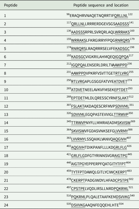
The subtype of the mAbs in the supernatants of the positive hybridomas was identified by Mouse Monoclonal Antibodies Isotyping Kit (Roche) according to the manufacturer's instructions. Epitopes were identified by peptide scanning. Briefly, based on the amino acid sequence of mature ROP18 (GenBank: CAJ27113.1, position: 98–554), a total of 24 overlapping peptides were designed and synthesized (Top-peptide Biotechnology, Shanghai, China) in sequences of 25 amino acid residues overlapping by six residues at each end of the polypeptide (Table 1). Peptides were suspended in sterile 0.9% NaCl to a concentration of 2 mg mL−1 and stored at −20 °C until use. The antigen epitopes of mAbs were identified by indirect ELISA with the synthetic peptides (50 μg mL−1) as coating antigens, the prepared A1 and T2 ascites (1:5000, section ‘SDS-PAGE and Western blotting’) as primary antibodies, and HRP-IgG as a secondary antibody.
Design of the synthesized peptide sequence

The underlined amino acids are repeated sequences with the end of the previous.
Preparation of mice ascites
After screened by ELISA, the two positive hybridomas, A1 and T2, were separately cultured in IMDM medium plus 10% FBS in 24-well plates at 37 °C under 5% CO2. Then, 1 × 10⁶ hybridomas were collected into a 1.5 mL centrifuge tube, washed with 1 mL preheated (37 °C) IMDM, centrifuged at 200 × g for 5 min and resuspended in 500 μL IMDM. The hybridomas were intraperitoneally injected into 8-week-old BALB/c mice, 1 week after they had been injected with 500 μL mineral oil (Solarbio). About 10 days after hybridoma injection, mice were euthanized and the formed ascites with anti-rROP18 mAbs were collected and centrifuged at 750 × g for 10 min. After removing the lipids in the upper layer, the ascites were centrifuged again at 12 000 × g for 30 min, and then purified by protein G agarose (Beyotime). Ultimately, the supernatants were collected, purified and stored at −80 °C until use.
SDS-PAGE and Western blotting
The specificity and sensitivity of the produced mAbs were assessed by Western blot. Briefly, rROP5, a protein member of the ROP2 family, and the purified rROP18 were separately mixed with an equal volume of protein loading buffer, and denatured for 10 min in boiling water. Then, samples were electrophoresed in 10% SDS-PAGE and transferred onto a nitrocellulose membrane (Millipore, Billerica, MA, USA). After the membranes were blocked with 5% skimmed milk at room temperature for 2 h and washed three times with PBS-T, purified mice ascites were added as the primary antibody (1:5000) and incubated at 4 °C overnight. Following another PBS-T wash, HRP-conjugated goat-anti-mouse Ig (1:5000, ZSGB-Bio, Beijing, China) was added as the secondary antibody. After incubation and wash as described for the primary antibody, an enhanced luminol-based chemiluminescent kit (Thermo Scientific) was used to detect the bands. Furthermore, to detect the ability of the mAbs to recognize the T. gondii strains, whole protein extracts from T. gondii (RH, TgCtwh3, TgCtwh6 and ME49 strains), P. falciparum and S. japonicum were also electrophoresed and assessed as described above. For comparative purposes, polyclonal anti-ROP18 antibodies produced in our laboratory were incubated as primary antibody in another set of Western blot assays.
Cloning and sequencing of the variable regions of heavy and light chains cDNAs from hybridomas
Total RNA was extracted from 1 × 10⁷ T2 hybridomas using RNeasy Mini Kit (Qiagen, Dusseldorf, Germany) and cDNA was synthesized using Transcriptor First Strand cDNA Synthesis Kit (Roche). The cDNA of the heavy and light genes were PCR-amplified using 30 pairs of primers (KMD Bioscience, Tianjing, China). Amplicons were visualized after electrophoresis in 1.5% agarose gel. Bands of interest were purified and ligated to pGEM-T vector for cloning (Promega, Madison, USA). After transforming competent cells, positive colonies were screened and subjected to nucleotide sequencing. Finally, CDRs of the mAbs were predicted using the IgBlast tool (https://www.ncbi.nlm.nih.gov/igblast/).
Indirect immunofluorescence assay
About 1 × 10⁴ HFF cells were grown on coverslips placed in wells of a six-well plate and incubated in 5% CO2 at 37 °C for 3 or 5 days. Then, cells were infected with T. gondii engineered RH-GFP strains for 12, 24 or 36 h. After washing with PBS twice, they were fixed with 4% paraformaldehyde (Sangon Biotech, Shanghai, China) for 30 min. After fixation, coverslips were washed three times, permeabilized with 0.1% Triton X-100 (Sigma) for 15 min, and blocked for 30 min at 37 °C with 5% sheep serum (ZSGB-Bio) in PBS. Cells were washed again and incubated with purified mice ascites (1:200 dilution) overnight at 4 °C, and then washed three times with PBS. Coverslips were then incubated with rhodamine-conjugated anti-mouse IgG (1:100 dilution, ZSGB-Bio) at 37 °C for 1 h. After that, cells were washed with PBS four times and stained with 4′, 6-diamidino-2-phenylindole (Sigma) for 5–10 min at room temperature to stain the nuclei. Lastly, images were recorded using a Zeiss LSM880 confocal microscope (Zeiss, Jena, Germany).
Statistical analysis
Data are presented as mean ± standard deviation. Statistical analysis was performed using GraphPad Prism Software and one-way ANOVA. Differences were considered statistically significant when P < 0.05.
Results
Expression, purification and identification of recombinant ROP18 protein
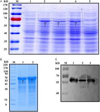
Under our experimental conditions, TgROP18 was successfully expressed by transformed competent cells after IPTG induction. Moreover, SDS-PAGE, as well as Western blotting analyses, revealed a single band and the estimated molecular size of the obtained recombinant protein was consistent with that calculated from the combined sizes of the protein (62.7 kDa) encoded by the rop18 gene and the N-terminal His-tag from the vector (Fig. 1A and C). The recombinant His-ROP18 protein was successfully purified by Ni2+-affinity chromatography, resulting in a purity of approximately 80% (Fig. 1B).
SDS-PAGE and Western blot analysis of the recombinant ROP18 protein (rROP18) expressed in Escherichia coli strain BL21. (A) Expression of pET28a-ROP18/BL21(DE3). Lane M: protein marker; Lane 1: proteins in pET28a-ROP18/BL21 before induction; Lane 2: proteins in pET28a-ROP18/BL21 after induction with isopropyl β-D-1-thiogalactopyranoside (IPTG); Lane 3: supernatant of pET28a-ROP18/BL21 bacteria after ultrasonication; Lane 4: precipitation of pET28a-ROP18/BL21 bacteria after ultrasonication; Lane 5: proteins in pET28a/BL21. (B) rROP18 purified by Ni2+-affinity chromatography. Lane M: protein marker; Lane 1–2: purified proteins. (C) Western blot analysis of the purified rROP18. Lane M: protein marker; Lane 1–3: rROP18 recognized by His monoclonal antibody.

Generation of monoclonal antibodies against rROP18
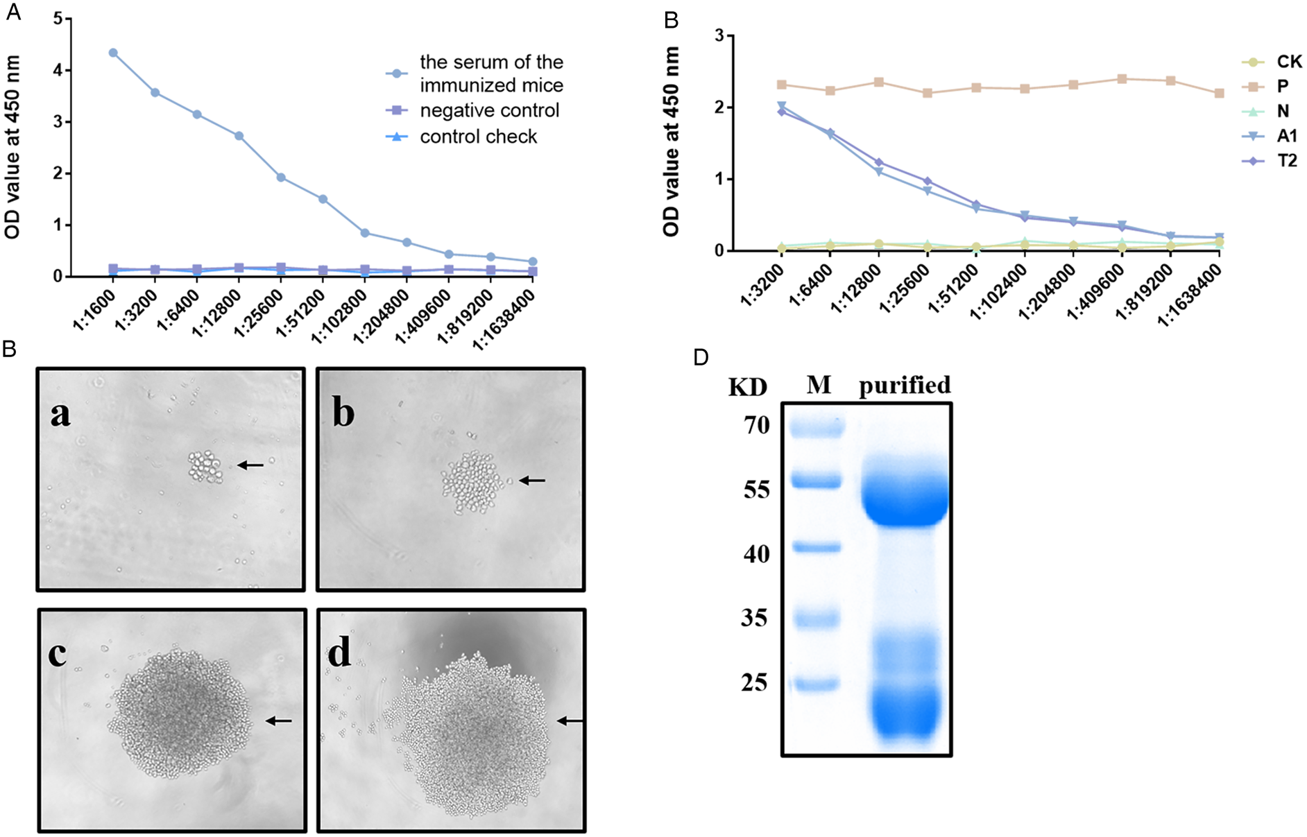
The presence of antibodies against TgROP18 was confirmed in the serum of immunized mice (up to 1: 204 800 dilution) before isolating the spleen cells (Fig. 2A). After screening and sub-cloning, two hybridomas secreting anti-TgROP18 mAbs were identified and named as A1 and T2 (Fig. 2B). Antibodies were detected in mice ascites produced by A1 and T2 hybridomas up to the 1: 102 400 dilution (Fig. 2C). This allowed their successful purification and the clear visualization of 25 and 45 kDa bands, corresponding to light and heavy chains, respectively (Fig. 2D).
Generation of monoclonal antibodies (mAb) against ROP18. (A) The presence of antibodies in the serum of immunized mice was detected up to the 1: 204 800 dilution by indirect ELISA. The serum of normal mice acted as negative control. The ratio of the positive group (P) to the negative group (N) was calculated, when P/N ⩾ 2.1 the result is positive, P/N < 2.1 and ⩾1.5 the result is uncertain, and P/N < 1.5 the result is negative. (B) Growth of subclones at different days: (a) 10 days; (b) 13 days; (c) 16 days; and (d) 20 days. (C) Levels of mice ascites produced by A1 and T2 hybridomas. (D) SDS-PAGE analysis of purified mice ascites. Lane M: protein marker; Lane purified: heavy and light chains of the purified mice ascites. Data represent three independent experiments, each with an intra-experiment duplicate.

Subtype and epitope identification of the anti-ROP18 antibodies
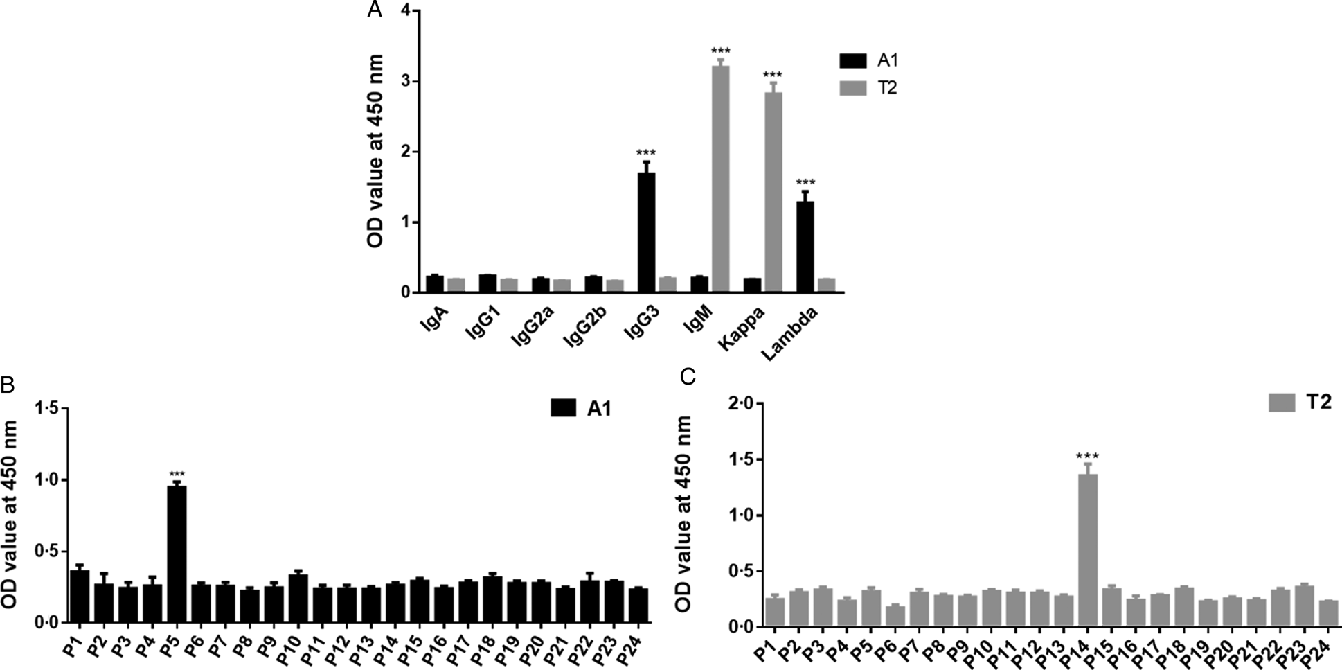
The mouse mAb isotyping kit revealed that the A1 and T2 mAbs belong to the IgG3 λ type and IgM κ type, respectively (Fig. 3A). Among the synthesized overlapping peptides, the A1 mAb had the highest activity against peptide 5, whereas T2 mAb reacted strongly against peptide 14. The core sequences of these two peptides are 180LRAQRRRSELVFE192 and 351NYFLLMMRAEADM363, respectively (Fig. 3B).
Subtype and epitope identification of the anti-ROP18 mAbs. (A) Subtype identification of A1 and T2 mAbs. Mouse monoclonal antibody isotyping kit indicated that the subtype of the A1 and T2 strains were IgG3 λ type and IgM κ type, respectively. ***P < 0.001. (B) Epitope identification of A1 and T2 mAbs by peptide scanning. The A1 and T2 mAbs recognized P5 and P14 peptides, respectively, which represented the core sequence of 180LRAQRRRSELVFE192 and 351NYFLLMMRAEADM363. **P < 0.01. Data represent three independent experiments, each with an intra-experiment duplicate.

Specificity of anti-ROP18 T2 mAbs
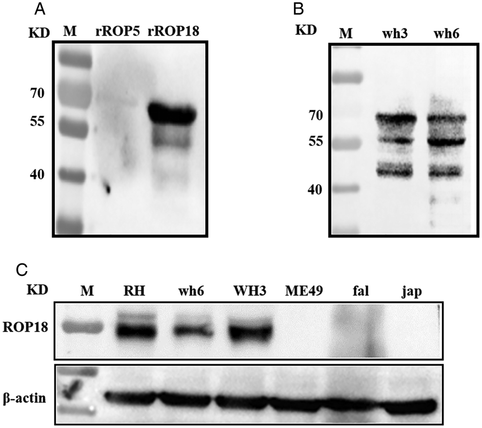
The T2 mAbs reacted specifically against rROP18 but not rROP5. A single band around 62.7 kDa was detected in membranes containing ROP18 (Fig. 4A). The mAbs produced by the T2 hybridoma could specifically recognize a 51 kDa protein in T. gondii RH (Type I), TgCtwh3 (Chinese I, high virulence) and TgCtwh6 (Chinese I, intermediate virulence) protein extracts, but did not react with that of T. gondii ME49 (Type II), P. falciparum and S. japonicum (Fig. 4C). We compared the specificity of the T2 mAb with that of a polyclonal antibody against ROP18 raised in rabbit. The polyclonal antibody reacted with whole protein extracts of T. gondii TgCtwh3 and TgCtwh6. However, in contrast to the produced mAb, Western blotting using the polyclonal antibody revealed protein bands at 45, 51 and 60 kDa positions (Fig. 4B). As A1 exhibited a lower stability than T2 during the passaging of hybridoma cells, A1 was not used for specificity analysis.
Specificity of the T2 mAbs evaluated by Western blot. (A) T2 mAbs reactivity against different recombinant proteins in ROP2 family. Lane M: protein marker; Lane 1: ROP5 recombinant protein; Lane 2: ROP18 recombinant protein. (B) Polyclonal antibody against ROP18 reactivity against Toxoplasma gondii strains. Lane M: protein marker; Lane wh3: whole protein extract from TgCtwh6; Lane wh6: whole protein extract from TgCtwh3. (C) T2 mAbs reacted with proteins from T. gondii strains, P. falciparum and S. japonicum. Lane M: protein marker, Lane RH: RH strain; Lane wh3: TgCtwh3 strain; Lane wh6: TgCtwh6 strain; Lane ME49: ME49 strain; Lane fal: P. falciparum; Lane jap: S. japonicum.

Sequence analysis of heavy chain and light chain variable regions in T2 antibodies
The sequencing results of heavy chain (H) and light chain (L) from T2 mAbs are presented in additional files (file S1 and file S2). After analysing the sequences using the IgBlast tool, nucleotide and amino acid sequences corresponding to H and L domains were predicted (Table 2).
Nucleotide and amino acid sequences of complementary determining regions (CDRs) of T2 monoclonal antibodies

Application of anti-ROP18 T2 antibodies in immunofluorescence
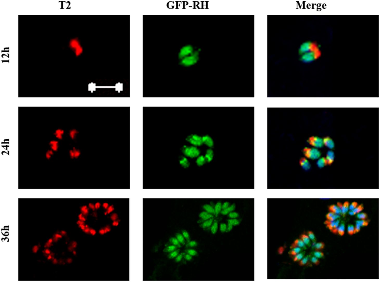
Confocal microscopy was used to localize the antigen recognized by T2 mAbs. The apexes of free tachyzoites and multiplying tachyzoites in the host cells were stained at 12, 24 h after infected T. gondii RH, while the host cells were not stained with T2 mAbs (Fig. 5). At 36 h post-infection, intense red fluorescence signals were observed at the apexes, and formed an orange petal arrangement.
Confocal laser scanning microscopy of the Toxoplasma gondii antigen recognized by T2 mAbs. Human foreskin fibroblast cells were infected with T. gondii GFP-RH strain tachyzoite for 12, 24 and 36 h, and T2 mAbs were used to detect the parasites. Toxoplasma gondii was double-stained with the T2 mAbs (red), GFP (green). Scale bar = 10 μm.

Discussion
The mAbs are monovalent antibodies that bind to a single epitope and are produced from a single B lymphocyte clone (Liu et al., Reference Liu, Chen, Shi, Sun, Zhang, Li, Xiao, Wang and Zhao2014). Since their generation in mice in 1975 – using a hybridoma technique – by Kohler and Milstein (Reference Kohler and Milstein1975), they have been widely used in biomedicine for different purposes, including the study of many apicomplexan parasites such as Plasmodium (Hakimi et al., Reference Hakimi, Goto, Suganuma, Angeles, Kawai, Inoue and Kawazu2015; Canepa et al., Reference Canepa, Molina-Cruz, Yenkoidiok-Douti, Calvo, Williams, Burkhardt, Peng, Narum, Boulanger, Valenzuela and Barillas-Mury2018; Carias et al., Reference Carias, Dechavanne, Nicolete, Sreng, Suon, Amaratunga, Fairhurst, Dechavanne, Barnes, Witkowski, Popovici, Roesch, Chen, Ferreira, Tolia, Adams and King2019), Babesia (Chiou et al., Reference Chiou, Kitoh, Igarashi and Takashima2014) and Eimeria (Liu et al., Reference Liu, Chen, Shi, Sun, Zhang, Li, Xiao, Wang and Zhao2014). Moreover, mAbs have become important tools for solving many problems in biology and medicine (Gao et al., Reference Gao, Huang, Zhu and Lv2018). Toxoplasma gondii is an important member of the Apicomplexa phylum. Although many mAbs targeting diverse antigens from different stages of T. gondii life cycle have been developed (Cunha-Junior et al., Reference Cunha-Junior, Silva, Silva, Souza, Souza, Prudencio, Pirovani, Cezar, Cascardo, Barbosa, Goulart and Mineo2010; Gondim et al., Reference Gondim, Wolf, Vrhovec, Pantchev, Bauer, Langenmayer, Bohne, Teifke, Dubey, Conraths and Schares2016), mAbs targeting TgROP18, the major virulence determinant between type I and III strains with respect to acute mortality, are not available (Saeij et al., Reference Saeij, Boyle, Coller, Taylor, Sibley, Brooke-Powell, Ajioka and Boothroyd2006; Taylor et al., Reference Taylor, Barragan, Su, Fux, Fentress, Tang, Beatty, Hajj, Jerome, Behnke, White, Wootton and Sibley2006; Sibley et al., Reference Sibley, Qiu, Fentress, Taylor, Khan and Hui2009). High expression of TgROP18 is closely related to the virulence of type I and type II strains, whereas its low expression is related to avirulence in type III strains (Saeij et al., Reference Saeij, Boyle, Coller, Taylor, Sibley, Brooke-Powell, Ajioka and Boothroyd2006; Taylor et al., Reference Taylor, Barragan, Su, Fux, Fentress, Tang, Beatty, Hajj, Jerome, Behnke, White, Wootton and Sibley2006; Chen et al., Reference Chen, Gao, Huo, Wang, Yu, Halm-Lai, Xu, Song, Hide, Shen and Lun2011). Indeed, TgROP18 has been an important target in investigations pertaining to diagnosis, pathogenesis, vaccine research and drug therapy of T. gondii (Sinai, Reference Sinai2007; Yuan et al., Reference Yuan, Zhang, Lin, Petersen, He, Yu, He, Zhou, He, Li, Liao and Zhu2011).
Considering the features of mAbs and the vital significance of TgROP18, in the present study, we developed two mAbs against TgROP18 using traditional hybridoma technology using purified rROP18 proteins expressed by E. coli as antigens. A previous report found that when immune antigens produced by prokaryotic expression systems lacking post-translational modification are used, antibodies against non-glycosylated and linearized epitopes are often produced, which often do not bind to native proteins (Jones et al., Reference Jones, McLoughlin, Connolly, Farquhar, MacGregor and Head2009). Our results demonstrated that the produced mAbs could recognize not only the recombinant TgROP18 but also the natural TgROP18 protein from the parasite. In terms of subtype of the two mAbs, we screened positive hybridomas using a secondary goat antibody against mouse IgG (H+L). Nevertheless, our further analysis revealed that one of the positive clones, T2, was in fact of the IgM κ type, and not IgG. One possible reason is that such secondary antibody can react not only with mouse IgG, but also with other immunoglobulin light chains of mice.
The specific binding reaction between antigen and antibody is directed by specific antigen epitopes rather than full-length molecules. Thus, the identification of an antigen epitope is of great significance in studying the whole process of immune response and understanding the structure and function of the antigen itself. The traditional methods of epitope identification include peptide scanning (Chen and Chang, Reference Chen and Chang2017), site-directed amino acid mutagenesis (Liu et al., Reference Liu, Mei, Hu, Yang, Liu, Li, Zhang, Cao and Liu2018), phage display (Liu et al., Reference Liu, Mei, Hu, Yang, Liu, Li, Zhang, Cao and Liu2018), immunoaffinity mass spectrometry (Purcell and Gorman, Reference Purcell and Gorman2004) and bioinformatic prediction technologies (Mei et al., Reference Mei, Li, Leier, Marquez-Lago, Giam, Croft, Akutsu, Smith, Li, Rossjohn, Purcell and Song2019). Among these methods, peptide scanning has the advantage to be convenient and accessible, but it can only identify linear epitopes. The two mAbs developed in the present study could specifically react with denatured recombinant and natural proteins, which showed that both of them recognize linear epitopes but not conformational epitopes of ROP18. The reason is that linear epitopes generally consist of consecutive linearly arranged amino acids that can still be identified even after denaturation of natural protein antigens. On the other hand, conformation epitopes consist of several amino acids arranged discontinuously but close to each other in space to form a specific conformation that is disassembled after denaturation. To identify the linear epitopes recognized by the two mAbs using peptide scanning, we designed 24 overlapping peptides with 25 amino acid residues overlapping by six residues at each end of the polypeptide. The analysis by indirect ELISA showed that the core sequences recognized by the A1 and T2 mAbs were 180LRAQRRRSELVFE192 and 351NYFLLMMRAEADM363, respectively. Both of them were located at the C-terminus of ROP18. Such identification of epitope recognized by anti-ROP18 mAbs supports the development of molecular vaccines and diagnostic tools in the future.
To verify the specificity of the two mAbs against TgROP18, we tested their reactivity against protein extracts from different parasite strains and recombinant ROP5, a pseudokinase in ROP2 family which acts together with ROP18 to modulate the virulence of the parasite. The data showed that the two mAbs recognize rROP18 but not ROP5; whether they cross-react with other ROP2 family proteins warrants further evaluation. Moreover, these mAbs react with both T. gondii Type I and Chinese I, but not with T. gondii Type II, P. falciparum and S. japonicum. Chinese I is the dominant genotype in China and accounts for about 65.2% of the isolates identified in this area (Chen et al., Reference Chen, Gao, Huo, Wang, Yu, Halm-Lai, Xu, Song, Hide, Shen and Lun2011; Chaichan et al., Reference Chaichan, Mercier, Galal, Mahittikorn, Ariey, Morand, Boumediene, Udonsom, Hamidovic, Murat, Sukthana and Darde2017; Zhu et al., Reference Zhu, Li, Pappoe, Shen and Yu2019). Thus, our findings indicate that these mAbs are good candidates to be used in detecting T. gondii infections in China. Unfortunately, these mAbs were not able to distinguish the high virulence strain from the intermediate virulence strain since they recognized both TgCtwh3 and TgCtwh6. Nevertheless, mAbs did not react with proteins from ME49 (Type II). Such selectivity may arise from substitutions in the amino acid sequence of ME49 ROP18. Actually, we found the V190F single point mutation in A1 and the A361T single point mutation in T2 epitopes in ME49. This is consistent with a previous report in which mAbs against the surface antigen p22 recognized different T. gondii strains based on amino acid substitutions in the epitope (Parmley et al., Reference Parmley, Gross, Sucharczuk, Windeck, Sgarlato and Remington1994). Altogether, these data showed that the mAbs against TgROP18 are not only species-specific but also strain-specific. As different types of T. gondii result in different clinical outcomes, scientists have been working towards the development of strain-specific diagnostic tools, over the past decades (Kong et al., Reference Kong, Grigg, Uyetake, Parmley and Boothroyd2003). Our results also indicate that the mAbs can be used in serotyping and in distinguishing Type I/Chinese I infections from those caused by Type II T. gondii.
Hybridomas can provide a continuous supply of mAbs with high affinity and specificity. However, the process of hybridoma cell preservation is associated with cases of loss of antibody gene, poor cell viability, low antibody production and even cell death. To overcome these limitations, the gene encoding the antibody of interest in hybridoma cells can be extracted and sequenced. Herein, we cloned and sequenced the genes of H chain and L chain from the T2 hybridoma, and predicted the amino acid sequences in their CDRs. Although this represents an important step towards the engineering of ROP18 mAbs and antibody derivatives, their application in the prevention, diagnosis and treatment of T. gondii infections, as well as the study of the parasite–host interaction, warrants further research.
Conclusion
We have successfully developed two new mAbs against TgROP18 with strain-specific reactivity. These mAbs are suitable for Western blotting, immunofluorescence and in vivo function analysis. Furthermore, the identification of the sequences of their CDRs allows the modification and design of mAbs against ROP18 proteins, which play critical roles in parasite growth and toxoplasmosis infection. Our mAbs may be useful for elucidating the roles of ROP18 in the interaction with the host cells, which might improve our understanding of the molecular mechanisms underlying T. gondii infection.
Supplementary material
The supplementary material for this article can be found at https://doi.org/10.1017/S0031182020000177
Acknowledgements
The authors would like to thank Dr Bo Wang (Anhui Medical University) for providing the cell culture of P. falciparum.
Authors' contributions
Yl designed the work; SR and ZF collected the data; ZF, SR, WY, LJ, HC, ZJ, LQ and SJ analysed the data; YL and ZF drafted the article; WY, LJ, HC, ZJ, SJ and LQ did critical revision of the article. All authors contributed to the discussion of the results followed by writing, read and approved the final manuscript.
Financial support
This work was supported by grants from the National Key R&D Program of China (to LY, grant no. 2017YFD0500400), Natural Science Foundation of China (to LY, grant no.81572022, 81871671, and JS, grant no. 81471983) and Key projects for supporting outstanding young talents in Universities in Anhui (to LY, grant no. gxyqZD2016044).
Conflict of interest
The authors declare that the research was conducted in the absence of any commercial or financial relationships that could be construed as a potential conflict of interest.
Ethical standards
This study was conducted strictly in accordance with the ‘Regulations on the Management of Experimental Animals of the People's Republic of China’ of the Ministry of Science and Technology of the People's Republic of China. Mice (BALB/c) were provided by the Experimental Animal Center of Anhui Medical University, and suffering of experimental animals was minimized. The authors assert that all procedures contributing to this work comply with the ethical standards of the Institutional Review Board of the Anhui Medical University Institute of Biomedicine on the care and use of laboratory animals.