Bovine mastitis, one of the most persistent, prevalent and expensive diseases of dairy cows (Viguier et al. Reference Viguier, Arora and Gilmartin2009; Derong & Zhao, Reference Derong and Zhao2010) is defined as ‘inflammation of the bovine udder’ (Fetrow, Reference Fetrow2000). This disease causes considerable distress to the animal, decreased milk production and major economic losses on dairy farms worldwide (Crispie et al. Reference Crispie, Alonso-Gómez, Collette, Klostermann, Flynn, Arkins, Meaney, Ross and Hill2008). Dairy cows are highly susceptible to mastitis, mainly during the dry period (Ryan et al. Reference Ryan, Flynn, Hill, Ross and Meaney1999) and antibiotics are currently administered at the drying period to help to eliminate subclinical cases and to prevent the establishment of new intramammary infections (Twomey et al. Reference Twomey, Wheelock, Flynn, Meaney, Hill and Ross2000).
The use of antibiotics for prophylactic treatment is being subjected to considerable debate all over the world because of its perceived connection with the emergence of antibiotic resistance in bacteria, particularly with the increased prevalence of organisms such as methicillin-resistant Staphylococcus aureus, which are prevalent in nosocomial infections in humans (David & Daum, Reference David and Daum2010). Moreover the use of antibiotics to control mastitis generates the appearance of residues in the milk of treated cows (Dalton, Reference Dalton2006). Such concerns have prompted the World Health Organization (WHO) and United Nations Organization for Food and Agriculture Organization (FAO), in the 34th session of the Codex Alimentarius Commission, to increase measures aimed at reducing antimicrobial resistance generated for antibiotic therapies in bovines (González-Gracia, Reference González-Gracia2011). Some researchers (Browning et al. Reference Browning, Mein, Barton, Nicholls and Brightling1990; Crispie et al. Reference Crispie, Flynn, Ross, Hill and Meaney2004a; Klostermann et al. Reference Klostermann, Crispie, Flynn, Ross, Hill and Meaney2008) have recommended that dry cow antibiotic therapy should not be used as a routine prophylactic measure but rather restricted only to the treatment of infected cows.
During recent years there has been increased interest in developing alternative approaches to the prevention of intramammary infections, particularly in dry cows (Crispie et al. Reference Crispie, Flynn, Ross, Hill and Meaney2004b, Reference Crispie, Twomey, Flynn, Hill, Ross and Meaney2005; Dallard et al. Reference Dallard, Ortega, Iguzquiza, Salvetti, Quaino and Calvinho2010). In this sense, there is a wide variety of proposals that include the application of vaccines (Giraudo et al. Reference Giraudo, Calzolari, Rampone, Rampone, Giraudo and Bogni1997; Pellegrino et al. Reference Pellegrino, Giraudo, Raspanti, Odierno and Bogni2010; Pereira et al. Reference Pereira, Oliveira, Mesquita, Costa and Pereira2011) and some immunomodulators (Larsen et al. Reference Larsen, Moser, Hoiby, Song and Kharazmi2004; Dallard et al. Reference Dallard, Ortega, Iguzquiza, Salvetti, Quaino and Calvinho2010). Other measures include the use of ‘organic’ products, such as the use of probiotic microorganisms (Klostermann et al. Reference Klostermann, Crispie, Flynn, Ross, Hill and Meaney2008) or some of the metabolic or bioactive compounds they are able to produce (Crispie et al. Reference Crispie, Twomey, Flynn, Hill, Ross and Meaney2005).
For the identification of the beneficial strains that can be used as probiotics, there are many criteria and laboratory assays, many of them recommended by international organizations (FAO & WHO, 2002; International Scientific Association for Probiotics and Prebiotics (ISAPP, 2009). One of the characteristics to be applied for the selection of probiotic strains is the capability of adhesion to epithelial cells (Otero & Nader-Macías, Reference Otero, Nader-Macías and Méndez-Vilas2007; Both et al. Reference Both, Gyorgy, Kibédi-Szabó, Tamás, Ábrahám, Miklóssy and Lányi2010). Adhesion is one of the strategies that promote the further colonization of the bacteria. Also the adhesion of the lactobacilli to the epithelium is the first step in the formation of a barrier or a biofilm to prevent undesirable microbial colonization (Lebeer et al. Reference Lebeer, Verhoeven, Perea-Vélez, Vanderleyden and De Keersmaecker2007). Adherence of Lactobacillus to epithelial surfaces has been studied in vaginal epithelial cells because of their competition with pathogens for the receptor sites for adhesion (Osset et al. Reference Osset, García, Bartolomé and Andreu2001; Nader-Macías et al. Reference Nader-Macías, Ocaña, Juárez Tomás, Silva de Ruiz and Pérez Martinez2007).
In earlier work, Lb. perolens CRL 1724 was isolated from bovine milk and characterized as a potential probiotic strain (Espeche et al. Reference Espeche, Otero, Sesma and Nader-Macias2009; Frola et al. Reference Frola, Pellegrino, Espeche, Giraudo, Nader-Macias and Bogni2011). Frola et al. (Reference Frola, Pellegrino, Espeche, Giraudo, Nader-Macias and Bogni2011) showed that in vitro Lb. perolens CRL 1724 was able to inhibit and co-aggregate with microorganisms considered as major bovine mastitis causing pathogens. Moreover Lb. perolens CRL 1724 showed a high efficacy in vitro to adhere to bovine teat canal epithelial cells and was recovered from all the mammary quarters in vivo. No clinical signs or teat damage were observed in lactating cows after the inoculation of 106 cfu/ml.
The aim of the present study was to evaluate whether intramammary inoculation of Lactobacillus perolens CRL 1724 in dry-period cows produced some type of structural modifications in the gland, using histological examination. The persistence of the strain in the udders 7 d post inoculation was also determined.
Materials and methods
Bacterial strain and culture conditions
Lb. perolens CRL 1724 (Centro de Referencia para Lactobacilos Culture Collection) used in this study was isolated and characterized as potentially probiotic from milk of healthy Holstein cows and characterized according to Espeche et al. (Reference Espeche, Otero, Sesma and Nader-Macias2009) and Frola et al. (Reference Frola, Pellegrino, Espeche, Giraudo, Nader-Macias and Bogni2011).
Lb. perolens CRL 1724, resistant to streptomycin, was grown in Man, Rogosa and Sharpe broth (MRS, Britania) at 37 °C for 18 h, and stored in milk yeast extract (MYE) (10 g low fat milk, 0·5 g yeast extract and 1 g glucose per 100 ml) with 12% glycerol at −20 °C. Before performing the experimental assays, bacteria were subcultured three times, every 12–14 h at 37 °C in MRS broth. The bacterial inoculum was prepared as follows: a culture of the strain (109 cfu/ml) incubated for 18 h at 37 °C in MRS broth was centrifuged and the bacterial pellet was washed twice with saline solution (0·8% NaCl). Cells were suspended in 5 ml of saline solution to obtain a concentration of 109 cfu/ml. The concentrated preparation was serially diluted in saline solution to 106 cfu/ml. This inoculum was fractionated and stored at 4 °C until inoculation was performed (a period no longer than 2 h) (Frola et al. Reference Frola, Pellegrino, Espeche, Giraudo, Nader-Macias and Bogni2011).
Intramammary inoculation of Lb. perolens CRL 1724 at drying off
The study was carried out on an experimental dairy farm in Córdoba, Argentina. The experimental farm operated under conventional management. Five clinically healthy non-lactating Holstein cows were used for the assays. One of the cows from the experimental dairy was used to evaluate the effect of Lb. perolens CRL 1724 after intramammary inoculation and the other four cows were used for histological examinations; one of them belonged to the experimental farm and the other three cows were selected from a commercial dairy farm. The animals were of parity 1 and 4 and were in late lactation. Cows were milked twice daily and produced an average of 14 kg milk/d before interruption of lactation. The animals were selected based on previous bacteriological studies and somatic cell counts (SCC). All the quarters used in this work were free of major mastitis-causing pathogens (MCPs) and with SCC in individual quarters <200 000 cells/ml. The animals were inoculated after evening milking. The cleaning of the udders before inoculation, and inoculation procedure were performed following the methodology described by Frola et al. (Reference Frola, Pellegrino, Espeche, Giraudo, Nader-Macias and Bogni2011). The unit of study was the mammary quarter. After intramammary inoculation, four of the animals (of parity 1 and showing reproductive problems) were removed from the herd and sent to slaughter at the end of the trial.
One cow was first used to evaluate whether intramammary inoculation of Lb. perolens CRL 1724 produced some type of effect on the udder, by evaluation of the clinical signs, milk appearance, SCC and recovery of viable lactobacilli in the milk. Taking in account previous results (Frola et al. Reference Frola, Pellegrino, Espeche, Giraudo, Nader-Macias and Bogni2011) three quarters were infused once on day 0 (D0) with 1 ml containing 106 cfu of Lb. perolens CRL 1724. One quarter was used as control. To minimize animal handling and to follow animal welfare best practices, no infusion was inoculated in the control quarter.
Three quarters of each one of the remaining four cows were infused with 1 ml of 106 cfu of Lb. perolens CRL 1724 for histological examination. One quarter was used as control and samples were taken 2 d after intramammary inoculation.
Sampling and bacterial recovery
Before inoculation, foremilk samples were collected from each quarter according to the National Mastitis Council procedure (National Mastitis Council, 2004) immediately before milking. Milk or dry secretion samples were transported refrigerated (a period no longer than 2 h) to the laboratory and immediately 10 μl was plated onto blood-agar (TSA with 5% of sheep blood) and incubated at 37 °C for 24 h. Bacteria were characterized by standard biochemical tests (Bergey & Holt, Reference Bergey and Holt1994). SCC was performed with a Somacount 300 (Bentley) according to the revised protocol of the 148A method C, fluoro-opto-electronic (International Dairy Federation Laboratory, 1995). In all cows, milk or dry secretion samples were obtained 2 d before infusion (D − 2), immediately prior to infusion (D0) and until 2 d (D2) post infusion. One cow, used to evaluate whether the intramammary inoculation of Lb. perolens CRL 1724 produces some type of effect on the udder, was sampled daily until day 7 (D7).
Serial dilutions of samples in saline solution were streaked on MRS agar plates in duplicate and incubated at 37 °C for 24–48 h under microaerophilic conditions (5% CO2, 95% air) for Lactobacillus isolation. Isolated colonies were identified as Lb. perolens CRL 1724 by phenotypic tests (Gram stain, morphology, catalase activity, nitrate reduction, indole production) and by determination of streptomycin resistance.
Clinical observations and animal care
Clinical signs were monitored throughout the experiment by a veterinarian, every 8 h during the first 24 h and daily until the end of the assay. General attitude and appetite of the cows were observed. The udders were palpated for soreness, swelling, hardness and heat, and the appearance of milk and dry secretion was assessed visually for clots and changes in colour or composition. Animals were cared for in accordance with The International Guiding Principles for Biomedical Research Involving Animals (1985).
Histological examination of the dry udder
Four inoculated cows were sacrificed and examined histologically before sending them to the slaughterhouse. Two representative samples of canal and cistern of bovine mammary gland were removed and fixed in 10% buffered formalin and 2·5% glutaraldehyde (in 0·1 mol/l phosphate buffer, pH 7·4) during 24 h at room temperature for histological examination and transmission electronic microscopy (TEM).
For histological examination, fragments fixed were embedded in paraffin and cut in 5-μm sections. Sections were stained with haematoxylin and eosin (H-E) according to Grignaschi et al. (Reference Grignaschi, Diaz, Alonso, Lardo and Licero1983), toluidine blue and Gram, placed on a glass slide and covered with cover-slip according to a method standardized in our laboratory. Samples were examined under high resolution optical microscopy (HROM) and pictures were taken with a Powershot G6, 7·1 megapixels (Canon INC, Japón) digital camera and the software AxioVision Release 4.6.3 (Carl Zeiss, Alemania). For TEM examination, fixed tissue fragments were fixed overnight in 1% osmium tetroxide and afterwards treated with an aqueous solution of 2% uranyl acetate for 40 min. After fixation, tissues were gradually dehydrated in a series of alcohol solutions of increasing strength, passed through acetone and embedded in Spurr resin. Ultrathin sections were stained with uranyl acetate and lead citrate and examined with Siemens Elmiskop 101 transmission electron microscope.
Results
Effect of intramammary inoculation of Lb. perolens CRL 1724
To evaluate the in-vivo performance of Lb. perolens CRL 1724, the tolerance of udder to the inoculation of 106 cfu/ml of lactobacilli was first determined in one non-lactating cow. The concentration used was tested previously in lactating cows (Frola et al. Reference Frola, Pellegrino, Espeche, Giraudo, Nader-Macias and Bogni2011). The inoculum was well tolerated by the animal: no clinical signs or teat damage were observed and the udders presented a normal aspect. There were no changes in the appearance of milk secretions. SCC in milk samples was 4·5 × 106 cells/ml after 24 h of intramammary inoculation and decreased to a normal value (1 × 10⁵ cells/ml) until the end of the assay (7 d post inoculation). Lb. perolens CRL 1724 was recovered until the end of the assay and from all the inoculated quarters. The highest bacterial recovery value (25 cfu/ml) was obtained 48 h after intramammary inoculations. All the quarters inoculated were negative for mastitis causing pathogen isolation during the whole trial period.
Histological examination
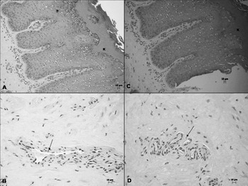
Intrammamary inoculation of Lb. perolens CRL 1724 evaluated by epithelial tissue sections from the teat canal of the udders (analysed by histological preparations) showed, in all cows, the presence of a stratified squamous epithelium (flat cells) with an important keratin layer (Fig. 1a, c). No difference was found between inoculated and non-inoculated quarters and both quarters presented scarce numbers of blood cells in blood vessels (Fig. 1b, d) in connective tissue. No bacteria were observed after H-E and Gram stains (data not shown).
High resolution optical microscopy showing haematoxylin and eosin stained from representative epithelial tissue sample of bovine mammary gland canal after intrammamary inoculation with Lactobacillus perolens CRL 1724. (a) and (b) non-inoculated with Lb. perolens CRL 1724. Presence of keratin superficial layer (K) and connective tissue with presence of empty blood vessel (black arrow) (20× and 40, respectively). (c) and (d) epithelial tissue samples and connective tissue from inoculated quarter without change compared with controls (20× and 40, respectively). The bar in each panel is equivalent to 10 μm.

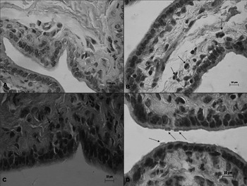
On the other hand, the epithelial tissue sections obtained from mammary gland cistern, in all samples analysed, were characterized by the presence of an epithelial lining composed of one or two layers of cuboidal or columnar cells without keratin layer. In all cows, mammary gland cistern showed differences between the inoculated and no-inoculated quarter (Fig. 2). The presence of some neutrophils was observed in the connective and epithelial tissue. In this sense, a higher number of neutrophils were observed near the epithelial zone (Fig. 2b, c). Another characteristic detected was a blood vessels hyperaemia which, together with neutrophils, denoted a mild inflammatory reaction (data not shown). Bacteria adhered to the surface of the cistern epithelial tissue were observed in some quarters inoculated with Lb. perolens CRL 1724 (Fig. 2d). This result was confirmed for Gram stain (data not shown).
High resolution optical microscope photographs showing haematoxylin and eosin stained from representative epithelial tissue sample of bovine mammary gland cistern after intrammamary inoculation with Lactobacillus perolens CRL 1724. (a) non-inoculated (100×). (b), (c) and (d) epithelial tissue of inoculated quarter. (b) and (c) presence of neutrophils near to the epithelial zone (black arrows) (100×). (d) bacterial adhered to the surface of epithelial tissue (greys arrows) (100×). The bar in each panel is equivalent to 10 μm.

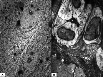
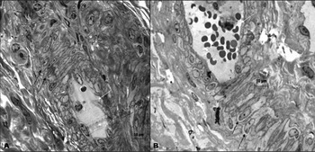
There were no ultrastructural modifications in the epithelial tissue cells of the canal and cistern of all mammary gland analysed after Lb. perolens CRL 1724 inoculation (Figs. 3 & 4, respectively). TEM and RHOM examinations did not show ultrastructural changes, necrosis or apoptosis of epithelial cells, which might be evidenced by the shrinkage of the nucleus and chromatin condensation (pyknosis), outbreak of the nucleus (cariorexis) or the appearance of small cytoplasm vacuoles due to water accumulation. No changes were observed in the nucleus, nucleolus, nuclear membrane and cytoplasmic membrane of the all samples assayed.
Transmission electron micrographs showing representative sample of teat canal epithelial cells after intrammamary inoculation with Lactobacillus perolens CRL 1724 (a) non-inoculated, nucleus (n), nucleolus (white arrows) and nuclear membrane (black arrows) of epithelial cells (4000×). (b) epithelial cells of inoculated quarter with nucleus (n), nucleolus (white arrows) and membrane nuclear (black arrows) without modifications (6000×). (K): Keratin.

High resolution optical microscope photographs showing toluidine blue stained of representative sample of tissue cells of bovine mammary gland cistern after intrammamary inoculation with Lactobacillus perolens CRL 1724. (a) non-inoculated, mammary cells (black arrows) and blood vessel with scarce presence of blood cells (white arrow) (100×). (b) mammary tissue cells of inoculated quarter without morphological modifications in nucleus, nucleolus or membrane nuclear (black arrows); full blood vessel (white arrow) (100×). The bar in each panel is equivalent to 10 μm.

Discussion
Bovine mastitis currently produces a serious health and economic problem in dairy farms around the world and antibiotics and disinfectants are routinely used to fight this disease. The generation of non-antibiotic formulations for the treatment and prevention of mastitis in cows has the potential to reduce the veterinary dependence on antibiotics in the control of this persistent and costly disease (Ryan et al. Reference Ryan, Flynn, Hill, Ross and Meaney1999). In this sense, the use of probiotic bacteria has been widely studied as a novel approach to prevent infections in animals, especially in the gastrointestinal and vaginal tract (Otero & Nader-Macías, Reference Otero, Nader-Macías and Méndez-Vilas2007; Walsh et al. Reference Walsh, Gardiner, Hart, Lawlor, Daly and Lynch2008).
Previous reports showed the isolation and characterization of Lb. perolens CRL 1724 to be a potential probiotic strain (Espeche et al. Reference Espeche, Otero, Sesma and Nader-Macias2009; Frola et al. Reference Frola, Pellegrino, Espeche, Giraudo, Nader-Macias and Bogni2011). Lb. perolens CRL 1724 was able to inhibit and co-aggregate in vitro with microorganisms considered as major bovine mastitis causing pathogens. Moreover Lb. perolens CRL 1724 showed a high efficacy to adhere to bovine teat canal epithelial cells in vitro. Intramammary inoculation of 106 cfu/ml of the strain in lactating cows showed no udder clinical signs or teat damage and bacteria could be recovered from milk until 15 d after challenge.
In this work the inoculation of 106 cfu/ml of Lb. perolens CRL 1724 was also well tolerated by the udders at the beginning of the dry period. No clinical signs or teat damage were observed in the inoculated quarters and the udders presented a normal aspect. There were no changes in the appearance of milk secretions. These results differ from those obtained by Beecher et al. (Reference Beecher, Daly, Berry, Klostermann, Flynn and Meaney2009) who reported that all the lactating cows inoculated with Lactococcus lactis at 108 cfu/ml experienced swollen udder quarters. Similar results were observed in lactating cows by Frola et al. (Reference Frola, Pellegrino, Espeche, Giraudo, Nader-Macias and Bogni2011), where a short-term significant increase in SCC was observed 1 d post inoculation as a normal reaction of the udder, returning to normal values at the end of the trial. In this sense, Crispie et al. (Reference Crispie, Alonso-Gómez, Collette, Klostermann, Flynn, Arkins, Meaney, Ross and Hill2008) and Klostermann et al. (Reference Klostermann, Crispie, Flynn, Ross, Hill and Meaney2008) reported increased values of SCC in the first 2 d after the inoculation of 109 cfu/ml of Lc. lactis which decreased on days 5 and 7 post inoculation, respectively.
In the present study, Lb. perolens CRL 1724 was recovered from the udders until the end of the assay. This could indicate that the strain persisted in the udders. Beecher et al. (Reference Beecher, Daly, Berry, Klostermann, Flynn and Meaney2009) observed the recovery of Lc. lactis for a period of 72 h post inoculation.
Different authors studied the effect of lactic acid bacteria in the control of mastitis utilizing, for example, a commercial probiotic for oral use in calves (Greene et al. Reference Greene, Gano, Smith, Hogan and Todhunter1991) or substances such as bacteriocins produced by lactic acid bacteria isolated from different niches (Crispie et al. Reference Crispie, Twomey, Flynn, Hill, Ross and Meaney2005; Cao et al. Reference Cao, Wu, Xie, Hu and Mo2007). In contrast, the host specificity and ecological niche specificity has been shown by different research groups (Ocaña et al. Reference Ocaña, Bru, Ruiz Holgado and Nader-Macías1999; Otero et al. Reference Otero, Morelli and Nader-Macías2006; Zoetendal et al. Reference Zoetendal, Vaughan and de Vos2006). In this work, the effect of intramammary inoculation of a lactobacilli strain isolated from bovine milk on the epithelial tissue of the udders was determined. The preliminary results obtained in this work show that Lb. perolens CRL 1724 did not cause damage in the epithelium cells.
Differences between epithelial tissue sections of the cistern of the udder from inoculated and non-inoculated quarters were observed. Neutrophils were observed close to the epithelium and together with hyperaemia of blood vessels denoted a mild inflammatory reaction. In this connection Crispie et al. (Reference Crispie, Alonso-Gómez, Collette, Klostermann, Flynn, Arkins, Meaney, Ross and Hill2008) showed that administration of the lactococcal culture into the mammary glands of uninfected animals elicited an immunomodulatory effect, with substantial recruitment of polymorphonuclear lymphocytes to the infused quarters and concluded that the mechanism by which the live culture can provide host protection against mastitis infection may be associated with its ability to elicit a rapid immune response. In addition, Beecher et al. (Reference Beecher, Daly, Berry, Klostermann, Flynn and Meaney2009) obtained a massive immune response, with an increase in pro-inflammatory genes as IL-1b, IL-8 and CXCR1, after inoculation of a strain of Lc. lactis in lactating cows and argued that this result may be one of the immunomodulatory mechanisms by which the bacterium confers its therapeutic effect. On the other hand, according to Sordillo & Streicher (2002) the mammary gland is protected by a variety of defence mechanisms and one of them is innate immunity. This mechanism is mediated by the physical barrier of the teat end, macrophages, neutrophils, natural killer (NK) cells, and certain soluble factors, and if these function adequately, most pathogens are readily eliminated within a short period of time and before the specific immune system is activated.
Adhesion to epithelial cells is an important step in both pathogenic infection and probiotic colonization of different mucosal surfaces such as gastrointestinal, urogenital and respiratory tracts. Adhesion of probiotic microorganisms to the mucosa and the antagonism against pathogens by interference mechanisms have been related to many of the health benefits attributed to probiotics. In consequence, the ability to adhere to epithelial cells is considered an important criterion for in-vitro selection of probiotics (Morelli, Reference Morelli2000).
Several studies have suggested that Lactobacillus adherence is mediated by proteins associated to the external protein S-layer (Henriksson et al. Reference Henriksson, Szewzyk and Conway1991; Frece et al. Reference Frece, Kos, Svetec, Zgaga, Mrsa and Suskovic2005) while others have suggested a role for lipoteichoic acid and carbohydrate (Chan et al. Reference Chan, Reid, Irvin, Bruce and Costerton1985). Zárate & Nader-Macias (Reference Zárate and Nader-Macias2006) argue that the adhesion to epithelial cells by pathogens is considered an important prerequisite for the onset of infections in the different tracts and that it is the first step in the colonization of the vaginal surface by probiotic microorganisms. Chan et al. (Reference Chan, Reid, Irvin, Bruce and Costerton1985) and Reid et al. (Reference Reid, Cook and Bruce1987) affirm that this ‘anti-infective’ mechanism may involve the blockage of pathogen adherence by both steric hindrance and competition for receptors in the urogenital tract. Bernet et al. (Reference Bernet, Brassart, Neeser and Servin1994) reported that adherent lactic acid bacterial strains may hinder the cell association and invasion by bacterial pathogens and explained such inhibitory effects of lactobacilli by a mechanism of non-specific steric hindrance on the receptors for pathogens. Other reports also showed that lactic acid bacterial strains with adhesion ability may hinder the contact between the epithelial cells and the pathogenic bacteria (Hudault et al. Reference Hudault, Lievin, Bernet-Camard and Servin1997; Coconnier et al. Reference Coconnier, Lievin, Lorrot and Servin2000). In consequence, when selecting lactobacilli for probiotic purposes, the adherent strains are preferred in order to form a film on the epithelial tract as a biological barrier against colonization of pathogenic bacteria.
In this work, bacteria on the epithelial tissue surface of inoculated cistern were observed, but no bacteria were observed in the epithelial tissue sections obtained from teat canal. This absence may be due to the keratin layer that coats the canal tissue and would prevent bacterial adherence to the epithelial surfaces as reported by Sandholm & Korhonen (Reference Sandholm, Korhonen and Sandholm1995). No inoculated samples of both cistern and canal were free of bacteria.
Lb. perolens CRL 1724 (106 cfu/ml) did not cause structural or morphological modifications, lesions or necrosis in the epithelial tissue cells of canal and cistern of bovine mammary gland. These observations suggest that intramammary inoculation of Lb. perolens CRL 1724 does not produce any damage to the epithelial cells covering the bovine mammary gland tissue studied. This condition, together with the mild inflammation observed at cistern tissue is a promissory result for inclusion of Lb. perolens CRL 1724 in a probiotic formulation.
Conclusions
In conclusion, our preliminary results show that Lb. perolens CRL 1724 could adhere in vivo to mammary gland cistern of non-lactating cows and generate a mild inflammatory reaction, characterized by the presence of neutrophils in the area close to the epithelia, without causing structural or ultrastructural modifications, lesions or necrosis in tissue cells of bovine mammary gland teat canal and cistern. This study provides important information for further studies directed towards including Lb. perolens CRL 1724 in the design of a probiotic product for preventing bovine mastitis in non-lactating dairy cows.
This work was supported by SECYT-UNRC, MINCYT Córdoba PID280. These are the results obtained from the project 'Design of a probiotic product for bovine mastitis prevention’ signed between CONICET and UNRC Res. 2907. Ignacio Daniel Frola is recipients of a fellowship from Consejo Nacional de Investigaciones Científicas y Técnicas (CONICET).