Introduction
A common and serious food-borne parasite disease called trichinellosis is brought on by eating raw, undercooked animal meat that contains infected Trichinella nematode larvae (Bai et al. Reference Bai, Hu, Liu, Tang and Liu2017; Luis Muñoz-Carrillo et al. Reference Luis Muñoz-Carrillo, Maldonado-Tapia, López-Luna, Jesús Muñoz Escobedo, Armando Flores-De La Torre and Moreno-García2019). Trichinella causes a wide range of illnesses, infecting over 150 animals as well as humans (Pozio et al. Reference Pozio2015). Human trichinellosis can be acute or chronic. The clinical symptoms of an acute stage include headache, fever, and gastrointestinal problems. The chronic stage may involve myositis caused by larval growth in skeletal muscles and can be severe, painful, or incapacitating. Furthermore, muscle dysfunction and discomfort might have a long-term impact on patients (Dupouy-Camet Reference Dupouy-Camet2014). Furthermore, encephalitis and secondary infections, such as bronchopneumonia or sepsis, can occur. Symptom duration depends on the infection dose and the severity of the illness (Gottstein et al. Reference Gottstein, Pozio and Nockler2009). Trichinellosis harms human health, raises concerns about food safety, and can result in significant financial losses. Consequently, finding a cure is vital. Trichinellosis infection is traditionally treated with albendazole (Gottstein et al. Reference Gottstein, Pozio and Nockler2009). Although it is fatal to Trichinella adults, its limited bioavailability reduces the likelihood of killing encapsulated larvae (Eid et al. Reference Eid, Ashour, Essa, El Maghraby and Arafa2020). Still, it can cause serious systemic reactions – for instance, encephalitis, seizures, drug allergies, and even fatalities (Yada vet al. 2012).
Acetazolamide is a sulfonamide that inhibits carbonic anhydrase. Most insect and nematode species, including Ancylostoma caninum, Ascaris spp., Entamoeba spp., Necator americanus, and T. spiralis, include beta-carbonic anhydrases (β-CAs), which are most likely mitochondrial enzymes (Zolfaghari-Emameh et al. Reference Emameh, Barker, Hytönen, Tolvanen and Parkkila2014). Inhibition of nematode β-CAs offers chances to treat or control many parasitic infections with negligible adverse effects on the hosts (Zolfaghari-Emameh et al. Reference Emameh, Kuuslahti, Vullo, Barker, Supuran and Parkkila2015). Acetazolamide, one of the CAs inhibitors used against T. spiralis, can interact with metabolic processes and potentially eliminate pathogens (Syrjänen et al. Reference Syrjänen, Tolvanen, Hilvo, Olatubosun, Innocenti, Scozzafava, Leppiniemi, Niederhauser, Hytönen and Gorr2010).
By combating poor cellular permeability, nonspecific distribution, low bioavailability, and the quick clearance of drugs against parasites out of the body, silver nanoparticles, a new emerging drug carrier, are helpful in treating a number of parasitic disorders. AgNPs displayed antiparasitic efficacy against a wide range of parasites, including Fasciola (Gherbawy et al. Reference Gherbawy, Shalaby, Abd El-Sadek, Elhariry and Banaja2013), filaria (Saini et al. Reference Saini, Saha, Roy, Chowdhury and Babu2016), Leshmania (Allahverdiyev et al. Reference Allahverdiyev, Abamor, Bagirova, Ustundag, Kaya, Kaya and Rafailovich2011), Cryptosporidium parvum, Entamoeba histolytica (Saad et al. Reference Saad, Soliman, Azzam and Mostafa2015), Blastocystishominis (Younis et al. Reference Younis, Abououf, Ali, Abd elhady and Wassef2020), Plasmodium, Toxoplasma, Giardia, and insect larvae (Elmi et al. Reference Elmi, Gholami, Fakhar and Azizi2013), and were also used as antimicrobial therapies (Sun et al. Reference Sun, Chen, Pan, Qu, Hao, Wang, Liu and Xie2019).
The total antioxidant capacity (TAOC) is a quantitative assay that indicates the cumulative impact of primarily non-enzymatic antioxidants found in plasma and body fluids (Ghiselli et al. Reference Ghiselli, Serafini, Natella and Scaccini2000). The TAOC measurement could offer details about the balance of oxidants and antioxidant systems (Collins Reference Collins2005). An insufficient antioxidant capability may contribute to excessive tissue damage. Improved antioxidant capacity may assist the host by reducing tissue damage caused by oxidative stress, reducing the severity of symptoms and the development of complications, and allowing the body to maintain a functioning immune system capable of eradicating the infection (Akaike et al. Reference Akaike, Suga and Maeda1998).
The goal of this study was to evaluate acetazolamide-loaded silver nanoparticles in the treatment of T. spiralis infection in mice by investigating the parasite load as well as histopathological and ultrastructural alterations in the gut and muscle phases of the parasite, in addition to measuring serum TAOC.
Materials and methods
Parasites and doses of infections
The Trichinella spiralis strain (Istituto Superiore di Sanità code: ISS6158) was obtained from the Parasitology Department, Faculty of Medicine, Zagazig University. The infective dose of 250 of larvae was given to each mouse (Chen et al. Reference Chen, Yang, Yang, Zhang and Zhu2012). Mice were starved for 12 hours before being infected and then fed the larvae.
Experimental design
Five groups of male Swiss albino mice of matched age (6 weeks) and weight (20–25 g) were used. Each group consisted of ten mice: Group I, control normal; Group II, infected with T. spiralis and not treated; Group III, infected, then treated with nanoparticles; Group IV, infected at that time and treated with acetazolamide; Group V, infected and subsequently treated with acetazolamide-loaded nanoparticles. Every group was subdivided into two subgroups of five mice each: group a, for detection of the intestinal phase (adults), mice were sacrificed six days post-infection (P.I.), and group b, for detection of the muscular phase (larvae), mice were sacrificed 45 days P.I. (AbouRayia et al. 2017).
Drugs
Acetazolamide in the form of 250-mg tablets (Cidamex, Chemical Industries Development (CID), Egypt, Giza) was dissolved in distilled water, and 100 mg/kg/day was orally given (Saad et al. Reference Saad, Ashour, Abou Rayia and Bedeer2016). About 50 mg/kg/day of acetazolamide-loaded nanoparticles were administered orally. Nearly 200 μg/mice of nanoparticles were orally given. Subgroup (a) received drugs on the second post-infection day for four consecutive days, whereas subgroup (b) received treatments on the 12th post-infection day for four days.
Nanoparticles preparation
An Erlenmeyer flask was filled with 30 L of 0.002M (NaBH4). For around 20 minutes, the flask was placed on a stir plate with a magnetic stir bar in an ice bath. A total of 2 mL of 0.001 M (AgNO3) was dripped into the swirling NaBH4 solution at a rate of about 1 drop per second, stopping after all of the AgNO3 was introduced. A few drops of 1.5 M NaCl solution were added to the suspension, causing it to darken yellow and eventually grey as the nanoparticles accumulated. In order to prevent aggregation, 0.3% PVP was added. To leave air bubbles and undissolved PVP in the beaker, the liquid was decanted into a mold. The liquid was then placed in a toaster oven for 30 minutes to evaporate (Mulfinger et al. Reference Mulfinger, Solomon, Bahadory, Jeyarajasingam, Rutkowsky and Boritz2007).
Characterization of silver nanoparticles
AgNP was characterized by its size and shape (Fisker et al. Reference Fisker, Carstensen, Hansen, Bødker and Mørup2000).
-
• Morphology and particle size determination
Scanning electron microscopy (SEM) analysis was performed after AgNP preparation by the LEO 1430 VP SEM machine. To prepare thin films of the sample on a carbon-coated copper grid, a small amount of the sample was added. Blotting paper was used to get rid of the extra solution. Afterward, the film was dried under a mercury lamp on the SEM grid (Ziel et al. Reference Ziel, Haus and Tulke2008).
-
• Fourier-transform infrared spectroscopy analysis
The synthesized Ag nanoparticle solution was centrifuged at 3000 rpm for 30 min to prepare AgNPs in powder form. The AgNPs-containing pellet was dispersed three times using sterile deionized water to remove biological contaminants and free proteins and enzymes, not capping ligands for AgNPs. The samples were dried overnight in an oven at 60°C, ground with KBr pellets, and evaluated on a Bruker Tensor 27 model at a resolution of 4 cm in the diffuse reflectance mode (Mulvaney Reference Mulvaney1996).
Preparation of acetazolamide-loaded nanoparticles
Acetazolamide-loaded nanoparticles were produced by adding silver nanoparticles to a solution containing 50 mg/ml acetazolamide concentrations. After centrifugation of the sample at 20,000 g at 14°C for 30 minutes, it was separated from the aqueous suspension. The Bradford protein assay spectrophotometric method was used to detect the protein concentration (free) in the supernatant at 595 nm. The loading capacity (LC) and encapsulation efficiency (EE) of nanoparticles were analysed as follows (Mulfinger et al. (Reference Mulfinger, Solomon, Bahadory, Jeyarajasingam, Rutkowsky and Boritz2007):
 $$ {\displaystyle \begin{array}{l}\%\mathrm{EE}=\left[\left(\mathrm{A}\hbox{-} \mathrm{B}\right)/\mathrm{A}\right] \times 100\\ {}\%\mathrm{LC}=\left[\left(\mathrm{A}\hbox{-} \mathrm{B}\right)/\mathrm{C}\right] \times 100\end{array}}, $$
where A is the total amount of drugs, B is the free amount of drugs, and C is the weight of nanoparticles.
Mortality rate (MR%)
At the 6th and 54th days post-infection, the mortality rate was estimated according to the present equation: MR% = number of dead mice at the sacrifice time/number of mice at the beginning of the experiment X100 (Eissa et al. Reference Eissa, El-Azzouni, Mady, Fathy and Baddour2012).
T. spiralis adults count in the intestine
The cleaned intestine was cut into 1-cm sections and incubated in 10 mL of saline for 2 hours at 37°C to allow the worms to move out of the tissue and gather in the container. The intestine was rinsed a number of times with pipetted saline. The fluid was gathered and centrifuged at 1500 rpm for 5 minutes. For counting the worms, the sediment was dissolved in drops of saline before being inspected drop by drop under a 10x microscope. (Issa et al. Reference Issa, El-Arousy and Abd EI-Aal1998).
Counting T. spiralis larvae
About 200 ml of distilled water with 1% pepsin hydrochloride was used to digest the dissected mice. The encysted larvae were collected using the sedimentation method after one hour of incubation at 37°C with continuous stirring with an electric stirrer (Denham Reference Denham 1965 ). A McMaster counting chamber was used for counting larvae in three samples of 0.1 mL (10x objective). To calculate the number of larvae per gram of tissue, the average of three counts was used (Abou Rayia et al. Reference Abou Rayia, Saad, Ashour and Oreiby2017). The efficacy of drugs was evaluated by comparing the number of larvae per gram retrieved from infected groups.
Assessment of serum TAOC
During the sacrification process, the collection of neck vein blood was done using the capillary tube on the 6th day of P.I. for subgroup a (intestinal phase) and on the 45th day of P.I. for subgroup b (muscular phase). The samples were left for 10 to 20 minutes to clot at room temperature. To get serum samples, these clots were removed using centrifugation for 20 minutes at 2000–3000 rounds per minute. TAOC was measured by an ELISA kit (Biospes Company, Jiulongpo Industry District, Chongqing, China) according to the manufacturer’s instructions (Wang et al. Reference Wang, Chen, Liu, Qiu, Wang, Chen and Xu2017).
Histopathological study
Fixation in formalin 10%, paraffin sectioning, and hematoxylin and eosin (H&E) staining were processed for the intestinal, lingual, and skeletal muscles of mice (Carleton et al. Reference Carleton, Drury, Willington and Cammeron1967). The presence of inflammatory reactions, nurse cells, and encapsulated T. spiralis larvae was blindly observed under a microscope (IX73, Olympus, Japan).
Scanning electron microscopy
The adults and larvae were incubated at 4°C overnight in a fixed solution (2.5% glutaraldehyde). Adults were washed for 5 minutes in 0.1 M sodium cacodylate buffer and fixed in 2% osmium tetroxide for 1 hour. The samples were dehydrated in ascending grades of alcohol and dried using a critical point of carbon dioxide drying. After sputter coating with gold, a scanning electron microscope (Hitachi SU8040, Japan) was used to observe the samples.
Statistical analysis
The data were analyzed by the SPSS (Statistical Package for the Social Sciences) version 26. The differences between the treated and control groups were analyzed using a one-way analysis of variance (ANOVA). Data are expressed as the mean ± standard deviation (SD) of at least three repeated experiments. Values with a p<0.05 were considered statistically significant. In all cases, p-values were expressed as #p<0.05, ##p< 0.01, and ###p<0.001.
Results
AgNPs morphology and particle size
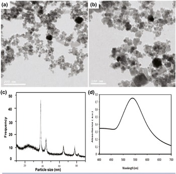
SEM revealed that the conjugation of nanoparticles with the drug (Figure 1a) had no impact on the morphology of nanoparticles alone. Most nanoparticles were spherical in shape with a smooth surface. The size distribution of nanoparticles was 40 nm for about 50%, 60 nm for 20%, and 60–90 nm for the remaining percentages. The concentration of nanoparticles was 2.33X10⁸ particles/ml as estimated by UV/visible spectrophotometry using Beer’s law. The surface plasmon resonance (SPR) peak can be seen as a symmetrical peak with a maximum of 520 nm.
The morphology and particle size determination of silver nanoparticles. a, b: SEM showed that the conjugation of nanoparticles with drugs did not affect the morphology of nanoparticles alone. c: FTIR spectra of nanoparticles show that 50% of nanoparticles were approximately about 40 nm and 20% were approximately about 60 nm, while the remaining percent ranged from 60 to 90 nm. d: UV/visible spectrophotometry determined the concentration of nanoparticles was to be 2.33X10⁸ particles/ml by using Beer’s law. The surface plasmon resonance (SPR) peak can be seen as a symmetrical peak with a maximum at 520 nm.

The mortality rate
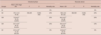
During the intestinal phase, the mortality rates revealed that mice in GI and GV survived the whole experiment, while 10% of mice in GII, GIII, and GIV died. In the muscular phase, no mortality was detected in GI and GV, but the mortality rate was 20% in GII, and GIII, while it was 10% in GIV (Table 1).
Adult and larval counts and the mortality during the intestinal and muscular phases of T. spiralis-infected groups

F: F for one-way ANOVA test, pairwise comparison between each of the two groups was done using post hoc test (Tukey)
HS: highly significant (p<0.01)
##: statistically significant at p ≤ 0.01 with infected control group (GII); R% = reduction percentage
The number of adult worms in the small intestine and larvae in the muscles
The lowest mean adult worms and larvae count were attained in GV, with the highest percentage reduction of 84.72% and 80.74%, respectively, with a statistically significant difference (P<0.001) compared to the control-infected group, followed by GIV, with a percentage reduction of 61.32% and 60.47%, respectively. GIII exhibited the highest mean adult worm and larvae count with a percentage reduction of 15.95% and 14.87%, respectively, with a statistically significant difference (P<0.01) compared to the control-infected group (Table 1).
Serum TAOC assay
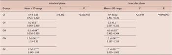
The highest mean blood TAOC levels in the intestinal and muscular stages were in GV, followed by GIV. There was also a statistically significant difference (P<0.001) in these groups compared to the control infected and the control healthy groups. There was a slight increase in the serum TAOC levels of GIII with a high statistically significant difference (P<0.001) compared to the control infected group (Table 2).
Serum levels of TAOC (pg/ml) during the murine intestinal and muscular phases of T. spiralis infection

F: F for One way ANOVA test, pairwise comparison between each of the two groups was done using post hoc test (Tukey)
HS: highly significant (p<0.01)
** : statistically significant at p ≤ 0.01 with healthy control group (GI)
## : statistically significant at p ≤ 0.01 with infected control group (GII)
Results of the histopathological study
Small intestine findings
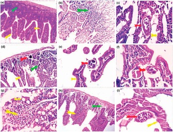
The small intestine of GVa exhibited mild mucosal invasion by degenerated Trichinella adult, normal intestinal villi, and mild mucosal and submucosal infiltrations by lymphocytes along with intact mucosa (Figure 2h, i), compared to the intense invasion by T. spiralis adult in GII, triggering pressure atrophy in the cells, and mucosal edema with an excessive inflammatory reaction – mostly lymphocytes in the surrounding intestinal mucosa (Figure 2b, c). Subsequently, GIVa revealed moderate mucosal invasion by degenerated Trichinella adults with moderate mucosal and submucosal infiltration by lymphocytes and eosinophils. Goblet cells appear hyperreactive (Figure 2e, g). However, GIIIa showed edematous intestinal villi, pressure atrophy of the mucosal epithelium, and severe mucosal infiltration by lymphocytes and eosinophils (Figure 2d, e).
Histopathological results of T. spiralis-infected mice treated throughout the intestinal phase (H&E) (X 200& X 400). a: The small intestine of the normal control GIa showing normal histological structures of villi (yellow arrow), crypt, glands (green arrow), and mucosa and muscle layers. b, c: The small intestine of GIIa shows intense invasion by T. spiralis adults (red arrows); pressure atrophy in the cells is seen (yellow arrow). The surrounding intestinal mucosa showed mucosal edema and an excessive inflammatory reaction, mostly lymphocytes (green arrows). d, e: The small intestine of GIIIa shows invasion by T. spiralis adults (red arrows). The adjacent villous and mucosal epithelium suffered pressure atrophy (yellow arrows) with excessive mucosal and submucosal infiltration by lymphocytes and eosinophils (green arrows). f, g: The small intestine of GIVa shows moderate intramucosal invasion by T. spirals adults (red arrows). Moderate mucosal and submucosal infiltration by lymphocytes and eosinophils, goblet cells appear hyperreactive (yellow and green arrows). h, i: The small intestine of the targeted GVa shows mild mucosal invasion by degenerated Trichinella adults (red arrows). The intestinal villi are apparently normal in most parts (yellow arrows). Mild mucosal and submucosal infiltrations by lymphocytes, together with intact mucosa, were seen (green arrows).

The skeletal and lingual muscle findings
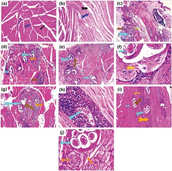
The skeletal and lingual muscles of GVb were infiltrated by a mild invasion of Trichinella larvae, which was accompanied by a mild inflammatory response of lymphocytes, macrophages, and eosinophils. Many of the larvae had entirely deteriorated and been replaced by inflammatory cells with normal muscle (Figure 3r, s), compared to GIIb, which had numerous encysted Trichinella larvae with a severe inflammatory hypersensitivity reaction and degenerative muscles surrounding it (Figure 3b, c). GIII showed severe invasion by encysted larvae surrounded by severe inflammatory reactions of eosinophils and round cells (Figure 3n, o). GIVb displayed moderate invasion by encysted Trichinella larvae surrounded by a moderate inflammatory reaction of eosinophils and round cells. Some larvae deteriorated and were replaced by inflammatory and transformed mesenchymal tissue (Figure 3p, q).
Histopathological results of T. spiralis-infected mice treated throughout the muscular phase (H, E) (X 200& X 400). j, k: muscles of normal control GIb showing normal histological structures with preserved longitudinal cross striations, peripherally situated multinuclear arrangement (blue arrows), interstitial tissue (black arrow), and tongue papillae. l, m: muscles GIIb show an invasion of encysted Trichinella larvae (light blue arrows). They were surrounded by an excessively inflammatory hypersensitivity reaction formed from eosinophils, lymphocytes, and macrophages (brown arrows). The surrounding muscle shows degenerative and necrotic changes besides the interstitial inflammatory edema (green). n, o: muscles of GIIIb showing severe invasion of the muscles by encysted larvae (light blue). They were surrounded by a severe inflammatory reaction of eosinophils and round cells (brown arrow) and transformed nuclei of the nurse cell (yellow arrow). The surrounding muscle denoted degenerative and necrotic changes, along with interstitial edema. p, q: muscles of GIVb showing moderate invasion by encysted Trichinella larvae (light blue arrow). They were surrounded by a moderate inflammatory reaction of lymphocytes, macrophages, and eosinophils (brown arrows). Some larvae were completely degenerated (q, blue arrows) and replaced by inflammatory cells. The surrounding muscle appears normal (orange arrow). r, s: muscles of the targeted GVb show mild invasion by encysted Trichinella larvae (light blue arrows). They were surrounded by a mild inflammatory reaction of eosinophils and round cells (brown arrows). Some of the larvae were degenerated and replaced by inflammatory and/or transformed mesenchymal tissue (fibrocartilagenous tissue) (orange arrows). The surrounding muscle showed mild degenerative changes (green arrow).

The ultrastructural changes of T. spiralis adults and larvae
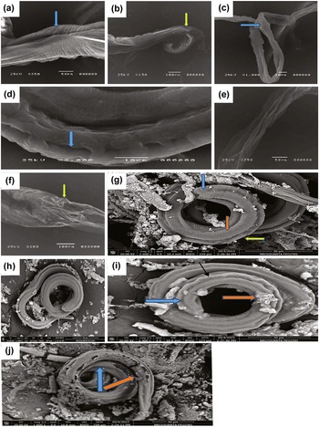
SEM of adult T. spiralis revealed a severely collapsed body with a sloughed cuticle, loss of continuity, and normal arrangement of annulations with damaged copulatory bursa in GVa (Figure 4e, f) compared to the normal adult in GIIa (Figure 4a, b). The adults had a moderately collapsed body with a sloughed cuticle, loss of continuity, and normal arrangement of annulations with the appearance of carrions in GIIIa (Figure 4c), whereas a mild degree of collapse, loss of continuity, and regularity of transverse creases with multiple depressions and widening of bacillary openings was detected in GIV (Figure 4d). T. spiralis larvae in GVb had a severely damaged body and an entire sloughed cuticle with the appearance of multiple blebs and carrion. The larva was completely dead, with a complete loss of the normal annulations observed (Figure 4j) compared to the normal ones in GIIb (Figure 4g). GIVb showed a corrugated cuticle, moderate loss of transverse annulations of the posterior end, and the appearance of carrions at the cuticle (Figure 4i), while mild loss of cuticle striation was observed in GIIIb (Figure 4h).
Scanning electron microscopy of T. spiralis adults and larvae.
a, b: Adults with GIIa show regular transverse creases with normal longitudinal ridges (blue arrow) and a normal copulatory bursa (yellow arrow). c: Adults of GIIIa show a moderately collapsed body with a sloughed cuticle, loss of continuity, and a normal arrangement of annulations with the appearance of carrion (blue arrow). d: Adults with GIVa have a mild degree of collapse and contraction without destruction. The cuticle also shows loss of continuity and regularity of transverse creases with multiple depressions and widening of bacillary openings (blue arrow). e, f: Adults of GVa show a severely collapsed body with a sloughed cuticle, loss of continuity, and normal arrangement of annulations with a damaged copulatory bursa (yellow arrow). g: GIIb showing the normal coiled appearance of the larva with normal annulations at the posterior end (blue arrow), normal transverse creases (brown arrow) along the body, and a normal longitudinal ridge with normal bacillary gland bands (yellow arrow). h: larva of GIIIb with mild loss of cuticle striation. i: larvae of GIVb show a corrugated cuticle (black arrows), moderate loss of transverse annulations of the posterior end (blue arrow), and the appearance of carrions at the cuticle (brown arrow). j: larva of GVb with severally damaged larvae and entirely sloughing of the cuticle, with the appearance of multiple blebs (blue arrows) and carrions (brown arrows). Additionally, a complete loss of the normal annulations was observed.

Discussion
As the therapeutic efficacy of Trichinella infection drugs has yet to be satisfactorily shown (Sun et al. Reference Sun, Chen, Pan, Qu, Hao, Wang, Liu and Xie2019), it is vital to develop novel, safe, and effective anti-Trichinella medicines. In this study, we evaluated the therapeutic efficacy of acetazolamide-loaded AgNPs on the intestinal and muscular phases of T. spiralis infection in mice.
For successful and early treatment of Trichinella infection, the eradication of intestinal forms represented by L1 larvae that within days develop into adults is substantial (Gottstein et al. Reference Gottstein, Pozio and Nockler2009). Furthermore, commencing the treatment during the muscular phase is the most difficult since the drug must be in sufficient concentration within the muscles for subsequent absorption into the capsules. Given that passive diffusion is the primary method used to track drug absorption through the T. spiralis capsule, the effectiveness of a treatment against encysted larvae increases with drug concentration in the muscle. In this study, with respect to the reduction percentage in the number of adults and larvae, the groups treated with acetazolamide-loaded AgNPs provided the highest reduction percentages (84.72%–80.74%), respectively, compared to the untreated mice, followed by groups treated with acetazolamide with 61.32% and 60.47%, respectively, and then groups treated with nanoparticles (15.95%–14.87%). Acetazolamide efficacy was comparable to that observed by other researchers of Trichinella infection (Saad et al. Reference Saad, Ashour, Abou Rayia and Bedeer2016). An explanation for these findings could be that the CA inhibitor disrupted the usual detoxification of cyanate, elevating the intracellular cyanate content to lethal levels and causing the parasite to die (Zolfaghari-Emameh et al. Reference Emameh, Kuuslahti, Vullo, Barker, Supuran and Parkkila2015). Another explanation could be that inhibiting β-CA in parasite cells alters mitochondrial metabolic cycles, potentially eliminating pathogens (McKenna and Supuran Reference Mckenna and Supuran2014). Additionally, Zolfaghari-Emameh et al. (Reference Emameh, Barker, Hytönen, Tolvanen and Parkkila2014) hypothesised that inhibiting CA activity might delay cellular metabolic pathways within parasites.
The superiority of acetazolamide-loaded AgNPs can be explained by the special characteristics of nanoparticles that enhance drug absorption, increase drug bioavailability, increase cellular permeability, and decrease drug excretion from the body (Sun et al. Reference Sun, Chen, Pan, Qu, Hao, Wang, Liu and Xie2019). Moreover, AgNPs had an antiparasitic impact as represented by the reduction in the ATP content of the cell, the damaging of mitochondria, and the increase in ROS production (Saini et al. Reference Saini, Saha, Roy, Chowdhury and Babu2016). Our findings agreed with Paredes et al. (Reference Paredes, Litterio, Dib, Allemandi, Lanusse, Bruni and Palma2018), who noticed an improvement in the pharmacological characteristics of albendazole-loaded AgNPs, and with El-Melegy et al. (Reference Elmelegy, Ghoneim, El Dien and Rizk2019), who stated that AgNPs improve the therapeutic effect of mebendazole in T. spiralis infection. Likewise, AgNPs coated with anti-seizure drugs kill brain-eating amoebae, which are always fatal parasites (Anwar et al. Reference Anwar, Rajendran, Siddiqui, Raza Shah and Mand Khan2019). Interestingly, acetazolamide-loaded AgNPs induced slightly higher percentage reductions in adults than in muscle larvae. This difference could be attributed to the timing of drug intake in relation to the affected T. spiralis stage. Besides, this effect may be due to some factors such as molting, changes in location, or basic biochemical differences in energy metabolism between the larval and adult stages (Yadav Reference Yadav and Temjenmongla2012).
Concerning the total antioxidant activity, the highest free-radical scavenging capacity in the sera of mice during the intestinal and muscular phases was observed in the targeted group given acetazolamide-loaded AgNPs, followed by the group given acetazolamide and AgNPs. These results highlighted the improvement in the host biochemical conditions in which Trichinella infection destroyed the antioxidant enzymatic systems and caused a disruption in metabolite homeostasis, as explained by a lower level of TAOC in the control infected group (Derda et al. Reference Derda, Boczoń, Wandurska-Nowak and Wojt2003). These findings could be explained by AgNPs acting as catalysts with antioxidant agents (Zhao et al. Reference Zhao, Zhou, Riaz Rajoka, Yan, Jiang, Shao, Zhu, Shi, Huang and Yang2018), as they can exist in two oxidation states (Ag+ and Ag2+) depending on the reaction conditions, and the formed AgNPs may be able to quench free radicals by giving or absorbing electrons, acquiring silver nanoparticles antioxidant properties (Shanmugasundaram et al. Reference Shanmugasundaram, Radhakrishnan, Gopikrishnan, Pazhanimurugan and Balagurunathan2013).
Pathological changes in the epithelium of the small intestine and skeletal muscles usually develop in trichinosis. Histopathological examination revealed that treatment with acetazolamide-loaded AgNPs ameliorated the T. spiralis-induced histopathological changes in the infected mice by reducing inflammatory cellular infiltration and restoring the normal villous structure. These findings were supported by Basyoni and El-Sabaa (Reference Basyoni and El-Sabaa2013), who suggested the higher effectiveness of different trichinicidal medications when given at an early stage. Our data also showed an improvement in the histopathological architecture of skeletal and lingual muscles in mice treated with acetazolamide-loaded AgNPs, including a decreased number of encysted larvae and their surrounding cellular infiltrates and increased regenerative muscles. Most larvae were degenerated and surrounded by mild inflammatory reactions, reflecting the relative concentration of the drug in the muscle after administration of the formula. Acetazolamide-loaded AgNPs showed higher effectiveness compared to acetazolamide or AgNPs. This can be explained by the fact that AgNPs increase the therapeutic efficacy of acetazolamide drugs against Trichinella infection. AgNPs have unique physiochemical properties as anti-inflammatory (Panáček et al. Reference Panáček, Kolář, Večeřová, Prucek, Soukupová, Kryštof, Hamal, Zbořil and Kvítek2009) and anti-angiogenesis (Rogers et al. Reference Rogers, Parkinson, Choi, Speshock and Hussain2008). Given that AgNPs have the ability to adhere to and be absorbed by the surface of targeted agents (Zayed et al. Reference Zayed, Guo, Lv, Zhang and Zhou2022), they can enter the intestinal intervillous spaces, providing a high local concentration of the drug on the surface of the membrane.
To confirm our results, T. spiralis adults and larvae of the targeted group given acetazolamide-loaded AgNPs were examined by SEM and proved to have severely damaged bodies, making them the most destroyed groups. Choi and Hu (Reference Choi and Hu2008) confirmed that nanoparticles disrupt the structure of glycoprotein and lipophosphoglycan molecules which are present on the surface of parasites and are responsible for the infection.
Conclusion
The results of our study showed that AgNPs are promising delivery systems for the oral administration of acetazolamide in the treatment of murine trichinellosis. Acetazolamide-loaded AgNPs had a lethal effect on adults and muscle larvae, generating significant in vivo damage, as proved histopathologically and ultrastructurally. None of the treatment strategies were able to completely eradicate the infection; however, acetazolamide-loaded AgNPs outperformed acetazolamide in the intestinal and muscle phases of T. spiralis infection.
Financial support
This research received no specific grant from any funding agency, commercial, or not-for-profit sectors.
Competing interest
None.
Ethics statement
The Zagazig University Institutional Animal Care and Use Committee authorized the animal experimental procedure (ZU-IACUC. 3/F/57/2020).