INTRODUCTION
The infestation of commercially important shellfish by several genera of the Polydora group (Polychaeta: Spionidae) has been well documented worldwide (see reviews by Martin & Britayev, Reference Martin and Britayev1998; Lleonart, Reference Lleonart2001). This field of research has, however, received scant attention in South Africa, but recent escalation in the mariculture of the abalone, Haliotis midae Linnaeus, 1758, and the oyster, Crassostrea gigas Thunberg, 1793 has led to renewed interest in this group of spionids. Polydora hoplura Claparède, 1870 has been recorded on both cultured oysters and abalone, while the latter are also infested by Boccardia proboscidea Hartman, 1940 and Dipolydora capensis (Day, Reference Day1955) (Nel et al., Reference Nel, Coetze and Van Niekerk1996; Simon et al., Reference Simon, Ludford and Wynne2006; Simon & Booth, Reference Simon and Booth2007), with the last species being the least problematic. To date, one and three species of the genera Dipolydora and Polydora, respectively, have been recorded on wild molluscs in southern Africa (Day, Reference Day1967; Schleyer, Reference Schleyer1991) suggesting that the number of genera and species of the Polydora group infesting wild molluscs in South Africa may have been underestimated.
A preliminary survey was conducted to gain a better understanding of the diversity of species of the Polydora group infesting wild molluscs on the south and south-east coasts of South Africa in order to identify any species that might pose future problems in aquaculture facilities. Here an account of the Pseudopolydora Czerniavsky, 1881 species is presented.
MATERIALS AND METHODS
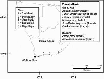
Molluscs were collected from the intertidal or shallow subtidal from five sites along the south and south-east coasts of South Africa in February and March 2005 and April 2006 (Figure 1). Worms were removed from the shells using a vermifuge, 0.05% phenol in seawater, for 3 hours to overnight. Once the worms abandoned their burrows, they were transferred to fresh seawater, relaxed with clove oil, preserved in 4% seawater–formalin and stored in 70% ethanol. Additional material from Sandbaai in Walker Bay was removed by dissolving the shells in which the worms were fixed (in 4% seawater–formalin and stored in 70% ethanol), in 5% HNO3 diluted in 70% ethanol.
Location of sampling sites and a list of potential molluscan hosts collected.

For scanning electron microscopy, the specimens were dehydrated in a series of ethanol of increasing concentration (80% to 100%), critical point dried and sputter coated. Specimens were viewed on Vega Tescan and Leo 1430 VP scanning electron microscopes. Specimens are lodged at the Iziko South African Museum, Cape Town, South Africa.
RESULTS AND DISCUSSION
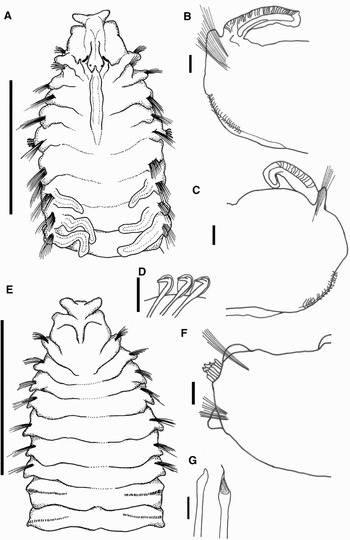
Pseudopolydora antennata. (A) Anterior region, dorsal view; (B) chaetiger 7, anterior view; (C) chaetiger posterior to 12, anterior view; (D) hooded hooks; (E) anterior region, ventral view; (F) chaetiger 5, anterior view; (G) modified spines of chaetiger 5. Scale bars: A & E = 1 mm; B, C, F & I = 100 µm; D, G = 35 µm.

Polydora antennata: Carazzi, Reference Carazzi1893: 25–26, Plate 2, figures 11 & 12;
Polydora (Carazzia) antennata: Fauvel, Reference Fauvel1927: 56–57, figure 19 i–m;
Polydora (Carazzia) antennata: Okuda, Reference Okuda1937: 237, figure 15;
Pseudopolydora antennata: Woodwick, Reference Woodwick1964: 149, figure 2.7 & 2.8;
Polydora antennata: Day, Reference Day1967: 473, figure 18.4 e–g;
Polydora (Pseudopolydora) antennata: Hartman-Schröder, Reference Hartman-Schröder1981: 87, figures 115–118;
Pseudopolydora antennata: Hutchings & Turvey, Reference Hutchings and Turvey1984: 16, figure 6.
MATERIALS EXAMINED
South Africa: Eastern Cape Province, Haga Haga, (32°45′41″S 28°14′30″E). No complete specimens found; anterior fragments were 7.5 mm for 39 chaetigers and 1 mm wide at chaetiger 5, 10.8 mm for 46 chaetigers and 1.17 mm wide at chaetiger 5, and 7.17 mm for 38 chaetigers and 0.8 mm wide (SAMC: SAM A21473), collected by C.A. Simon, 25 February 2005. Two specimens were gravid, with eggs visible in chaetigers 15–24.
DESCRIPTION
Prostomium incised with lateral lobes giving it a T-shaped appearance; caruncle extends to about mid-chaetiger 5 (Figure 2A). 1 or 2 pairs of eyes present, anterior pair 3 times size of, and closer together than, posterior pair; short occipital tentacle (Figure 2A).
Chaetiger 1 with small notopodial lobes, chaetae absent (Figure 2A). On chaetigers 3–12 notochaetae spread out with a wide post-chaetal lobe (Figure 2B), chaetae on chaetigers 3–4, 6–12 arranged in 3 rows, first row with geniculate winged chaetae, second row with short spear-shaped chaetae and final row with long simple capillary chaetae; from chaetiger 13 onwards chaetae extend from a bottle-shaped lobe (Figure 2C). Number of notochaetae decrease towards posterior, with spear-shaped and capillary chaetae, and finally only capillary chaetae. Neurochaetae from chaetigers 1–7 a single row of spear-shaped chaetae. Hooded hooks start on chaetiger 8, main tooth of the hooded hooks < 90° to the shaft with the secondary fang closely applied, shaft has a constriction (Figure 2D), 13 hooded hooks per anterior fascicle, increasing to 26 in posterior chaetigers of fragment. No accompanying neurochaetae (Figure 2B, C & E).
Chaetiger 5 with well developed post-chaetal lobes (Figure 2A, E, & F); in ventral view chaetiger 5 similar to preceding and succeeding chaetigers in terms of size and appearance (Figure 2E). Single row of winged notochaetae and spear-shaped neurochaetae present. Two types of spines arranged in a U-shape, inner spines longer with an angled distal end and rounded point, outer spines spoon-shaped with bristles within concavity and tapering point (Figure 2G) which may be worn or broken off.
Branchiae start on chaetiger 7, from chaetiger 8 branchiae overlap along midline of body. On two larger specimens they continue to end of fragment.
DISTRIBUTION
Day (Reference Day1967) recorded Ps. antennata from Saldanha Bay and Strandfontein on the west coast of South Africa. In the present study it was found only at Haga Haga on the east coast.
ECOLOGY
Associated with the oyster, Saccostrea cuccullata Born, 1778. The burrows were visible as indentations on the external surface of the valve of the oyster that was attached to the rock and it is therefore doubtful that they had specifically bored into the shells. This supposition is supported by the fact that although Day (Reference Day1955) had found a specimen in algal mats, they were more numerous in dredgings off the Saldanha Bay coast (Day, Reference Day1961).
REMARKS
Recorded descriptions demonstrate a degree of intraspecific variation with regards to caruncle length and shape of the modified spines of chaetiger 5. In the specimens examined here, the caruncle extends to mid-chaetiger 5, but has been described as extending to the posterior of chaetiger 2 (Day, Reference Day1967, original material misplaced; Hartman-Schröder, Reference Hartman-Schröder1981), from the anterior of chaetiger 5 to the posterior margin of chaetiger 6 (Woodwick, Reference Woodwick1964), middle of chaetiger 6 (Hutchings & Turvey, Reference Hutchings and Turvey1984) and to the posterior margin of chaetiger 6 (Okuda, Reference Okuda1937). The modified spines of chaetiger 5 of the specimens in the present study most closely resemble those of Ps. antennata as described by Okuda (Reference Okuda1937) and Hartman-Schröder (Reference Hartman-Schröder1981). Day (Reference Day1955, Reference Day1967) describes only the spoon-shaped spine on chaetiger 5. The spines in the outer row lack the second tooth described by Woodwick (Reference Woodwick1964). The South African specimens differ from those described by Hutchings & Turvey (Reference Hutchings and Turvey1984) in several ways; in the former the modified spines of chaetiger 5 are arranged in a tight U-shape (not a J-shape), the inner spines are not hirsute, and the worms are shorter, for similar numbers of chaetigers. Day (Reference Day1955) suggested that the local specimens may represent a separate variety of Ps. antennata, and the current study supports this. However, a final decision can only be made once more, and better, material have been examined.
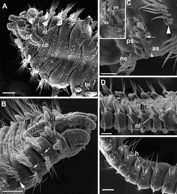
Pseudopolydora dayii sp. nov. Dorsal (A) and dorso-lateral (B) view of the anterior end showing the occipital tentacle (ot), well-developed notopodial lobes (nl), ciliary bands (cb) along the sides of the caruncle, lateral organs (small arrows) and J-shaped arrangement of the anterior row of chaetae on chaetiger 4 (large arrow); (C) spines of chaetiger 5 showing J-shaped arrangement of the anterior row of pennoned spines (as) and falcate posterior spines (ps), with broken (arrow) and complete spines (small arrowhead) in anterior row. Note the J-shapped arrangement of the geniculate notochaetae in the anterior row of chaetiger 4 and the placement of the neuropodial lobe (ne) on chaetiger 5. Insert shows a close-up of the modified spines of chaetiger 5; (D) chaetigers 6–11, showing the change in structure of the post-chaetal notopodial lobes and branchiae (br); (E) lateral view of posterior chaetigers showing the hooks (h). Scale bars: A, B, D–E = 100 µm; C = 40 µm; insert = 20 µm.

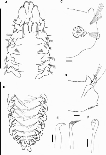
Pseudopolydora dayii sp. nov. (A) Dorsal anterior and (B) dorsal posterior chaetigers (holotype: SAM A21468); (C) chaetiger 6 showing glandular sacs; (D) transverse section of a posterior chaetiger showing the positions of the hook, dorsal cirrus, neuropodial lobe and hooded hooks; (E) modified falcate (left) and pennoned (right) spines of chaetiger 5; (F) hooded hooks. Scale bars: A & B = 1 mm; C = 70 µm; D = 50 µm; E = 35 µm; F = 14 µm.

MATERIAL EXAMINED
Holotype: South Africa, Eastern Cape Province, Haga Haga (32°45′41″S 28°14′30″E), collected by C.A. Simon, 25 February 2005, associated with the abalone Haliotis midae (SAMC, accession number SAM A21468).
Paratypes: 1, same data as holotype, (SAMC, accession number SAM A21469); 3, Western Cape Province. Struisbaai, 34°45′19″S 20°03′02″E, collected by C.A. Simon, 7 March, 2005, associated with H. midae, Perna perna (mussel) and Scutellastra longicosta (limpet) (SAMC, accession numbers SAM A21470-A21472); 2, Sandbaai, 34°25′60″S 19°11′60″E, collected by C.A. Simon, September 2003, SAM A21474 SEM preparations. SAM A21472 includes light microscope slides and remaining fragments.
Additional material examined: Eastern Cape Province: Grootbank, 33°59′14″S 23°32′36″E, collected by C.A. Simon, 9 March 2005; Western Cape Province: Mossel Bay, 34°10′56″S 22°07′20″E, collected by C.A. Simon, 8 March 2005, Gansbaai (34°36″13′S 19°21″5′E) abalone farm, April 2007.
ETYMOLOGY
This species is named in honour of John Day, South Africa's most prominent polychaete taxonomist.
DESCRIPTION
Holotype 13.25 mm for 74 chaetigers and 0.8 mm wide at chaetiger 5. Longest paratype measures 14 mm for 79 chaetigers, and 0.8 mm wide at chaetiger 5. Prostomium strongly incised, caruncle extending to posterior margin of chaetiger 2, nuchal organ ciliary band along lateral margins of caruncle (Figures 3A & B, 4A); finger-like occipital tentacle present between notopodial lobes of chaetiger 1, occipital tentacle and notopodial lobes of similar size and twice as long as they are wide (Figures 3A & B, 4A). Holotype with 1 eye, paratypes with 1 or 2 pairs, anterior pair up to 3 times larger than second. Only one specimen with intact palps; extend to approximately the ninth chaetiger with pigment at base of palp. Live specimens cream-coloured, 1 or 2 pairs of black spots on dorsal surface of chaetiger 5 (Figure 4A).
Chaetiger 1 separated from the peristomium, with prominent post-chaetal notopodial lobes approximately 1.5 times the length of those on succeeding chaetigers; post-chaetal notopodial lobes of chaetigers 2 to 8 broad, from chaetiger 9 the lobes become more cirriform (Figure 3A, B & D); notopodial post-chaetal lobes of chaetigers 2–4 similar in length to branchiae on chaetiger 6 (Figure 3A & B). Chaetiger 1 with approximately seven capillary notochaetae up to ~213 µm long inserted anteriorly to notopodial lobes; winged neurochaetae present (Figures 3A & B, 4A). Notochaetae of chaetigers 2–4, 6 and subsequent chaetigers arranged in 2 rows, but posterior chaetigers have 1 row. On chaetigers 2–4, capillary chaetae up to 125 µm, approximately twice as long as winged chaetae in anterior row. Notochaetae in anterior row of chaetigers 3, 4 and 6 geniculate and arranged in weak J-shape, but most marked in chaetiger 4 (Figure 3B & C), with winged and capillary chaetae in the posterior row. Notopodia on posteriormost 14 to 22 chaetigers with stout hooks (usually 1, but sometimes 2 hooks per notopodium) accompanied by up to 4 capillaries (Figures 3E, 4B & D). Chaetigers 1–8 with 2 rows of neurochaetae; anterior row of chaetigers 1–5 with winged chaetae, which are shorter than capillary chaetae in posterior row; chaetigers 6–8 have mainly short winged chaetae with approximately 3 inferior capillary chaetae. Hooded hooks start on chaetiger 9 (Figure 3D); about 17, 12 and 8 hooks per series on anterior, median and posterior chaetigers of holotype, in paratypes about 16 hooks per series in anterior and mid body, about 10 on posterior chaetigers. Hooks bidentate on all chaetigers, with slightly constricted shaft, main fang about 45° to shaft with small angle (Figure 4F) between teeth. No other chaetae associated with hooded hooks (Figure 3D & E, 4D). On posterior chaetigers (~last 25% of the body) lobe placed lateroposteriorly to hooded hooks (Figure 4D).
Chaetiger 5 moderately modified and about 1.5 times the length of preceding chaetigers, 2 types of modified spines arranged in 2 vertical rows. Anterior row spines pennoned with subdistal constriction, forms weak J-shape with 1 pennoned spine present in posterior row; posterior row with falcate, weakly bristled spines (Figures 3C, 4E); numbers of spines variable: holotype has 4 spines in anterior row and 5 in posterior row; paratypes may have 5:5, 4:6, or 5:4 anterior:posterior arrangement; 2 to 3 dorsal capillaries present and about 5 winged neurochaetae and approximately 7 straight lanceolate chaetae inserted anteriorly to postchaetal lobe; postchaetal neuropodial lobe the same size as those of preceding and subsequent chaetigers.
Branchiae start on chaetiger 6 (Figurs 3A & D, 4A) and extend for ½ to ⅔ of body; longest branchiae on chaetigers 12–16, but never overlap in mid-dorsum, reduce in size posteriorly. Pygidium reduced, with 4 lobes; the 2 ventral lobes largest, dorsal pair small and difficult to see (Figure 4B).
Paired glandular pouches composed of few large sacks visible in chaetigers 6–8 (Figure 4C). Lateral organs visible on chaetigers 2, 3 (Figure 3B), 6 and subsequent chaetigers.
Two specimens, collected in September 2003 and April 2007, were brooding planktic, 4-chaetiger larvae with long swimming chaetae. No nurse eggs present.
DISTRIBUTION
South and south-east coasts of South Africa.
ECOLOGY
This worm is a surface fouler of live gastropods including the abalone Haliotis midae, the limpet, Scutellastra longicosta (Lamarck, 1819) and the mussel, Perna perna Linnaeus, 1758, in the intertidal and shallow subtidal regions. When present, there were a maximum of four per shell. These worms have been found mainly on wild molluscs, but a few individuals have been found on badly infested cultured abalone from a farm in Gansbaai in the Western Cape Province.
REMARKS
Pseudopolydora dayii bears a close resemblance to the genus Carazziella Blake & Kudenov, 1978—the falcate spines on chaetiger 5 are weakly bristled, the modified spines on chaetiger 5 are arranged in a line and not in a U- or J-shape, and the pygidium is reduced with 4 lobes (see Blake & Kudenov, Reference Blake and Kudenov1978; Blake, Reference Blake1979, Reference Blake, Blake, Hilbig and Scott1996). However, owing to the presence of the neuropodial lobe and a modified short pennoned spine on chaetiger 5, the weak J-arrangement of the anterior spines on chaetigers 4 and 5 and the high number of hooded hooks per ramus compared to most Carazziella species, the new species was placed within Pseudopolydora.
Pseudopolydora dayii is unique for this genus in that the hooded hooks first appear on chaetiger 9, the angle between the shaft and the main fang of the hooded hooks is closer to 45° than 90° and it possesses neuropodial lobes ventral to the hooded hooks on posterior chaetigers. It resembles Ps. gibbsi Light, 1974 and Ps. pigmentata Woodwick, Reference Woodwick1964 most closely with respect to the distribution of the branchiae (starting on chaetiger 6), the absence of notopodial lobes on chaetiger 5 (only 6 of the 17 species of this genus lack a notopodial lobe on chaetiger 5), the arrangement of the modified spines on chaetiger 5 in a straight, vertical or gently curved line (instead of the J- or U-shaped arrangement common in this genus) and a strongly curved falcate spine and habitat (associated with calcium substrates: gastropod shells, sponge and coral rock, respectively). In addition, Ps. dayii and Ps. gibbsi both have stout hooks on the posterior chaetigers and a reduced pygidium (only anterior fragments of Ps. pigmentata were described). The only other species of Pseudopolydora with modified notochaetae on the posterior chaetigers is Ps. corallicola Woodwick, Reference Woodwick1964, which has spines. The new species differs from Ps. gibbsi and Ps. pigmentata in having an occipital tentacle, a strongly bilobed prostomium and more hooded hooks per series (8–16 compared to 6–8 in Ps. dayii and Ps. gibbsi, respectively).
ACKNOWLEDGEMENTS
Thanks are due to Pat Hutchings (Australian Museum, Sydney) and Andy Mackie (National Museum of Wales) for confirming the generic placement of the species, providing valuable comments on the manuscript and finding elusive references; 2 anonymous referees for providing valuable input; the staff of South Africa National Parks and the Shell Museum and Aquarium, Mossel Bay; Anthony Bernard and Felicia Keulder for field assistance; abalone farmers who provided infested abalone; the staff of the Electron Microscopy Units at Rhodes and Stellenbosch Universities and Dylan McGarry for the diagrams. Funding was provided by Marine and Coastal Management, the Abalone Farmers' Association of South Africa, a Rhodes University Joint Research Committee grant and a National Research Foundation post-doctoral grant.