Introduction
Research in community samples of older adults has found that vascular lesions seen on brain magnetic resonance imaging (MRI) are associated with functional impairment in late life (Pantoni et al., Reference Pantoni2006; Zheng et al., Reference Zheng2012a). These age-related white matter changes, a biomarker indicating cerebrovascular disease, are linked cross-sectionally to gait and motor disturbance, falls, and poor balance (Baezner et al., Reference Baezner2008; Blahak et al., Reference Blahak2009; Masdeu and Wolfson, Reference Masdeu and Wolfson2009). Longitudinally, increased baseline volume of these white matter hyperintensities (WMH) has been linked to incident falls and physical impairment (Rosano et al., Reference Rosano, Kuller, Chung, Arnold, Longstreth and Newman2005; Srikanth et al., Reference Srikanth2009; Wakefield et al., Reference Wakefield2010; Zheng et al., Reference Zheng2012b) and becoming dependent in a short period of time due to motor and cognitive deterioration (Inzatari et al., Reference Inzatari2007).
The association between geriatric depression, particularly late-onset depression, and significant cerebrovascular disease has been well-documented, leading to the “vascular depression” hypothesis that proposed a subtype of late-life depression (Alexopoulos et al., Reference Alexopoulos, Meyers, Young, Campbell, Silbersweig and Charlson1997; Krishnan et al., Reference Krishnan, Hays and Blazer1997), with some older depressed patients exhibiting increased hyperintensities in both subcortical white and gray matter (Steffens and Krishnan, Reference Steffens and Krishnan1998). Greater progression of WMH volume has been associated with poorer outcomes in late life depression. For example, Taylor et al. (Reference Taylor2003) reported that over a two-year period older depressives who achieved and sustained remission from major depression had significantly less increases in WMH volume than did the group that did not achieve remission.
Functional impairment has been well established as both a correlate and consequence of late life depression (Alexopoulos et al., Reference Alexopoulos1996; Steffens et al., Reference Steffens, Hays and Krishnan1999; Bruce, Reference Bruce2001). Among older depressed patients, an increased volume of subcortical WMHs is associated with functional impairment (Steffens et al., Reference Steffens, Bosworth, Provenzale and MacFall2002), but it is not known whether this relationship differs from that observed among nondepressed older adults.
These relationships among vascular lesions, late-life depression and functional impairment are complex. Katz (Reference Katz2004) suggested each of these conditions could be a cause or consequence of the other, and may also be similar syndromes. Not only could these conditions share common risk factors, but they also may be parallel processes. For example, WMHs have been shown to predict three major geriatric syndromes: voiding, mobility, and cognition (Wakefield et al., Reference Wakefield2010).
The purpose of these cross-sectional analyses was to examine as a first step whether associations between WMH volume and overall functional status differed in the presence of late life major depression. We hypothesized that greater WMH volume would be associated with more functional limitations, and that the association would be more significant among patients with depression. A secondary exploratory goal was to examine these relationships among different domains of function – limitations in basic activities of daily living (ADL), instrumental ADL activities (IADLs) and mobility.
Methods
Study sample
A total of 237 depressed patients (67% female) and 140 never depressed older adults (70% female) aged 60 years or older made up the study sample. The depressed participants were inpatients and outpatients who met DSM-IV criteria for major depression and were enrolled in the Neurocognitive Outcomes of Depression in the Elderly (NCODE) study at Duke University (Steffens et al., Reference Steffens2007), a guideline-based naturalistic treatment study which began in 1994 and had ongoing enrollment until 2011. Both new (incident) and recurrent (prevalent) cases were recruited through the psychiatry and primary care clinics. To be eligible to enroll in NCODE, participants had to be free of dementia or suspected dementia. Other exclusion criteria included the presence of another major psychiatric illness such as schizophrenia or bipolar disorder; active alcohol or drug abuse or dependence; any primary neurological illness; or metal in the body which precluded brain imaging. Patients with comorbid anxiety disorders were included if the anxiety disorder was not the primary psychiatric diagnosis. The same exclusion criteria were applied to the never depressed older adults who were recruited from the Duke Center for Aging subject registry. Participants have been followed for up to 19 years. Measures used in these analyses were those obtained at the time of study enrollment. All study procedures were reviewed and approved by the Duke University Institutional Review Board.
Study variables
depression status
All participants were administered the Duke Depression Evaluation Schedule (DDES) (Blazer and Hughes, Reference Blazer and Hughes1991) at baseline, a composite questionnaire that included the Center for Epidemiologic Studies – Depression Scale (CES-D) (Radloff, Reference Radloff1977) to screen for clinically significant depression symptoms as well as questions concerning health and functioning. A geriatric psychiatrist confirmed a current diagnosis of DSM-IV major depression for study entry into the patient group. The DDES also included sections of the NIMH Diagnostic Interview Schedule (DIS) (Robins et al., Reference Robins, Helzer, Croughan and Ratcliff1981) which was used to rule out a history of depression in the never depressed group. Depression status was coded as 1 = Yes (depressed patient) and 0 = No (never depressed comparison participant).
functional status
The dependent variable in these analyses was functional status at baseline based on responses to 16 items in the DDES assessing basic ADLs, IADLs, and mobility. ADL tasks included items modified from Katz et al. (Reference Katz, Downs, Cash and Grotz1970): eating, dressing, grooming, walking, bathing, using the toilet and bending down while standing to pick up objects on the floor. IADL tasks included items modified from Fillenbaum et al. (Reference Fillenbaum1988): getting around in the neighborhood, shopping for groceries or household articles, preparing meals, cleaning house, doing yard work or gardening, and keeping track of money and bills. Mobility tasks were modified from Rosow and Breslau (Reference Rosow and Breslau1966): walking one-fourth of a mile, walking up and down one flight of stairs, and taking care of or watching children. Participants were asked for each task “Can you. . .” and responses were coded as yes, yes but with difficulty, or no. For these analyses, each task was coded 0 = no difficulty and 1 = some difficulty or unable to do. Summary scores were developed for ADL tasks (range 0–7), IADL tasks (range 0–6) and mobility limitations (range 0–3) as well as a score reflecting the total number of limitations across the three domains (range 0–16).
Control variables included age as a continuous variable, sex (1 = female, 0 = male), race (1 = White, 0 = non-White), years of education, and cognitive status based on the total score from the Mini-Mental State Examination (Folstein et al., Reference Folstein, Folstein and McHugh1975). Self-reported high blood pressure (1 = Yes, 0 = No) was coded in response to the question “Do you have high blood pressure or hypertension?”
lesion volume
The independent variable of interest was the total volume of cerebral white matter lesions. Participants were imaged with a 1.5 Tesla whole-body MRI system (Signa, GE Medical Systems, Milwaukee, WI). The pulse sequence parameters have been previously described (Payne et al., Reference Payne, Fetzer, MacFall, Provenzale, Byrum and Krishnan2002). The images were processed for lesion volumes by analysts blinded to all identifying information including depression diagnosis and physical function status.
A dual-echo fast spin-echo axial acquisition was used for volumetric measurement of brain structures, including gray and white matter lesions. The image processing procedures have been previously described (Payne et al., Reference Payne, Fetzer, MacFall, Provenzale, Byrum and Krishnan2002). The method is a supervised, semi-automated method that uses the multiple magnetic resonance contrasts available to identify different tissue classifications through a “seeding” process wherein a trained analyst manually selects pixels in each tissue type that are to be identified (such as gray matter, white matter, cerebrospinal fluid, lesions, background). Gray and white matter lesion areas are then selected based on a set of rules that allowed analysts to reliably select lesion regions. For the analyses presented in this manuscript, only white matter lesion volume, which included both periventricular and deep white matter lesions, was of interest. Once the brain was segmented and the lesions identified, left and right cerebral hemispheres were manually traced. Lesion volumes were derived by multiplying the lesion area on each slice by the slice thickness (3 mm) and then summing lesion volumes from all slices. The measure of total cerebral white matter volume was converted to a log transformation because of the variable distribution.
Statistical analysis
We first compared the characteristics of the sample by depression status using t-statistics for continuous variables and χ2 statistics for categorical variables. We used regression models to estimate the associations between white matter volume and the total number of functional limitations as well as by domain of function (IADL and mobility). We did not do analyses specific to the number of ADL limitations because the sample of never depressed comparison participants did not have any ADL limitations.
Our primary research hypothesis required a test of whether the association between WMH volume and functional limitations differed among the depressed. Relevant theoretical (VanderWeele and Robins, Reference VanderWeele and Robins2007) and methodological (Rothman et al., Reference Rothman, Greenland and Lash2008) work indicates that these effects should be estimated on an additive scale (effects on a rate or count) rather than on a multiplicative scale (effects expressed as rate ratios). From several available models, we chose the simplest and most commonly employed – ordinary least squares regression with a WMH*depression product term used to test whether the association between WMH volume and functional impairment differed in the presence of depression. To address any possible misspecification of the distribution of the dependent variable and its residuals and associated heteroscedasticity due to the vagaries of the distribution of the predictor and outcome, we employed bootstrapping (n = 2,500 iterations) and report the mean regression coefficient, adjusted standard error, and significance level for each parameter in the models. Standard errors were calculated by standard methods, controlling for both the estimated variance and the variance of the bootstrap (Efron and Tibshirani, Reference Efron and Tibshirani1994). Because the bootstrap procedure could yield samples that slightly differ if the models were re-estimated, we also report the percent of the 2,500 iterations that were significant (p < 0.05) for each product term computed as part of the bootstrapping. The descriptive and regression analyses were conducted using SAS Version 9.3 software (SAS Institute, 2011)
Results
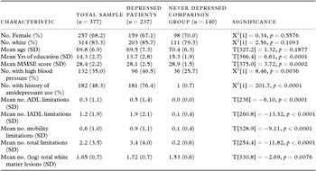
The characteristics of the sample are shown in Table 1. The depression patients had fewer years of education and on average lower MMSE scores than never depressed comparison participants. A higher proportion of patients reported they had high blood pressure. Depression patients were more likely to have a history of antidepressant use, and had on average more limitations in function and a greater volume of white matter lesions at baseline.
Characteristics of the sample

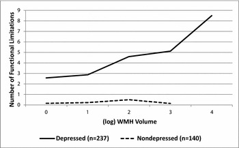
Figure 1 shows the mean number of functional limitations for each level of WMH volume by depression group. The mean values from these uncontrolled analyses suggested WMH volume was associated with functional status only among the depressed patients.
The mean number of functional limitations associated with each level of WMH volume by depression group (n = 377).

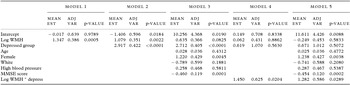
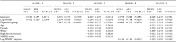
As shown in Table 2, in uncontrolled analyses (Model 1) and analysis controlling for depression status (Model 2), the volume of white matter lesions was significantly associated with the number of functional limitations. When the demographic and health variables were added to the model, the association between WMH volume and functional status was reduced (Model 3). As shown in Models 4 and 5, the association between WMH volume and functional status differed between the depressed and never depressed groups. The difference between the slopes was significant in both the uncontrolled model (p = 0.0204 with p < 0.05 in 88.6% of the iterations), as well as when the covariates were included (p = 0.0289 with p < 0.05 in 83.2% of the iterations). In the never depressed group, for each unit increase in (Log) WMH volume controlling for demographic and health variables, there was a −0.25 mean change in the total number of functional limitations (p = 0.5833 with p < 0.05 in 0.7% of the iterations), whereas in the depressed group there was on average a 1.03 mean increase in total limitations (p = 0.0207 with p < 0.05 in 92.2% of the iterations).
The association between (log) white matter lesion volume and the total number of functional limitations by depression status (n = 377)

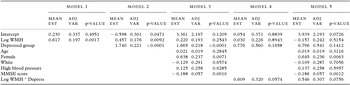
As shown in Table 3, WMH volume was associated with the number of IADL limitations in the uncontrolled model and when depression status was controlled (Models 1 and 2). When the demographic and health variables were added to the model (Model 3), the association between WMH and IADL limitations was not significant. In the fully controlled model (Model 5) among the never depressed, for each unit increase in (log) WMH there was a mean −0.16 change in the number of IADL limitations (p = 0.5154 with p < 0.05 in 0.4% of the iterations). In the depressed group, there was a mean increase of 0.39 IADL limitations (p = 0.0991 with p < 0.05 in 69.2% of the iterations). These group differences were, on average, not significant in either the uncontrolled (p = 0.0574) or controlled (p = 0.0756) models (Models 4 and 5).
The association between (log) white matter lesion volume and the number of IADL limitations by depression status (n = 377)

Table 4 shows the association between WMH volume and mobility limitations. WMH volume was significantly associated with the number of mobility limitations in the uncontrolled model, controlling for depression status and when the demographic and health variables were added to the model (Models 1–3). As shown in Models 4 and 5, the effect of WMH volume on mobility significantly differed by depression status. The difference between the slopes for the groups was significant in both the uncontrolled model (p = 0.0202 with p < 0.05 in 88.2% of the iterations) and controlling for covariates (p = 0.0289 with p < 0.05 in 84.4% of the iterations). In the fully controlled model (Model 5), a one-unit increase in (log) WMH volume was associated with a mean −0.01 decrease in the number of mobility limitations in the never depressed group (p = 0.9425 with p < 0.05 in 0.2% of the iterations). In the depressed group, an increase in WMH volume was associated with a mean 0.39 increase in mobility limitations (p = 0.0038 with p < 0.05 in 98.0% of the iterations).
The association between (log) white matter lesion volume and the number of mobility limitations by depression status (n = 377)

Discussion
We report new findings that suggest the relationship between vascular lesions and functional status differs in the presence of late-life depression. Within our sample, the relationship between WMH volume and the total number of functional limitations was only significant among those diagnosed with late life depression. That is, we did not find a significant association between WMH volume and functional status outside of depression, supporting our research hypothesis. When we examined these associations by domain of functional status in our exploratory analyses, we found that these differences by depression status were observed for mobility but not IADL limitations. The relationship between WMH and the overall number of limitations as well as the number of mobility limitations remained significant among the depressed patients when the demographic and health variables were controlled. These findings extend earlier findings reported by Steffens et al. (Reference Steffens, Bosworth, Provenzale and MacFall2002), which estimated the probability of one or more functional limitations association with greater WMH volume among depressed patients, and suggest the pattern is different among those without concurrent depression. Previous research has shown that lesions among those with late life depression are primarily ischemic (vascular) in origin, while lesions among the non-depressed are not always ischemic (Thomas et al., Reference Thomas2002), which could explain differential associations of WMH volume between the two groups.
Our findings across domains of function also provide new information. The significant modifying role of depression in the association between WMH and mobility was not unexpected. Mobility limitations are often the first sign of functional decline, and may account for much of the relationship between depressive symptoms and functional status. This may be due, in part, to decreased ability to initiate movement (psychomotor retardation) often seen in depression and in other subcortical disorders such as parkinsonism. That is, someone who is psychomotor slowed from depression may report some limitations in mobility. Previous research in this sample reported that white matter lesions were associated with smaller caudate volumes, particularly among the depressed patients compared to the never depressed comparison adults. The authors speculated that lesions may lead to caudate atrophy (Hannestad et al., Reference Hannestad2006). The caudate is associated with movement regulation, suggesting a possible mechanism why the association between WMH volume and mobility may differ in the presence of depression. Putamen volume is also associated with the melancholic subtype of depression, including psychomotor changes (Tupler et al., Reference Tupler, Krishnan, McDonald, Dombeck, D'Souza and Steffens2002), and may contribute to the observed group differences.
Depression did not significantly modify the association between WMH and IADL function, activities affected in part by cognition. The interrelationships among white matter lesions, cognition, and depression are complex. Depressed itself is associated with increased lesion volume. Depression may also affect cognition. White matter lesions may affect some cognitive processes outside of depression, which may account for less marked differences by depression status between WMH volume and IADL tasks.
It is not fully known why some patients with major depression develop functional decline as a consequence of the disease. These preliminary cross-sectional findings do not support the hypothesis that depression and disability share a common risk factor in WMHs, but rather that depression is an important modifier in the association between WMH volume and functional status, particularly mobility. Patients with greater volumes of WMH and/or vascular risk factors who are also depressed may be particularly at risk for functional decline. Also, while vascular damage may be impossible to repair, depression is a potentially modifiable risk factor to reduce the worsening of white matter lesion burden. Depressed older adults with functional limitations, particularly in mobility, may have worse cerebrovascular disease that might require management of vascular risk factors.
Our research has some limitations. The analyses presented here are cross-sectional and temporality and causality cannot be established. The same risk factors that lead to the vascular changes may also have led to the late-life depression. We were limited in these analyses to magnetic resonance acquisition technology that was available in 1994 when our study started. Our global method of lesion volume included lacunar infarcts and perivascular spaces but did not include brain microbleeds. These would likely account for only a very small percentage of the volume, however, and should not impact the findings.
Our comparison sample was particularly healthy, and these contrasts between the depressed and non-depressed groups may be less evident in a more representative sample. Future research could examine the association between lesion volume and functional status within a sample of depressed and nondepressed participants paired on lesion volume. Though self-report of hypertension was not optimal, previous analyses of these data (Taylor et al., Reference Taylor2005) showed this measure to be correlated with lesion volume, which supports the validity of self-report. We may have had insufficient power to detect a difference in the association between WMH volume and the number of IADL limitations. Our study, however, has multiple strengths including a large sample of depressed patients and never depressed comparison participants with available MRI data on vascular lesions. Future research will examine these patterns between groups over time.
Conflict of interest
None.
Descriptions of authors’ roles
Dr. Hybels and Dr. Pieper conducted the data analysis. Dr. Landerman participated in all the statistical discussions. Dr. Payne provided expertise in brain imaging, while Dr. Steffens provided clinical expertise in late life depression. While Dr. Hybels prepared the first draft of the paper, all of the authors provided input to the paper and have approved the final version.
Acknowledgments
This research was supported by NIMH grants R03 MH 095917, R01 MH54846, and K24 MH70027. The content is solely the responsibility of the authors and does not necessarily represent the official views of the National Institutes of Health.