Introduction
Cholesterol granulomas of the petrous apex are rare. Numerous surgical approaches have been used to treat such granulomas.Reference Brackmann and Toh1 They are usually accessed via a transtemporal or middle fossa approach (i.e. using a translabyrinthine, infracochlear, infralabyrinthine or supralabyrinthine approach).Reference House2–Reference Haberkamp4 Five cases of endoscopic removal of petrous apex cholesterol granuloma have been reported.Reference Samadian, Vazirnezami, Moqaddasi, Rakhshan, Khormaee and Ashraf5–Reference Michaelson, Cable and Mair9
We present a sixth case, which differed from previously reported cases in that our patient presented with symptoms suggestive of Ménière's disease. Our patient was safely and successfully managed using an endoscopic trans-sphenoid approach to the petrous apex.
We also outline the use of the endoscope in dealing with skull base lesions.
Case report
A 63-year-old man was referred with a history suggestive of early Ménière's disease. He reported continual imbalance and a ‘muzzy’ feeling, along with tinnitus and decreased hearing in the right ear. He also reported headaches in the occipital region. He was otherwise fit and well.
The ENT examination was normal.
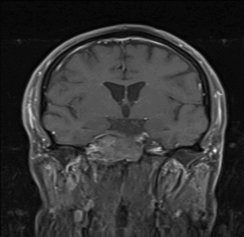
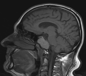
Audiography was also normal. Computed tomography (CT) and magnetic resonance imaging (MRI) showed a large lesion (58 × 32 × 27 mm) in the region of the right petrous apex, eroding the clivus and touching the brainstem (Figures 1 to 3). A provisional diagnosis of cholesterol granuloma was made.
Axial computed tomography scan demonstrating the isodense cholesterol granuloma encroaching on the posterior wall of the right sphenoid sinus.

Coronal, T1-weighted magnetic resonance imaging scan demonstrating the varied nature of the constituents of the cholesterol granuloma.

Sagittal, T1-weighted magnetic resonance imaging scan demonstrating the hyperintense nature of the cholesterol granuloma.

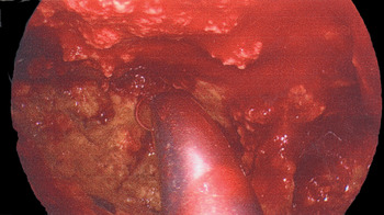
The lesion was approached endoscopically via bilateral sphenoidotomies joined by removing the posterior part of the septum. The posterior wall of the sphenoid was removed to reveal a huge cholesterol granuloma, which was evacuated (Figure 4).
Intra-operative endoscopic image showing the cholesterol granuloma being evacuated; the suction catheter can be seen in the resultant cavity.

The patient made an uneventful recovery.
Discussion
Cholesterol granulomas are rare. They were initially described in 1893, in the peritoneum.Reference Meyer10 They have been subsequently reported in numerous other locations, including the pneumatic pathways of the temporal bone.Reference Manasse11, Reference Wagner12 A cholesterol granuloma arises in the pneumatised spaces of the temporal bone, presumably as a result of negative pressure due to occlusion of the air cell system. This causes haemorrhage into the air cells, which leads to formation of cholesterol crystals and a subsequent foreign body reaction and progressive granuloma formation.Reference Beaumont13, Reference Hiraide, Inoye and Miyakogama14
Cholesterol granulomas are most commonly found in the maxillary sinus. Patients may present with a clear yellow discharge, in some cases containing evidence of bleeding. The diagnosis is usually made after imaging.Reference Chao15 Cholesterol granulomas of the petrous apex are uncommon, because this site is pneumatised in only 30 per cent of temporal bones; the incidence is less than 0.6 cases per million population per year.Reference Thedinger, Nadol, Montogomery, Thedinger and Greenberg16, Reference Lo, Solti-Bohmann, Brackmann and Gruskin17 Petrous apex cholesterol granulomas tend to remain clinically silent, but as the lesion expands, headaches and cranial neuropathies may arise, impairing hearing, balance, speech and swallowing.Reference Goldofsky, Hoffman, Holliday and Cohen18 Contralateral involvement of cranial nerves may be seen.
The treatment for symptomatic petrous apex cholesterol granuloma is surgical drainage and permanent aeration to prevent recurrence. Due to the lack of a true epithelial lining, total surgical excision is not essential. The goal is to open the granuloma widely, create a well aerated cavity and connect it to ventilated spaces. Numerous surgical approaches have been described for petrous apex cholesterol granuloma removal: translabyrinthine, infracochlear, infralabyrinthine, supralabyrinthine and trans-sphenoidal. Determination of the appropriate approach depends on the patient's hearing status, as well as the location of the lesion relative to adjacent neurovascular structures.Reference Brackmann and Toh1
Patients with good hearing whose lesions abut the posterior wall of the sphenoid sinus can undergo drainage via the trans-sphenoidal approach.Reference Sabin, Vordi and Symon19 This approach was first described by Montgomery in 1977, and involved an external procedure performed via an incision near the medial canthus.Reference Montgomery20 In 1994, Fucci adapted this approach to enable the use of a nasal endoscope, gaining access to the petrous apex via a transnasal–trans-sphenoid approach.Reference Fucci, Alford and Lowry21 The procedure is appropriate for lesions that contact or invade the sphenoid sinus and are accessible via the posterior wall, as in our case. The technique is far less invasive compared with an intracranial procedure. Facial weakness is a well described complication of lateral temporal and middle fossa approaches, but is highly unlikely with an endonasal approach. Endoscopic visualisation reduces the risk of creating a cerebrospinal fluid (CSF) leak; it also enables identification of important structures lying adjacent to the sphenoid sinus, and allows wide marsupialisation.Reference Casiano22–Reference Zinreich, Tebo, Long, Brem, Mattox and Loury24
• Cholesterol granulomas of the petrous apex are rare
• The diagnosis is based on high resolution computed tomography and magnetic resonance imaging
• The presented case of petrous apex cholesterol granuloma was unusual as the patient presented with symptoms suggestive of Ménière's disease
• The trans-sphenoidal approach to the petrous apex is rarely used for the drainage and ventilation of cholesterol granuloma, but is preferred when the lesion lies in the medial petrous apex, abutting and/or prolapsing into the posterior sphenoid sinus wall
• The trans-sphenoidal approach is highly conservative and spares cochlear and vestibular function (unlike other lateral approaches); it also facilitates follow up and the treatment of any recurrence
Although there is a theoretical risk of injury to the optic nerves or the internal carotid artery, as well as the possibility of a CSF leak, the wide field of vision helps to avoid vital structures. Moreover, the trans-sphenoidal approach can be safely performed in individuals with a high jugular bulb (damage of which is a concern with the subcochlear approach), and when there is any risk of damage to the inner ear and facial nerve.Reference Haberkamp4
Traditional surgical treatments of cholesterol granuloma have the shortcoming of producing a relatively narrow isthmus through which permanent drainage must occur, and are associated with high rates of restenosis with cyst reaccumulation.Reference Brackmann and Toh1
Long-standing cholesterol granulomas frequently contain haemosiderin sediment and concretions, which may be difficult to clean and evacuate through the relatively small opening obtained. Endoscopic visualisation allows more complete removal of such debris. The trans-sphenoidal approach also facilitates follow up by enabling simple, adequate endoscopic examination as an out-patient, and easier treatment of recurrence.
Conclusion
Trans-sphenoidal access to the petrous apex represents an alternative route for the drainage and ventilation of cholesterol granulomas. This approach is the technique of choice when the cholesterol granuloma abuts the posterior wall of the sphenoid sinus. The trans-sphenoid approach, unlike other lateral approaches to the petrous apex, spares cochlear and vestibular function and allows post-operative endoscopic follow up.