Cambay amber is found in working lignite mines in the northwest of India, in the state of Gujarat, about 30 km northeast of Surat. Age determination of the amber-bearing sediments is based on findings of benthic foraminifera (Sahni et al. Reference Sahni, Saraswati, Rana, Kumar, Singh, Alimohammadian, Sahni, Rose, Singh and Smith2006; Punekar & Saraswati Reference Punekar and Saraswati2010), dinoflagellates (Garg et al. Reference Garg, Ateequzzaman, Prasad, Tripathi, Singh, Jauhri and Bajpai2008) and data from strontium isotopes and stable carbon isotopes, as well as mammal fossils (Clementz et al. Reference Clementz, Bajpai, Ravikant, Thewissen, Saravanan, Singh and Prasad2011; Smith et al. Reference Smith, Kumar, Rana, Folie, Solé, Noiret, Steeman, Sahni and Rose2016). The data suggest an Early Eocene age (54 Ma), indicating that the time of sediment deposition took place just as India entered the equatorial humid belt, shortly after the Palaeocene Eocene Thermal Maximum (PETM) and at the beginning of the Early Eocene Climatic Optimum (EECO) (Zachos et al. Reference Zachos, Dickens and Zeebe2008). The amber has been deposited in a near-shore environment in fluctuating conditions ranging through lacustrine, swamp, marshy and deltaic environments, with coastal mangrove forests, palm-dominated zones and an angiosperm evergreen forest (e.g., McCann Reference McCann2010; Rao et al. Reference Rao, Sahni, Rana and Verma2013). The reconstruction of the amber forest has revealed a tropical forest growing under warm and humid conditions and with diverse aquatic habitats ranging from marine to brackish to fresh waters.
Investigation of the inclusions from Cambay amber has shown to facilitate understanding of India's geological history, as demonstrated by the analysis of distributional patterns of the inclusions (e.g., Rust et al. Reference Rust, Singh, Rana, McCann, Singh, Anderson, Sarkar, Nascimbene, Stebner, Thomas, Solórzano Kraemer, Williams, Engel, Sahni and Grimaldi2010). Moreover, Cambay amber has revealed a highly diverse fauna of eusocial insects, such as corbiculate bees, rhinotermitid termites and modern ants (Formicidae), indicating that these groups diversified before the formation of the amber, probably during the PETM (summarised in Rust et al. Reference Rust, Singh, Rana, McCann, Singh, Anderson, Sarkar, Nascimbene, Stebner, Thomas, Solórzano Kraemer, Williams, Engel, Sahni and Grimaldi2010).
So far, only few dipteran inclusions have been described from Cambay amber (Solórzano Kraemer & Evenhuis Reference Solórzano Kraemer and Evenhuis2008; Solórzano-Kraemer & Wagner Reference Solórzano-Kraemer and Wagner2009; Grimaldi & Singh Reference Grimaldi and Singh2012; Stebner et al. Reference Stebner, Szadziewski, Rühr, Singh, Hammel, Kvifte and Rust2016, Reference Stebner, Szadziewski, Singh, Gunkel and Rust2017; Zakrzewska et al. Reference Zakrzewska, Stebner, Mateusz, Singh and Giłka2017 (this volume)). Within the present work, a fossil limoniid is described from Cambay amber. The fossil belongs to the genus Dicranomyia and is part of large amber flow from the Valia mine. The flow has revealed nearly 200 arthropod inclusions that are currently under systematic investigation, and which might eventually add interesting information about temporal and spatial diversity in the former amber forest.
The genus Dicranomyia Stephens, Reference Stephens1829 is numerous and distributed worldwide today (Oosterbroek Reference Oosterbroek2016). Fossil representatives of this genus are frequent (Evenhuis Reference Evenhuis2015). Over 40 known fossil species of Dicranomyia are known from different periods, mainly from the Eocene but also from younger epochs such as the Oligocene or Miocene, and also from different parts of the world.
Thirty two of them are known from the Eocene: four species are known from the Early Eocene (Ypresian) (after Grimaldi & Ross Reference Grimaldi, Ross, Fraser and Sues2017) of Denmark (Krzemiński Reference Krzemiński2001); four from the Ypresian–Lutetian of the USA (Scudder Reference Scudder1877; Cockerell Reference Cockerell1908); 14 from Baltic amber (Meunier Reference Meunier1899, Reference Meunier1906, Reference Meunier1916; Alexander Reference Alexander1931; Savchenko Reference Savchenko1967; Krzemiński Reference Krzemiński2000a; Kania et al. Reference Kania, Penar and Krzemiński2013; Kania Reference Kania2014; Krzemiński et al. Reference Krzemiński, Kania and Durak2015); and ten known as compression fossils from the Priabonian (Scudder Reference Scudder1894; Cockerell & Haines Reference Cockerell and Haines1921; Cockerell Reference Cockerell1922) of the USA and England.
Three species are known from the Oligocene of the USA (Brown Reference Brown1985), one from France (Théobald Reference Théobald1937) and one from Russia (Krzemiński Reference Krzemiński2000b). Nine species are known from the Miocene, only one from Italy (Krzemiński & Gentlini Reference Krzemiński and Gentlini1992), two from Germany (Meunier Reference Meunier1906), four from Mexico and two from the Dominican Republic (Podenas & Poinar Reference Podenas and Poinar1999).
Dicranomyia is numerous in Baltic amber, but has not previously been described from Early Eocene Cambay amber.
1. Material and methods
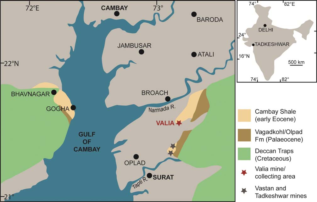
The biotic diversity and biogeographical significance of Cambay amber was reviewed by Rust et al. (Reference Rust, Singh, Rana, McCann, Singh, Anderson, Sarkar, Nascimbene, Stebner, Thomas, Solórzano Kraemer, Williams, Engel, Sahni and Grimaldi2010). The specimen was collected from the Valia lignite mine (N 21°24′41.37″, E 073°05′56.31″) (Fig. 1) during January 2015 and is part of a large amber flow, with 199 arthropod inclusions. The amber is deposited in the Birbal Sahni Institute of Palaeosciences, Lucknow, India (BSIP).
Map showing location of the Valia mine (N 21°24′41.37″; E 073°05′56.31″) – red asterisk – where the specimen was found (modified after Sahni et al. Reference Sahni, Saraswati, Rana, Kumar, Singh, Alimohammadian, Sahni, Rose, Singh and Smith2006).

The amber was ground using a Buehler Phoenix Beta grinding machine and the specimen was studied using a Nikon SMZ 1500 stereomicroscope equipped with a Nikon DS-Fi1 camera. The drawings were made from studying the specimen and photographs. The digital format made it possible to take measurements with NIS-Elements D 3.0 software. The measurements were taken only when the relevant structures were not distorted. The measurements of the length of the discal cell was taken from its posterior edge to the point of connection of cross-vein vein m–m with vein M3 (Kania Reference Kania2014). The term “d-cell base” is used following Krzemiński (Reference Krzemiński1993). The drawings were made by IK.
2. Systematic palaeontology
Order Diptera Linnaeus, Reference Linnaeus1758 Infraorder Tipulomorpha Rohdendorf, Reference Rohdendorf1961 Family Limoniidae Speiser, Reference Speiser1909 Subfamily Limoniinae Speiser, Reference Speiser1909 Genus Dicranomyia Stephens, Reference Stephens1829 Subgenus Dicranomyia Stephens, Reference Stephens1829
Type species. Limnobia modesta Meigen, Reference Meigen1818; by subsequent designation of Coquillett Reference Coquillett1910, p. 533.
Dicranomyia (Dicranomyia) indica sp. nov. (Figs 2–5)
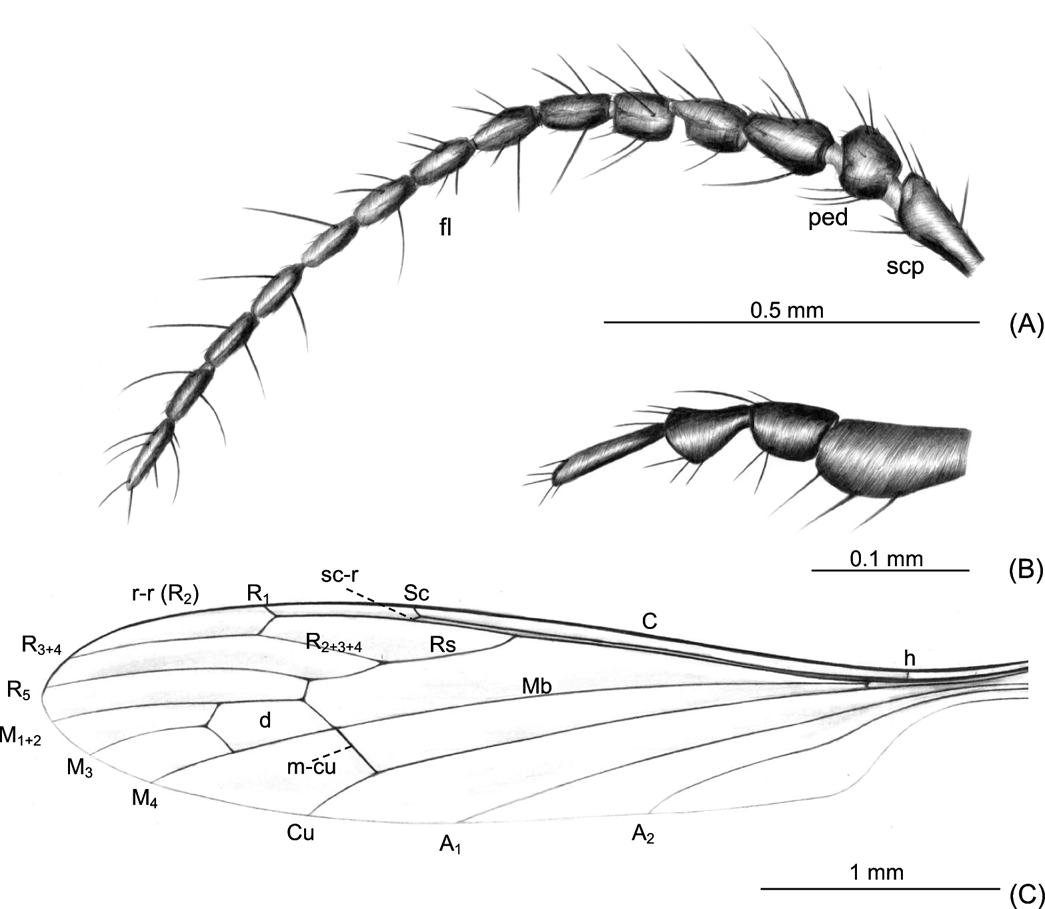
Dicranomyia (D.) indica sp. nov., BSIP Val-3.1 (female), holotype: (A) antenna, lateral view; (B) palpus, lateral view; (C) wing (reconstruction) Abbreviations: fl = flagellomeres; ped = pedicel; scp = scape.

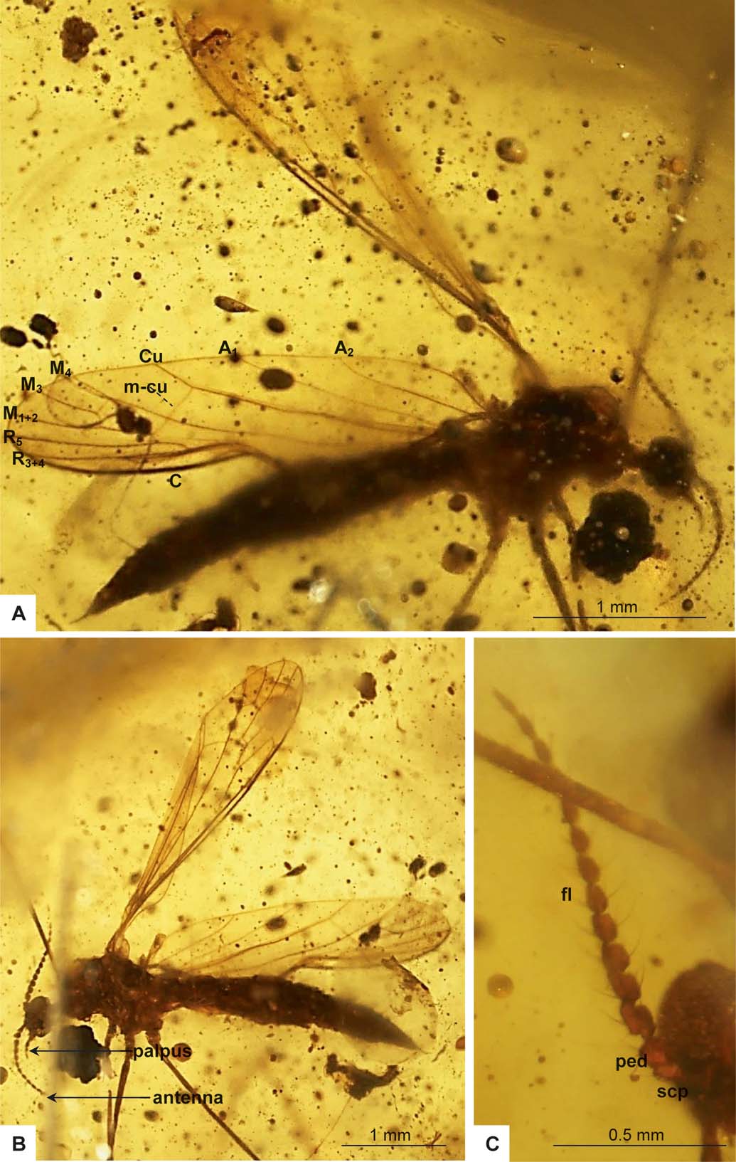
Dicranomyia (D.) indica sp. nov., BSIP Val-3.1 (female), holotype: (A) body, lateral view; (B) body, latero-ventral view; (C) antenna, ventral view. Abbreviations: fl = flagellomeres; ped = pedicel; scp = scape.

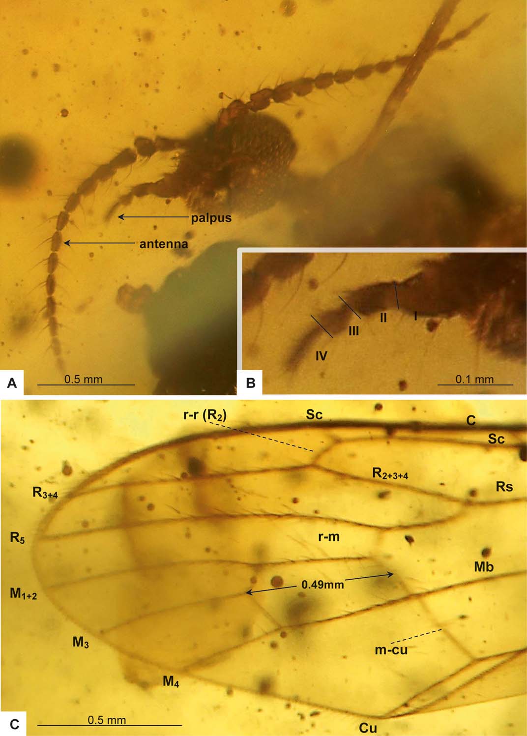
Dicranomyia (D.) indica sp. nov., BSIP Val-3.1 (female), holotype: (A) head, fronto-ventral view; (B) palpus, lateral view; (C) part wing venation.

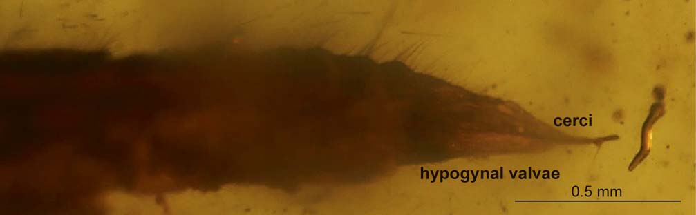
Dicranomyia (D.) indica sp. nov., BSIP Val-3.1 (female), holotype: ovipositor, lateral view.

Diagnosis. Flagellomeres 1–4 very asymmetrically dilated, with the inner surface well developed, wide; first flagellomere twice as long as wide, the second and third almost as long as wide; flagellomeres 5–14 are similar in shape but narrower and elongated, becoming narrower, more slender and cylindrical distally; last flagellomere tapered at the end, almost as long as the penultimate one; vein Sc comparatively elongated, ending behind mid-length of Rs, opposite distal two third of vein Rs; d-cell elongated, twice as long as wide, almost as long as M3; cross-vein m–cu straight, in or just before fork of Mb; R1 ending beyond the level of cross-vein r–m, opposite the half the length of d-cell, approximately two-fifths of the length of R2+3+4 and R3+4 measured from Rs fork; Rs is as long as R2+3+4.
Etymology. The species is named after the country of origin – India.
Material examined. Holotype specimen No. BSIP Val-3.1 (female) (deposited in the BSIP); Cambay amber, Surat, India.
Description. Body dark-brown, 4.71 mm long.
Head not large, 0.35 mm long, 1.49 mm wide, but with huge, distinctly separate eyes; antenna (Figs 2A, 3A, B, C, 4A) 14-segmented, c.1.17. mm long, the morphology of the antenna is very characteristic with scape elongated, pedicel wide, 0.07 mm long; first flagellomere 0.10 mm long; flagellomeres 2–13 c.0.07–0.08 mm long, the last one 0.09 mm long; flagellomeres 1–4 very asymmetrically dilated, with the inner surface strongly developed, almost as long as wide; the marked development of some flagellomeres gives a serrulate appearance of the antennae; flagellomeres 5–14 are a similar shape but narrower, elongated, becoming narrower, more slender and cylindrical distally, two penultimate flagellomeres almost oval, the last flagellomere elongated, almost half longer than penultimate one, distinctly narrow at apex. The apex of each segment with a short glabrous pedicel; outer edge of flagellomeres with three or even four elongated setae, much longer than the length of the flagellomere on which they occur; palpus (Figs 2B, 3A, B, 4A, B): four-segmented, 0.26 mm long, segments I–III not very elongated, widened in distal part; length of segments: II – 0.07 mm, III – 0.06 mm, IV – 0.08 mm.
Thorax (Fig. 3A, B): wing (Figs 2C, 3A, B, 4C) 4.06 mm long, approximately 0.95 mm wide; pterostigma not very visible, vein Sc relatively elongated, ending behind mid-length of Rs, opposite distal two third of vein Rs; cross-vein sc–r about 1.5 times its length from the tip of Sc; vein R3+4 longer than Rs; R1 ending beyond the level of cross-vein r–m, opposite half the length of the d-cell, approximately 2/5 of the length of R2+3+4 and R3+4 measured from Rs fork; d-cell elongated, 0.49 mm long, twice as long as wide, almost as long as M3 (0.50 mm); cross-vein m–cu straight, 0.27 mm long (in left wing), 0.30 mm (in right wing), in or just before the fork of Mb; vein M1+2 approximately one-third longer than d-cell; A1 and A2 almost straight.
Abdomen (Fig. 3A, B): ovipositor (Figs 3A, B, 5), 0.66 mm long, short, about one-tenth of body length.
Comparisons. The feature that separates D. (D.) indica sp. nov. from other species is the morphology of the antenna. The species is very like D. (D.) alexandri Kania, Reference Kania, Penar and Krzemiński2013 from Baltic amber, especially in the shape of the flagellomeres. In D. (D.) alexandri, flagellomeres 1–7 are very asymmetrically dilated, with the inner surface well developed, wide, almost as long as wide. In D. (D.) indica sp. nov., the flagellomeres are also asymmetrically dilated, but only flagellomeres 1–4, and not as much as in D. (D.) alexandri. These species differ in their wing venation: in D. (D.) alexandri, vein Sc is short, ending before the midlength of Rs; in D. indica sp. nov., this vein ends beyond half the length of Rs; also, the d-cell in D. indica sp. nov. is elongated, twice as long as wide, and approximately as long as M3; whereas in D. (D.) alexandri, the d-cell is comparatively short, approximately as long as wide, distinctly shorter than M3; R1 ending beyond the level of cross-vein r–m in D. indica sp. nov., opposite half the length of the d-cell; in D. (D.) alexandri, opposite the level of cross-vein r–m, at one-third the length of the d-cell.
In all other species known from the Baltic amber, antennae are without strongly asymmetric dilating flagellomeres; instead, flagellomeres are rather elongated or the first flagellomeres are short and oval, as in Dicranomyia (D.) lobata Meunier, Reference Meunier1906. The new species also differs from other species of the subgenus Dicranomyia known from the Eocene in the wing venation.
In D. (D.) graciosa Meunier, Reference Meunier1916, D. (D.) grandis Meunier, Reference Meunier1899, D. (D.) kalandyki Krzemiński, Reference Krzemiński2000b and D. (D.) perpendicularis Savchenko, Reference Savchenko1967, vein Sc is very short, ending before the mid-length of Rs; whereas in D. indica sp. nov., this vein ends beyond the mid-length of Rs, opposite the distal two-thirds of vein Rs. In D. (D.) baltica Kania, Reference Kania2014 and D. (D.) lobata, cross-vein m–cu is situated distinctly before fork of Mb; in D. indica sp. nov., cross-vein m–cu is situated at the fork of the Mb or just before; whilst in D. (D.) ewa Kania, Reference Kania2014, D. (D.) graciosa and D. (D.) grandis, it is positioned beyond the bifurcation of the Mb. In D. indica sp. nov. vein Rs is as long as R2+3+4; in all other species of the subgenus Dicranomyia known for Baltic amber, this vein is shorter than R2+3+4. The d-cell in D. indica sp. nov. is approximately as long as M3; in D. (D.) baltica, D. (D.) gorskii Kania, Penar & Krzemiński, Reference Kania, Penar and Krzemiński2013, D. (D.) grandis, D. (D.) lobata, D. (D.) meunieri Alexander, Reference Alexander1931, D. (D.) perpendicularis and D. (D.) sinuata Meunier, Reference Meunier1916, this vein is longer in comparison to the d-cell length.
In contrast to D. indica sp. nov., wings with pterostigma occur in D. (D.) alexandri, D. (D.) baltica, D. (D.) ewa, D. (D.) gorskii, D. (D.) graciosa, (D.) grandis, D. (D.) kalandyki, D. (D.) lobata, D. (D.) meunieri, D. (D.) sinuata and D. (D.) succinica Kania, Reference Kania2014. In D. indica sp. nov., R1 ends beyond the level of cross-vein r–m, opposite half the length of the d-cell; whilst in D. (D.) gorskii, R1 ends opposite the level of cross-vein m–m.
In D. indica sp. nov., cross-vein sc–r is positioned at about 1.5 times its length from the tip of Sc; in D. (D.) baltica, D. (D.) graciosa, (D.) grandis, (D.) kalandyki and D. (D.) meunieri, about twice its length from the tip of Sc; in D. (D.) ewa, D. (D.) gorskii, D. (D.) perpendicularis and D. (D.) sinuata, about its length from the tip of Sc; and in D. (D.) succinica, about three times its length from the tip of Sc.
Moreover, in contrast to Dicranomyia (s. lato) thybotica Henriksen, Reference Henriksen1922, where vein R1 is elongated, ending beyond the level of cross-vein r–m, far beyond d-cell, in D. indica sp. nov. this vein is shorter, ending beyond the level of cross-vein r–m, but opposite half the length of the d-cell. In Dicranomyia indica sp. nov., Rs bifurcation is positioned before the fork of Mb; in D. (s. lato) thybotica, it is positioned approximately at the fork of Mb.
In contrast to Dicranomyia (s. lato) speciosa Krzemiński, Reference Krzemiński2001, where cross-vein m–cu is positioned distinctly before the fork of Mb, this vein is in or just before the fork of Mb in D. indica sp. nov. In D. (D.) speciosa, Rs is elongated, almost as long as both R2+3+4 and R3+4. In D. indica sp. nov., this vein is shorter than R3+4. In Dicranomyia (s. lato) bellissima Krzemiński, Reference Krzemiński2001, vein Sc is very short, ending opposite one-quarter the length of Rs; whilst in D. indica sp. nov., this vein is longer, ending beyond the mid-length of Rs. In Dicranomyia (Dicranomyia) freiwaldi Krzemiński, Reference Krzemiński2001, Sc is very elongated, reaching beyond the fork of Rs, cross-vein m–cu is situated before fork of Mb and the distal portion of the wing is covered with numerous macrotrichiae; in D. indica sp. nov., this vein ends beyond the mid-length of Rs, opposite the distal two-thirds of vein Rs, cross-vein m–cu is situated at or just before bifurcation of Mb and macrotrichiae are absent from the wing.
In the new species, the d-cell is elongated and twice as long as wide, almost as long as M3; in Dicranomyia excavata Cockerell & Haines Reference Cockerell and Haines1921, vein M3 is longer, approximately 1.5 times longer than the d-cell and the d-cell is about 1.5 times as long as wide. The fork of Rs is situated opposite the Mb bifurcation in D. excavata; in D. indica sp. nov., the fork of Rs is situated distinctly before the fork of Mb.
Dicranomyia rhodolitha Cockerell, Reference Cockerell1908 is characterised by the occurrence of small dark spots on the costa, 2.6 mm and 4 mm from the base; in D. indica sp. nov., there are no spots on the wing.
In contrast to Dicranomyia longipes Scudder, Reference Scudder1894, D. indica sp. nov. is smaller, with wings about 4 mm long. The length of the wing of D. longipes is about 7 mm. Moreover, the discal cell in D. longipes is closed, but much narrower apically than at base by the length of the third posterior cell; while in D. indica sp. nov., the d-cell is not narrowed. In D. longipes and Dicranomyia stagnorum Scudder, Reference Scudder1894, the distal part of the marginal cell, according to Scudder, is almost as long as the proximal; in other species known from the Eocene, such as Dicranomyia inferna Scudder, Reference Scudder1894, Dicranomyia fragilis Scudder, Reference Scudder1894, Dicranomyia stigmosa Scudder, Reference Scudder1894, Dicranomyia primitiva Scudder, Reference Scudder1894, Dicranomyia fontainei Scudder, Reference Scudder1894 and Dicranomyia rostrata Scudder, Reference Scudder1894, the distal part of the marginal cell is much shorter than the proximal; in D. indica sp. nov., it is longer. Moreover, the d-cell of D. stagnorum is open, whilst the d-cell of D. indica sp. nov is closed.
3. Conclusions
The PETM was an abrupt global warming event at 55 Ma, which was accompanied by a rapid global increase of deep-sea water temperatures (≈6–8°C) and globally higher temperatures (≈3–5°C) (e.g., Katz et al. Reference Katz, Pak, Dickens and Miller1999; Jardine Reference Jardine2011). This rapid warming may have been caused by a mass release of methane from the sediments in the sea floor, the decomposition of organic matter in terrestrial settings, or volcanic activity, which in turn led to a mass release of carbon into the atmosphere (e.g., Katz et al. Reference Katz, Pak, Dickens and Miller1999; Svensen et al. Reference Svensen, Planke, Malthe-Sørenssen, Jamtveit, Myklebust, Eidem and Rey2004; Zachos et al. Reference Zachos, Dickens and Zeebe2008; Jardine Reference Jardine2011). Whatever the reason, the results gave rise to the increased radiation of vertebrates and angiosperms, as indicated by the sudden appearance of modern mammalians (Gingerich Reference Gingerich2006; Smith et al. Reference Smith, Kumar, Rana, Folie, Solé, Noiret, Steeman, Sahni and Rose2006) and the spread of the first Shorea forests (Rust et al. Reference Rust, Singh, Rana, McCann, Singh, Anderson, Sarkar, Nascimbene, Stebner, Thomas, Solórzano Kraemer, Williams, Engel, Sahni and Grimaldi2010). In western India, these warming events – together with India's geographical location at the equatorial humid-belt – resulted in an extremely hot and humid climate, with the rate of mean annual precipitation exceeding the rate of evaporation (e.g., Kent & Muttoni Reference Kent and Muttoni2008), and with annual mean temperatures of ca.32°C (Saraswati et al. Reference Saraswati, Rangaswami and Navada1993). In this context, the first record of the dipteran family Limoniidae is of importance, since it adds further data to the diversity of a tropical forest at the time of expanding modern diversity.
Furthermore, Cambay amber was formed ca.80 Ma after India's separation from Gondwana and ca.30 Ma after India's separation from Madagascar (e.g., Storey Reference Storey1995). The “Biotic ferry” model postulates that India was an isolated continent for at least 30 Ma (after India's separation from Madagascar and before its collision with Asia), allowing a highly endemic fauna to develop. However, insects in Cambay amber show worldwide faunal affinities (e.g., Rust et al. Reference Rust, Singh, Rana, McCann, Singh, Anderson, Sarkar, Nascimbene, Stebner, Thomas, Solórzano Kraemer, Williams, Engel, Sahni and Grimaldi2010; Engel et al. Reference Engel, Grimaldi, Nascimbene and Singh2011, 2013; Stebner et al. Reference Stebner, Szadziewski, Singh, Gunkel and Rust2017), suggesting that the Indian subcontinent was not highly isolated at the time of the amber formation. Faunal connections to Baltic amber and Fushun amber from China have been observed within the Diptera (Grimaldi & Singh Reference Grimaldi and Singh2012; Stebner et al. Reference Stebner, Szadziewski, Singh, Gunkel and Rust2017).
Recent representatives of the subgenus Dicranomyia in India are reported from the northeastern or eastern areas, such as Assam, Sikkim, Uttarakhand and Bengal and Uttar Pradesh, and also from the valley between the Great Himalayas and the Pir Panjal mountain range (Kashmir, northwestern India – Himachal Pradesh). Some species are known from southeastern India (e.g., Tamil Nadu) and from midwestern India (e.g., Maharashtra and Goa) (Oosterbroek Reference Oosterbroek2016).
However, Dicranomyia indica sp. nov. seems to have its closest relative in Baltic amber. The species is similar to Dicranomyia alexandri Kania, Reference Kania, Penar and Krzemiński2013 from Baltic amber (Kania et al. Reference Kania, Penar and Krzemiński2013). Shared characters between the two species are the antennal segments, which are asymmetrically dilated. In other fossil representatives from Baltic amber, the flagellomeres are cylindrical. Thus, Dicranomyia indica sp. nov. adds further evidence to the already reported faunal connections between both deposits. It must be emphasized, however, that Dicranomyia is a cosmopolitan genus today and, thus, further findings of limoniids in Cambay amber are needed in order to get a more detailed analysis of the distribution patterns of this group in the Eocene.
4. Acknowledgements
We are deeply indebted to Dr Andrew Ross and an anonymous reviewer for valuable comments and remarks on this manuscript. FS was supported by funding to JR (Steinmann Institute, Universität Bonn, Germany) – No. RU665/10-1 from the German Research Foundation (DFG). HS thanks the Director of the Birbal Sahni Institute of Palaeosciences, Lucknow, India for his support.