Introduction
Sudden unilateral or bilateral sensorineural hearing loss due to infarcts of the anterior inferior cerebellar artery,Reference Lee, Whitman, Lim, Lee and Park1–Reference Hausler and Levine8 posterior inferior cerebellar arteryReference Lee9, Reference Lee and Yoon10 or basilar arteryReference Muttikkal, Kesser and Mukherjee11–Reference Lee and Baloh13 have previously been reported in the literature. The majority of cases involve elderly patients with a past medical history of a cerebral infarct, arrhythmia, hypertension or diabetes, predisposing them to a cerebrovascular event. To the best of our knowledge, no cases of unilateral sensorineural hearing loss due to a cerebellar infarct in young, fit and well patients have been described. We report a case of sensorineural hearing loss in a young male with no traditional cardiovascular risk factors but with known misuse of anabolic steroids.
Case report
A 32-year-old male visited our accident and emergency department complaining of a 6-hour onset of dizziness, unsteadiness, nausea, headache and reduced hearing in his right ear. He also reported blurred vision and photophobia. He was otherwise fit and well, working full-time in a gym and powerlifting as a hobby. He was a non-smoker, with no significant medical history. On direct questioning, he admitted that he had taken anabolic steroids at regular intervals for the last three years (testosterone enanthate, Oral Turinabol™ and oxymetholone).
On examination, the patient was cardiovascularly stable, with a Glasgow coma scale score of 15. His pupils were equal and reactive to light. Initial cranial nerve examination findings were normal. Further neurological examination showed normal tone, power, reflexes, co-ordination and sensation in all four limbs.
The patient was admitted to the acute medical unit with a working diagnosis of subarachnoid haemorrhage. He underwent an unenhanced computed tomography scan, which ruled out the presence of intracranial haemorrhage or a space-occupying lesion. The cerebral parenchyma, ventricles and basal cisterns were also found to be normal. He underwent a lumbar puncture, with normal findings. Blood tests revealed an elevated white blood cell level of 13.85 × 10⁹ per litre and a high red blood cell count of 6.63 × 10¹² per litre. His haemoglobin value was also raised to 18.2 gm/dl. The remaining blood parameters, including clotting, renal and liver function, were unremarkable. He was kept in hospital overnight for observation and given analgesics to treat the headache, with a working diagnosis of migraine.
The next morning, the patient complained of complete right-sided deafness and tinnitus, with increased unsteadiness. As a result, an urgent ENT opinion was sought. ENT examination revealed intact ear drums bilaterally, with normal ear canals. The tuning fork tests showed Weber's test lateralising to the left side and a positive Rinne test on the left ear. On the right side, the patient could not detect any sound via air conduction on tuning fork testing. Grade 1 horizontal nystagmus was detected on leftward gaze. Repeat examination of the remaining cranial nerve was unremarkable. Neurological assessments for ataxia, Romberg's sign and dysmetria were negative. A working diagnosis of lateral pontine syndrome was made in light of interrupted blood supply of the anterior inferior cerebellar artery.
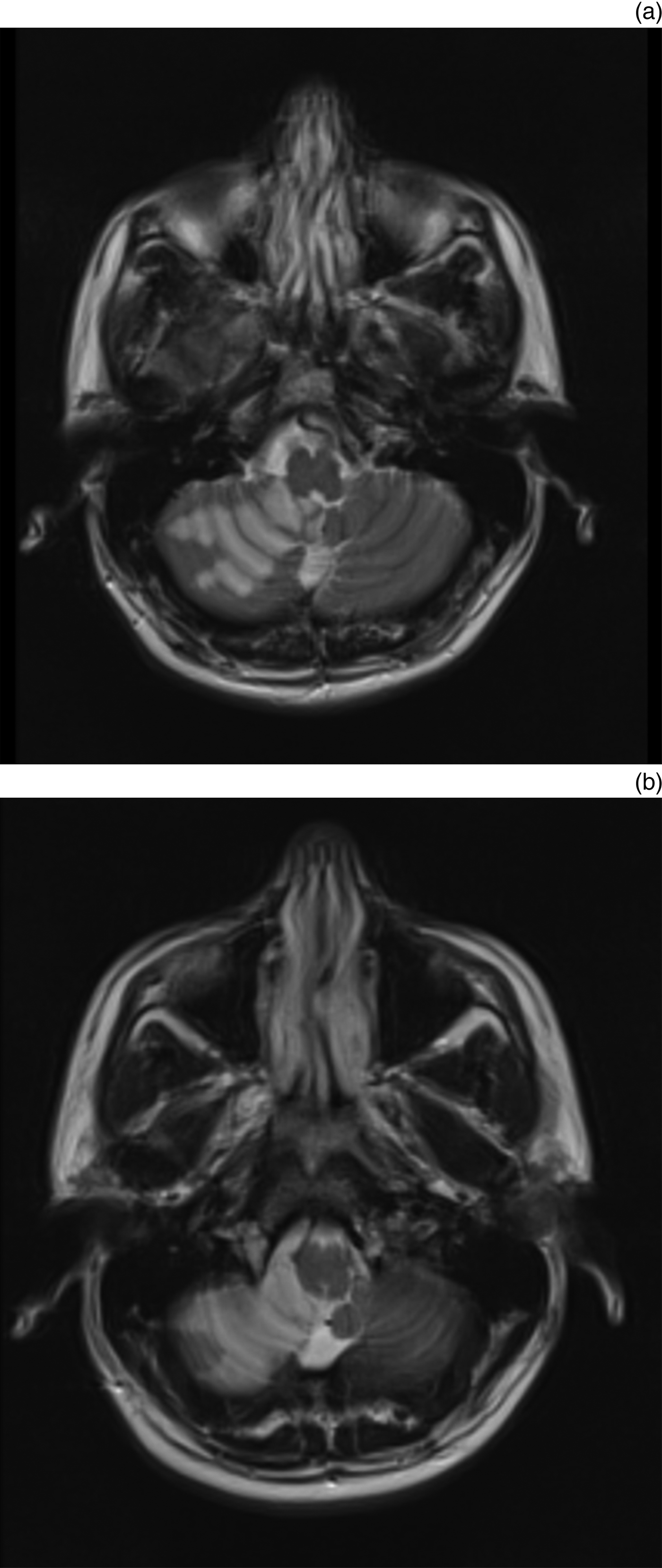
An urgent magnetic resonance imaging scan of the brain and brain stem showed a well-defined focal area of signal change involving the inferior aspect of the right cerebellar hemisphere that exhibited diffusion restriction (Figure 1). The occlusion was in the region of the right anterior inferior cerebellar artery, in keeping with an acute anterior inferior cerebellar artery infarction. There was no evidence of haemorrhage. The remainder of the brain and brain stem was normal, with patent basilar arteries.
Axial, T2-weighted magnetic resonance imaging scans of the patient (a & b), which show a diffuse high signal intensity in the region of the right anterior inferior cerebellar artery, suggesting acute infarction.

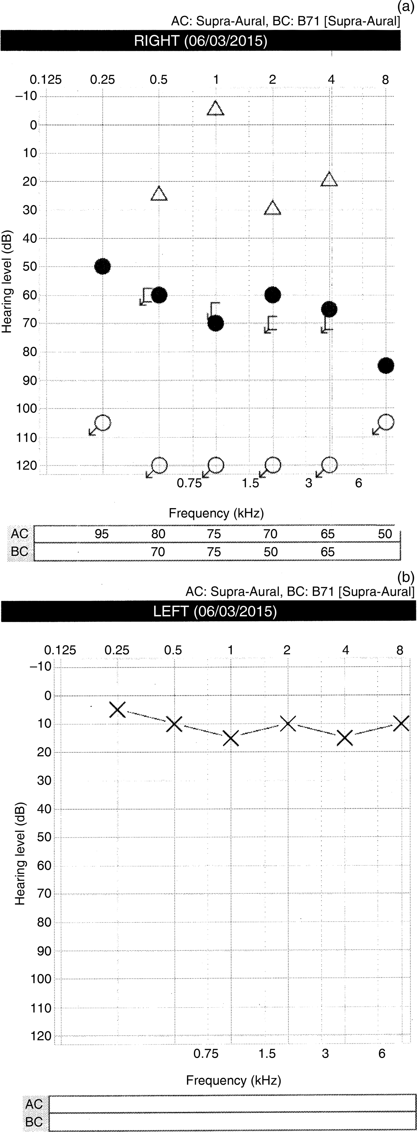
The pure tone audiogram showed hearing within normal limits on the left ear, and a profound hearing loss on the right side with no response to the limit of the equipment with air conduction (Figure 2). Unmasked bone conduction was within normal limits. Findings were consistent with a right sensorineural hearing loss due to a right anterior inferior cerebellar artery infarct.
Pure tone audiogram of the patient (a, right ear; b, left ear), showing profound sensorineural hearing loss in the right ear (a) on day 1 following acute presentation with hearing loss. ∆ = Bone conduction (unmasked) right ear; • = air conduction (masked) right ear; [ = bone conduction (masked) right ear; ↙ = no detectable sound; ○ = air conduction (unmasked) right ear; AC = air conduction; BC = bone conduction; × = air conduction (unmasked) left ear.
The patient was commenced on antiplatelet medications in the form of 300 mg aspirin daily for two weeks, followed by clopidogrel 75 mg daily. He was also started on simvastatin 40 mg once per day and was advised to stop taking the anabolic steroids.
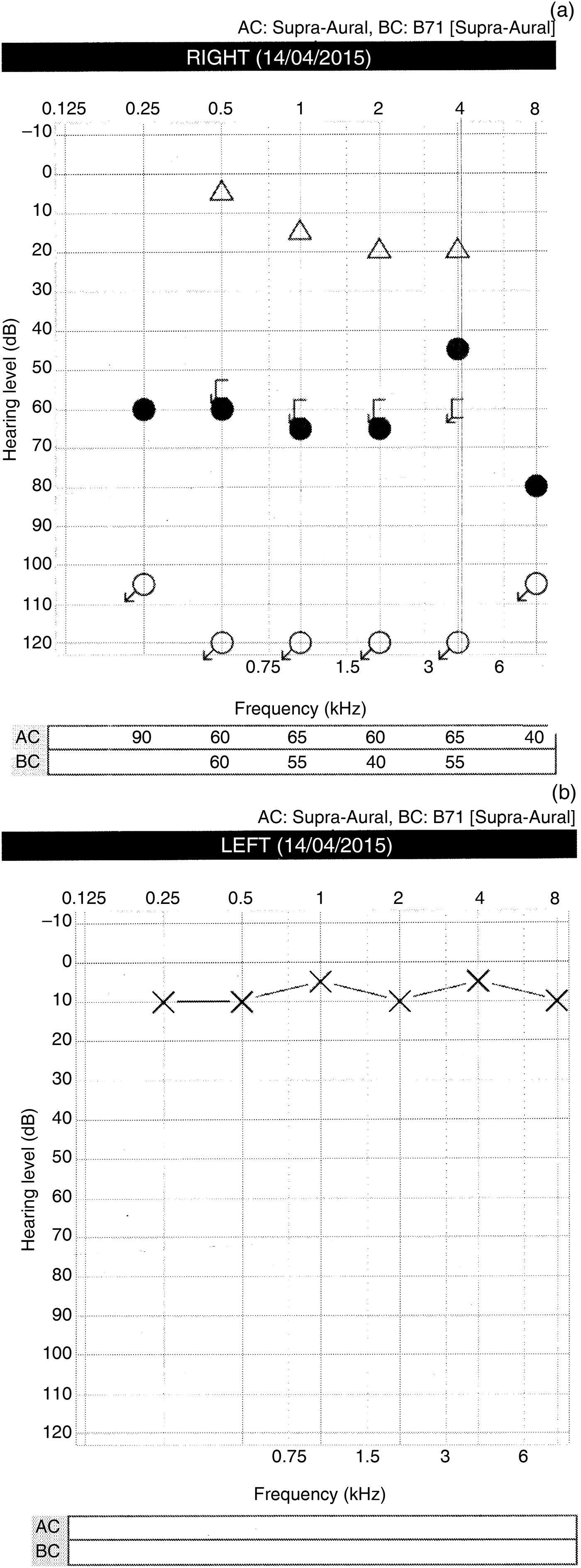
The patient was followed up six weeks after the initial onset of symptoms. He was not complaining of dizziness anymore, but he reported occasional tinnitus in the right ear. On examination, he had no nystagmus. His blood test results were normal. A follow-up pure tone audiogram did not show any improvement in thresholds in the right ear (Figure 3).
Pure tone audiogram of the patient (a, right ear; b, left ear), showing no improvement in right-sided hearing loss six weeks following the acute presentation, after antiplatelet treatment. ∆ = Bone conduction (unmasked) right ear; • = air conduction (masked) right ear; [ = bone conduction (masked) right ear; ↙ = no detectable sound; ○ = air conduction (unmasked) right ear; AC = air conduction; BC = bone conduction; × = air conduction (unmasked) left ear.
Discussion
Many theories have been proposed to explain the mechanism of sensorineural hearing loss, although its cause remains unknown in the majority of cases.Reference Screiber, Agrup, Haskart and Luxon14 Inflammation, most commonly due to a viral infection or ischaemia of the vertebrobasilar system due to circulatory compromise, is the most well known cause.Reference Screiber, Agrup, Haskart and Luxon14–Reference Mosnier, Stepanian, Baron, Bodenez, Robier and Meyer18
The main blood supply to the inner ear originates from terminal branches of the anterior inferior cerebellar artery, and in a small proportion of the population it originates from the posterior inferior cerebellar artery or directly from the basilar artery.Reference Flint, Haughey, Lund, Niparko, Richardson and Robbins19 Many cases have been reported of isolated infarctions of these arteries resulting in unilateral sensorineural hearing loss. Lee and Yoon reported two patients who developed sensorineural hearing loss. Both patients had a background of atrial fibrillation, and the cause of their hearing loss was attributed to small emboli causing isolated infarctions of the posterior inferior cerebellar artery.Reference Lee and Yoon10 Muttikkal et al.Reference Muttikkal, Kesser and Mukherjee11 presented a patient with hearing loss after an infarct of the lateral lemniscus, and Martines et al.Reference Martines, Dispenza, Gagliardo, Martines and Bentivegna5 identified a patient with sensorineural hearing loss after an infarct of the anterior inferior cerebellar artery. In both cases, the patients’ medical history included all the traditional cardiovascular risk factors of a cerebrovascular accident. The same pattern was found in all case reports of cerebellar infarcts linked with sensorineural hearing loss.Reference Lee, Whitman, Lim, Lee and Park1–Reference Lee and Baloh13 A recent systematic review and meta-analysis confirmed that acquired and inherited cardiovascular risk factors are associated with an increased risk of sensorineural hearing loss.Reference Lin, Krall, Westerberg, Chadha and Chau20
We present a case of sudden hearing impairment secondary to polycythaemia in a patient taking anabolic steroids with no independent risk factors currently linked with sensorineural hearing loss. It is known that anabolic steroids induce polycythaemia by increasing the production of erythropoietin of the stem cells on the bone marrow.Reference Kopera21–Reference Viallard, Marit, Mercie, Leng, Reiffers and Pellegrin23 In this case, the patient had been using a combination of testosterone enanthate, Oral Turinabol and oxymetholone at regular intervals over the previous three years. The erythrogenic effect of testosterone propionate and nandrolone caproate, androstanolone, oxymetholone, nandrolone phenylpropionate, and nandrolone decanoate have been confirmed in in vivo studies in polycythaemic mice. Increased erythropoiesis was measured by the incorporation of 59Fe (iron) into newly formed red cells.Reference Bozzini and Alippi24 Further comparative studies have confirmed the positive erythropoietic effect of androgenic and anabolic steroids.Reference Gorshein, Murphy and Gardner25 Androgenic steroids – in particular oxymetholone – have been used in the treatment of anaplastic anaemia. Low-Beer and Scott described a case of symptomatic polycythaemia following treatment of anaplastic anaemia with oxymetholone.Reference Low-Beer and Scott26 Whenever oxymetholone was discontinued, relapse of pancytopenia was observed.
The use of anabolic steroids has increased over the last three decades, despite evidence of their adverse effects. There are published case reports and small case–control studies that describe patients previously fit and well who presented with acute myocardial infarction and polycythaemia attributed to anabolic steroid use.Reference Stergiopoulos, Brennan, Mathews, Setaro and Kort27–Reference Nieminen, Rämö, Viitasalo, Heikkilä, Karjalainen and Mäntysaari32 In a previous case, a cerebral infarct causing a sensorimotor transient ischaemic attack was attributed to anabolic steroid use in a 40-year-old male weightlifter who had no other significant past medical history.Reference Harston, Batt, Fan, Okell, Sheerin and Littlewood33 As in our case, the patient had increased haemoglobin levels and red blood cell count, causing an aggregation of red blood cells and an increase in blood viscosity, resulting in a focal impairment of cerebral function. A transcranial Doppler examination revealed a global reduction in flow velocities.Reference Harston, Batt, Fan, Okell, Sheerin and Littlewood33
• Unilateral sudden sensorineural hearing loss due to cerebral infarction has been described in the literature
• Traditional coronary risk factors are related to these cerebrovascular episodes
• This paper described a case of sensorineural hearing loss secondary to anabolic steroid use causing polycythaemia
• Misuse of anabolic steroids should be considered in the differential diagnosis of sensorineural hearing loss
Doctors working in the emergency department, general practitioners and ENT doctors should be aware of the link between anabolic steroids and polycythaemia, which on rare occasions might manifest with a cerebral infarct affecting hearing. This should be especially suspected in young male individuals with no other cardiovascular risk factors. Including a question about the use of anabolic steroids in such patients who present with sensorineural hearing loss could help in attaining a prompt working diagnosis of a cerebellar infarct affecting hearing. In our case, the correct diagnosis was made almost 24 hours after initial presentation of the patient to the emergency department, resulting in a delay of starting the appropriate treatment. Cerebral infarct secondary to anabolic steroid use should be considered in the differential diagnosis of sensorineural hearing loss.
Conclusion
We observed a case of sudden unilateral sensorineural hearing loss due to an infarct of the anterior inferior cerebellar artery in a young male with no known risk factors predisposing him to a stroke. A link between the use of anabolic steroids and cerebellar infarct was made. Doctors should be aware of the possible association between anabolic steroids and cerebral infarcts when assessing young patients presenting with acute unilateral sensorineural hearing loss and include it in their differential diagnosis.