Introduction
The term amyloidosis is used for a group of diseases associated with the deposition of amyloid fibril protein in certain organs and tissues of humans and animals. According to the Nomenclature Committee of the International Society of Amyloidosis (Sipe et al., Reference SIPE, BENSON, BUXBAUM, IKEDA, MERLINI, SARAIVA and WESTERMARK2014), 31 extracellular fibril proteins have been reported in humans, ten of which have been described in animals. In domestic animals, in general, and in mammals and birds, in particular, the most common form of amyloidosis is AA amyloidosis, which derived from the acute phase reactant serum amyloid A (SAA), with serum amyloid A apolipoprotein (apoSAA) as its precursor protein (Woldemeskel, Reference WOLDEMESKEL2012).
In chickens, amyloid arthropathy was first described by Landman et al. (Reference LANDMAN, GRAYS and DWARS1994) as a complication of bacteraemia derived chronic (poly) arthritis. These authors observed that the first signs of AAA in chickens are multi-organ amyloid deposits and joint amyloidosis associated with growth depression and lameness, which occur mainly during the rearing period from five to six weeks of age onwards. Lame birds have difficulties accessing food and water, and consequently can become dehydrated and die. Therefore, AAA compromises the welfare of chickens and has a significant economic impact since the disease affects about 20-30% of all European chicken flocks (Landman et al., Reference LANDMAN, GRUYS and GIELKENS1998a).
The incidence of AAA has been reported in field cases in brown layer breeds (Peperkamp et al., Reference PEPERKAMP, LANDMAN, TOOTEN, ULTEE, VOORHOUT and GRUYS1997; Bautista et al., 2009) and occasionally in broiler breeder flocks (Gregersen et al., Reference GREGERSEN, PETERSEN, CHRISTENSEN and BISGAARD2010). AAA has never been reported in broiler chicks in field cases probably because they are slaughtered at an early age of seven weeks. AAA has never been reported in field cases in white layer breeds and they are more resistant to induced AAA than brown layers (Ovelgönne et al., Reference OVELGÖNNE, LANDMAN, GRUYS, GIELKENS and PEETERS2001).
Enterococcus faecalis (E. faecalis) is the most frequently isolated bacterium in field cases of amyloid arthropathy, which has only been described in association with this disease in chicken and not in humans or other species so far (Landman et al., Reference LANDMAN, MEKKES, CHAMANZA, DOORNENBAL and GRUYS1999a). It is therefore considered to have a significant role in amyloid arthropathy pathogenesis (Landman et al., Reference LANDMAN, GRUYS and GIELKENS1998a). However, not only E. faecalis but also other agents like Staphylococcus aureus, Escherichia coli and Salmonella enteritidis are able to develop induced AAA in chickens five to six days after inoculation (Landman et al., Reference LANDMAN, VAN DEN BOGAARD, DOONENBAL, TOOTEN, ELBERS and GRUYS1998b).
In the last twenty years, major progress in the understanding of amyloid arthropathy has been gained. However, several topics related to amyloid arthropathy remain unclear, such as the higher susceptibility of brown layers compared to white layers, the biochemical mechanism of the pathogenesis and the transmission route as well as a possible effective treatment to prevent or cure the disease. The present review summarises the knowledge collected in the literature during the last 20 years.
Amyloidosis
Amyloidosis is a general term that refers to diseases associated with the deposition of amyloid fibril protein, mainly in the extracellular spaces of organs and tissues of animals and humans. These amyloid fibril proteins are the main component of the amyloid and are deposited as insoluble fibrils due to the sequential changes in protein folding under certain conditions.
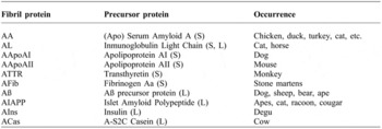
Amyloidosis can be classified based on the tissue distribution of amyloid deposits (local or systemic amyloidosis), the absence or presence of pre-existing disease (primary or secondary amyloidosis) and the chemical type of amyloid protein fibril. To date, there are 31 amyloid fibril proteins characterised in humans (Sipe et al., Reference SIPE, BENSON, BUXBAUM, IKEDA, MERLINI, SARAIVA and WESTERMARK2014), which differ in primary amino acid sequence (Landman et al., Reference LANDMAN, GRUYS and GIELKENS1998a). Ten of these 31 fibrils proteins have been described in domestic and wild cage animals (Sipe et al., Reference SIPE, BENSON, BUXBAUM, IKEDA, MERLINI, SARAIVA and WESTERMARK2014) ( Table 1 ).
Due to the great number of recognised amyloid types, the pathogenesis of amyloidosis is extremely variable and, therefore, research has focused on fibrillogenesis (Landman et al., Reference LANDMAN, GRUYS and GIELKENS1998a). Amyloid fibril formation begins with abnormal folding of an amyloidogenic precursor protein. Depending on the precursor protein, the misfolding can result from an inadequate proteolytic cleavage (e.g., Islet Amyloid Polypeptide (IAPP)), an amino acid substitution (e.g., transthyretin (TTR)) or an excessive production of serum concentrations in response to acute phase (e.g., SAA) (Dember, Reference DEMBER2005). According to Woldemeskel (Reference WOLDEMESKEL2012) all resulting fibrils are arranged in a characteristic ß-pleated sheet fibrillar structure and they are morphologically indistinguishable, despite the great diversity in structure and function of amyloidogenic protein precursors.
Amyloid fibril proteins and their precursors in animals*.

* Modified from Sipe et al., Reference SIPE, BENSON, BUXBAUM, IKEDA, MERLINI, SARAIVA and WESTERMARK2014. S: systemic; L: local.
Besides the fibril protein that defines the type of amyloidosis, all types of amyloid deposits contain several common components such as, proteoglycans (PG´s), serum amyloid P (SAP) and glycosaminoglycans (GAGs). The latter is a promoter of fibrillogenesis (Dember, Reference DEMBER2006).
AA amyloidosis
AAA, also referred to as systemic amyloid according to the distribution of amyloid deposition, occurs as a consequence of chronic inflammatory conditions. This leads to a dramatic increase in the levels of SAA to fight the inflammation (Westermark, Reference WESTERMARK2005). It should be noted that SAA is an acute phase protein synthesised by the liver (Gruys et al., Reference GRUYS, TOUSSAINT, NIEWOLD and KOOPMANS2005).
According to Landman et al. (Reference LANDMAN, GRUYS and GIELKENS1998a) amyloid fibrils are formed from SAA prior to C-terminal proteolytic cleavage and transformation into mature AA fibrils, which are produced at a different site from the sites of deposition. Among the chronic infections and inflammatory diseases that lead to the development of AAA in humans are tuberculosis, rheumatoid arthritis and leprosy (Dember, Reference DEMBER2005).
In chickens, AAA is characterised by AA amyloid depositions in the joints (mainly in knee and hock joints and with a higher incidence in the left leg) (Steentjes et al., Reference STEENTJES, VELDMAN, MEVIUS and LANDMAN2002; Bautista et al., 2009). Interestingly, it occurs mainly associated with chronic infections induced by arthropathic and amyloidogenic E. faecalis strains (Landman et al., Reference LANDMAN, PEPERKAMP, KOCH, TOOTEN, CRAUWELS and GRUYS1997). This form of amyloidosis is the only amyloid type found in chickens so far (Landman et al., Reference LANDMAN, GRUYS and GIELKENS1998a).
Aetiology of amyloid arthropathy
There are various bacteria, which may induce amyloid arthropathy. Landman et al. (Reference LANDMAN, VAN DEN BOGAARD, DOONENBAL, TOOTEN, ELBERS and GRUYS1998b) observed that brown layers inoculated intravenously with doses between 102 and 108 colony-forming units (CFU) of Enterococcus faecalis, Staphylococcus aureus, Escherichia coli and Salmonella enteritidis showed joint amyloidosis. However, the most severe joint amyloidosis was observed in layers infected with E. faecalis. Landman and Feberwee (Reference LANDMAN and FEBERWEE2001) observed that brown layers developed joint amyloidosis after intra-articular inoculation of Mycoplasma synoviae. However, E. faecalis is the bacterium which has been isolated mostly in amyloidogenic joints of field cases of amyloid arthropathy in brown layers (Landman et al., Reference LANDMAN, GRAYS and DWARS1994) and which has been showed to produce a large amount of amyloid deposits when inoculated (Landman, Reference LANDMAN1999).
According to the results of the analysis of routine post-mortem conducted by Landman et al. (Reference LANDMAN, VAN DEN BOGAARD, DOONENBAL, TOOTEN, ELBERS and GRUYS1998b) in field cases, 12.9% (249/1930) of the necropsied brown layers during 1996 and 1997 suffered from AAA. E. faecalis were isolated in 76.6% of the affected birds. S. aureus and E. coli were only isolated in 8.3% and 5% of the affected brown layers respectively. In the case of the broiler breeders, only 2.9% (53/1815) suffered from AAA in the same period of time. S. aureus, E. faecalis and E. coli were isolated in 73.4%, 25% and 1.5% of the affected birds, respectively. Steentjes et al. (Reference STEENTJES, VELDMAN, MEVIUS and LANDMAN2002) examined 30 birds from five affected broiler breeder flocks by unilateral (left side) lameness. They found 83% (25/30) amyloid-positive hock joint of broiler parent stock. E. faecalis was isolated in 77% (23/30) from the hock joints of the affected birds, and S. aureus was isolated from the joint in only one affected bird (1/30).
Additionally, E. faecalis is a trigger of the acute phase response in chickens and, therefore, is considered to be a causative agent of amyloid arthropathy (Landman et al., Reference LANDMAN, GRAYS and DWARS1994). For this reason, it has been deemed important to underline the role of E. faecalis in amyloid arthropathy and its possible routes of transmission in the following two sections.
enterococcus faecalis
Although E. faecalis constitutes a part of the endogenous intestinal microflora of birds and humans, it has been associated with high numbers of poultry diseases (Gregersen et al., Reference GREGERSEN, PETERSEN, CHRISTENSEN and BISGAARD2010). E. faecalis has been isolated from pulmonary hypertension syndrome in broilers (Tankson et al., Reference TANKSON, THAXTON and VIZZIER-THAXTON2001), septicaemia and endocarditis in a broiler parent flock (Chadfield et al., Reference CHADFIELD, CHRISTENSEN, CHRISTENSEN and BISGAARD2004). According to Gregersen et al. (Reference GREGERSEN, PETERSEN, CHRISTENSEN and BISGAARD2010), E. faecalis were isolated in 5.4% (167/3100) of the necropsied broiler breeders affected by different lesion types such as arthritis, amyloidosis, valvular endicarditis, salpingitis, peritonitis and septicaemia.
Landman et al. (Reference LANDMAN, VAN DEN BOGAARD, DOONENBAL, TOOTEN, ELBERS and GRUYS1998b) and Steentjes et al. (Reference STEENTJES, VELDMAN, MEVIUS and LANDMAN2002) observed by Pulsed-Field Gel Electrophoresis (PFGE) that several E. faecalis strains associated with amyloidosis from different origins showed similar Restriction Endonuclease Digestion (RED) patterns. Therefore, Landman et al. (Reference LANDMAN, VAN DEN BOGAARD, DOONENBAL, TOOTEN, ELBERS and GRUYS1998b) suggested a wide distribution of a single virulent clone of E. faecalis causing AAA in chickens. Petersen et al. (Reference PETERSEN, CHRISTENSEN, PHILIPP and BISGAARD2009) observed the same tendency through Multilocus Sequence Typing (MLST). With this technology, they showed the specific association between AAA in poultry and the Sequence Type (ST) 82 of E. faecalis, which has been related with this disease worldwide (Petersen et al., 2007). Later, Petersen et al. (2010) developed a Real-Time Polymerase Chain Reaction (RT-PCR) method for the rapid and specific detection of the MLST 82 of E. faecalis, based on a so-called kinetic PCR approach.
According to the database from the Multi Locus Sequence Typing website (http://www.mlst.net), ST36, ST59, ST82, ST170, ST171, ST172 and ST174 have been associated with AAA. Additionally, Gregersen et al. (Reference GREGERSEN, PETERSEN, CHRISTENSEN and BISGAARD2010) observed that ST32, ST176, ST177 and ST249 have the capacity of inducing amyloidosis in broiler breeders. However, of all the STs cited above, only ST36, ST59 and ST82 have, until now, been identified in both poultry and humans (http://www. mlst.net). Other STs have been associated with first week mortality in layers (Olsen et al., Reference OLSEN, FRANTZEN, CHRISTENSEN and BISGAARD2012b) and with septicaemia and/or salpingitis in broilers (Gregersen et al., Reference GREGERSEN, PETERSEN, CHRISTENSEN and BISGAARD2010; Olsen et al., Reference OLSEN, CHRISTENSEN and BISGAARD2012c), as well as in connexion with human infections (http://www.mlst.net). Therefore, Freitas et al. (Reference FREITAS, NOVAIS, RUIZ-GARBAJOSA, COQUE and PEIXE2009) suggested that isolated strains in animals and humans might have the same origin which could be spread through the food chain. Furthermore, Hayes et al. (Reference HAYES, ENGLISH, CARTER, PROESCHOLDT, LEE, WAGNER and WHITE2003) argued that poultry meat in particular, might be a route of transmission of E. faecalis to humans. In regards to its zoonotic potential, E. faecalis is a serious challenge due to its antimicrobial resistance and is therefore gaining great attention.
E. faecalis possess the ability to acquire antimicrobial resistance and virulence factors which are not exclusively associated with a disease (Olsen et al., Reference OLSEN, SCHØNHEYDER, CHRISTENSEN and BISGAARD2012a). E. faecalis have a natural intrinsic resistance towards several antimicrobials such as chloramphenicol, clindamycin, erythromycin, tetracycline, high-level aminoglycosides, beta-lactamase and vancomycin (McBride et al., Reference MCBRIDE, FISCHETTI, LEBLANC, MOELLERING and GILMORE2007). Many virulence genes related with enterococcal infections have been identified: aggregation substance (asa1 or agg), endocarditis antigen (efaA), cytolisine (cylA), gelatinase (gelE), collagen-binding protein (ace), hyaluronidase (hyl) and extracellular surface proteins (esp). The research done by Ciftci and Diker (Reference CIFTCI and DIKER2009) showed that the virulence genes gelE, cylA and asa1 (or agg) could have an effect on the pathogenesis of amyloid arthropathy. The chickens inoculated (intraarticular and intravenous inoculations) with gelE negative strains did not show any pathological signs whereas 65.2% of gelE positive strains caused amyloid arthropathy. Additionally, the chickens inoculated with asa1 (or agg) or cylA strains (both negative and positive strains) showed amyloidosis. In a study done by Olsen et al. (Reference OLSEN, SCHØNHEYDER, CHRISTENSEN and BISGAARD2012a), the virulence genes ace, efaA and gelE were present in all isolates obtained from poultry and humans. However, these virulence genes cannot be the only cause for the pathogenicity of E. faecalis in poultry since these genes were also present in all commensal isolates. The esp gene was the only gene not detected among any poultry isolates. Additionally, Olsen et al. (Reference OLSEN, SCHØNHEYDER, CHRISTENSEN and BISGAARD2012a) did not observe any difference on the virulence profile among isolates from clinical birds and cloacal isolates from healthy birds. Furthermore, these authors did not find major differences in the virulence gene profile from E. faecalis isolates of human and poultry which would confirm the possible zoonotic potential of this bacterium.
transmission of e. faecalis
Several experimental investigations to demonstrate the possible routes of transmission of E. faecalis in chickens have been reported in the literature (Landman et al., Reference LANDMAN, MEKKES, CHAMANZA, DOORNENBAL and GRUYS1999a; Reference LANDMAN, FEBERWEE, MEKKES, VELDMAN and MEVIUS1999b; Reference LANDMAN, VELDMAN, MEVIUS and DOORNENBAL2000; Reference LANDMAN, FEBERWEE, VELDMAN and MEVIUS2001 and 2003; Fertner et al., Reference FERTNER, OLSEN, BISGAARD and CHRISTENSEN2011; Olsen et al., Reference OLSEN, CHRISTENSEN and BISGAARD2012c).
Landman et al. (Reference LANDMAN, FEBERWEE, MEKKES, VELDMAN and MEVIUS1999b) found that the same E. faecalis strain which infected the parent stock was also present in an analysed offspring population. But the reproductive organs from the infected birds were not affected. Therefore, they concluded that vertical transmission is possible. Landman et al. (1999a and 1999b) discarded the theory that E. faecalis is spread by the transovarial route because yolk sac infections of chicken embryos with E. faecalis resulted in a massive mortality within a few days of incubation. However, after inoculation of egg albumen with low doses of E. faecalis, some chicks were successfully hatched. This suggests that an infection via the oviduct is a possible way of vertical transmission (Landman et al., Reference LANDMAN, MEKKES, CHAMANZA, DOORNENBAL and GRUYS1999a; Reference LANDMAN, FEBERWEE, MEKKES, VELDMAN and MEVIUS1999b).
Landman et al. (Reference LANDMAN, VELDMAN, MEVIUS and DOORNENBAL2000) observed that some hatchery air samples (up to 103 CFU/l air), Marek's disease vaccine suspensions (up to 106 CFU/ml vaccine suspension) and injection needles (9,500 to 61,000 CFU/needle) of two hatcheries were contaminated with an arthropathic and amyloidogenic E. faecalis strain during processing and vaccination of the newly hatched layer chickens. Besides, three isolates showed a similar RED pattern to E. faecalis 6085.94 (amyloidogenic E. faecalis strain used as control) and the other seventeen isolates were close or unrelated to control E. faecalis strain. Although the authors could not explain the high concentration of E. faecalis found in Marek vaccine suspensions, they suggested that this could be a possible iatrogenic infection route in chickens.
Fertner et al. (Reference FERTNER, OLSEN, BISGAARD and CHRISTENSEN2011) have carried out studies on the transmission of E. faecalis among chickens during a hatch in two hatchers. They sampled a cloacal swab from each single chicken and assumed that an immediate massive growth of E. faecalis in pure culture (at 0 h), would be an indicator for vertical transmission. Not immediate but massive growth within the next 24 hours indicated either a horizontal or vertical transmission due to the interaction of the chicks in the hatchery, as well as from infected or contaminated eggs. Fertner et al. (Reference FERTNER, OLSEN, BISGAARD and CHRISTENSEN2011) observed the same tendency for ST82 and ST249. A massive increase of the bacterial growth from 0 to 24 h in brown layers suggests horizontal transmission - orally and cloacally by means of the ‘drinking’ route between chicks. In white layers, only the ST177 was isolated at 0 h and no later than 24 h. However, other STs like ST82 and ST249 were isolated only after 24 h suggesting the horizontal transmission of these STs.
In accordance with the results of Fertner et al. (Reference FERTNER, OLSEN, BISGAARD and CHRISTENSEN2011), Olsen et al. (Reference OLSEN, CHRISTENSEN and BISGAARD2012c) observed an increment of the growth of E. faecalis in broiler chickens during the hatch. E. faecalis colonised 15% of all the chickens at the beginning of the hatch and after 24 hours was detected in 70% of them. This suggests a vertical and horizontal transmission. Besides, all of the STs of E. faecalis that were isolated at the time of hatch were also present after 24 h. The authors therefore suggest that the intestine of broiler chickens is a possible route for the horizontal transmission of E. faecalis from the hatchery to the farm.
On the basis of the information obtained, both vertical and horizontal transmissions of E. faecalis seem possible through different transmission routes. Moreover, Landman et al. (Reference LANDMAN, VELDMAN, MEVIUS and DOORNENBAL2000) suggested that AAA in chickens could be an iatrogenic infection after intramuscular inoculation of Marek vaccine suspensions contaminated with arthropathic and amyloidogenic E. faecalis strains.
Pathogenesis of amyloid arthropathy
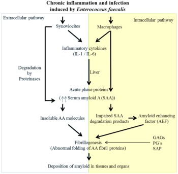
AAA is associated with chronic inflammation and infection, which appears to be induced by E. faecalis in chickens (Westermark, Reference WESTERMARK2005). The acute phase response is a complex systemic early-defence mechanism activated to fight the inflammation or infection. Pro-inflammatory cytokines play an important role in initiating the acute phase response (Sevimli et al., Reference SEVIMLI, MISIRLIOĞLU, YALÇIN, BÜLBÜL, YILMAZTEPE and ALTUNBAS2008). The increase in the levels of interleukin (IL) -1 and IL-6 during an infection is induced by the synoviocytes (type B cells) and macrophages (type A cells) as a local response, as shown in Figure 1 . Although there is very little literature on the role of cytokines in amyloid arthropathy, Sevimli et al. (Reference SEVIMLI, MISIRLIOĞLU, YALÇIN, BÜLBÜL, YILMAZTEPE and ALTUNBAS2008) reported that IL-1 appears to have an important role in the amyloid pathogenesis due to its positive association with high SAA levels in blood and the severity of amyloidosis. However, the role of IL-6 is still unclear.
SAA is mainly synthesised by the liver during the acute phase response. However, the extrahepatic production of SAA has been reported in different human tissues such as ovary, brain or kidney (Upragarin et al., Reference UPRAGARIN, LANDMAN, GAASTRA and GRUYS2005). Ovelgönne et al. (Reference OVELGÖNNE, LANDMAN, GRUYS, GIELKENS and PEETERS2001) reported that SAA is synthesised in the synoviocytes and in the vessel walls of the synovium in chickens affected by amyloid arthropathy while the healthy chickens did not express SAA mRNA in the synoviocytes. According to Kisilevsky and Manley (Reference KISILEVSKY and MANLEY2012), SAA is gaining more attention not only because it is the precursor protein of AAA, but also as its plasma concentration may increase 1000-fold during the acute phase response, its influence on high-density lipoprotein (HDL) structure and function, and on HDL's role in reverse cholesterol transport. However, the physiological role for SAA still remains contradictory among the different proposals of the current literature.
According to Upragarin et al. (Reference UPRAGARIN, LANDMAN, GAASTRA and GRUYS2005) and Abbas (Reference ABBAS2005), SAA can be degraded by intra- and extracellular pathways ( Figure 1 ). Through the intracellular pathway, SAA degradation products may act as a source of the AA fibrin precursor (Upragarin et al., Reference UPRAGARIN, LANDMAN, GAASTRA and GRUYS2005), that is to say, like an amyloid enhancing factor (AEF). According to Landman et al. (Reference LANDMAN, GRUYS and GIELKENS1998a), AEF is considered to be a material with high content of ß-pleated sheets that acts as nest or template for the generation and growth of AA amyloid fibrils. Through the extracellular pathway, SAA is degraded by proteinases (Upragarin et al., Reference UPRAGARIN, LANDMAN, GAASTRA and GRUYS2005) and a possible defect in the cells of monocyte origin that degrade SAA could generate insoluble rather than soluble AA molecules (Abbas, Reference ABBAS2005).
According to Upragarin et al. (Reference UPRAGARIN, LANDMAN, GAASTRA and GRUYS2005), a persistent acute phase response will provide an elevated precursor protein SAA necessary for SAA and AA fibril formation. However, an elevated production of SAA by itself is not sufficient for the deposition of amyloid since it is common to inflammatory states that do not necessarily lead to amyloidosis (Abbas, Reference ABBAS2005). Furthermore, other factors are involved in the development of amyloidosis since the amyloid formation does not occur in all individuals in the same way (Upragarin et al., Reference UPRAGARIN, LANDMAN, GAASTRA and GRUYS2005). Thus, factors such as the isoform or proteolytic cleavage site of SAA (amyloid fibrils are transformed from SAA prior to C-terminal proteolytic cleavage), the SAP and GAGs are thought to contribute to amyloid pathogenesis (Landman, Reference LANDMAN1999).
Speculative illustration of pathogenesis of amyloid arthropathy.

Pathology of amyloid arthropathy
The published literature of Landman et al. (1994 and 1998b), Peperkamp et al. (Reference PEPERKAMP, LANDMAN, TOOTEN, ULTEE, VOORHOUT and GRUYS1997), Ovelgönne et al. (Reference OVELGÖNNE, LANDMAN, GRUYS, GIELKENS and PEETERS2001), Petersen et al. (Reference PETERSEN, CHADFIELD, CHRISTENSEN, CHRISTENSEN and BISGAARD2008) and Bautista et al. (2009) describes field cases of avian amyloidosis and agrees that the gross features of amyloid arthropathy are characterised by deposits of orange-coloured amyloid materials in the joints (mainly in knee joints and with a higher incidence in the left knees) and a turbid orange-yellow amyloid material in the synovial fluid.
Landman et al. (Reference LANDMAN, GRAYS and DWARS1994) observed the first signs of amyloid arthropathy in field cases in brown layer breeds between five and six weeks of age. Affected chickens were smaller than healthy ones. The chickens had ruffled feathers and chirped like younger chickens, showing reduced weight gain, uni- and bilateral swelling of tibio-metatarsal and femorotibial joints, mainly on the left side. Petersen et al. (Reference PETERSEN, CHADFIELD, CHRISTENSEN, CHRISTENSEN and BISGAARD2008) observed erosion of the articular cartilage as well as in bone tissue in commercial layers between 24 and 37 weeks of age. Bautista et al. (2009) reported that 5% of the commercial layers showed lameness and inflammation of the knee joints after 17 weeks of age, though no significant mortality was observed.
Additionally, avian amyloidosis is often accompanied by hepatomegaly and renomegaly, and sometimes by hydropericardium and ascites. The most affected organs are the liver, spleen and kidneys. At necropsy, deposits of amyloid can be seen in the liver, located in Disse's space, in the kidney, located in the interstitium of cortex and medulla, and in the spleen, located in the white pulp. The liver usually shows a pale brownish hue and the spleen appears pale (Bautista et al., 2009). Amyloid deposits occasionally appear in the heart, and can be found in the digestive tract, in the proventriculus, and the small intestine, located in the duodenal or jejunal section, the pancreas, being the least affected organ (Peperkamp et al., Reference PEPERKAMP, LANDMAN, TOOTEN, ULTEE, VOORHOUT and GRUYS1997). Rarely brain, lungs and skin are affected (Landman et al., Reference LANDMAN, GRUYS and GIELKENS1998a).
Regarding the reproductive system, Landman et al. (Reference LANDMAN, FEBERWEE, MEKKES, VELDMAN and MEVIUS1999b) observed that four out of six 27-week-old brown layer breeds with arthritis, which were inoculated intravenously with 109 CFU of E. faecalis, had productive ovaries and remained in production (from 50 to 100%) although with oscillations. Landman et al. (Reference LANDMAN, FEBERWEE, VELDMAN and MEVIUS2001) examined 18 brown layer breeds that displayed lameness. At necropsy, it was observed that ten of them suffered from avian amyloidosis, of which seven had active reproductive organs (70%) and only one bird stopped laying eggs (10%). This means a diminished number of eggs in affected birds and, therefore, an important financial loss for the poultry industry.
Field cases of AAA in broiler parent stocks have been reported by Landman et al. (Reference LANDMAN, VAN DEN BOGAARD, DOONENBAL, TOOTEN, ELBERS and GRUYS1998b), Ovelgönne et al. (Reference OVELGÖNNE, LANDMAN, GRUYS, GIELKENS and PEETERS2001) and Steentjes et al. (Reference STEENTJES, VELDMAN, MEVIUS and LANDMAN2002), and the symptoms were very similar to those previously described in layers.
Peperkamp et al. (Reference PEPERKAMP, LANDMAN, TOOTEN, ULTEE, VOORHOUT and GRUYS1997) observed that amyloid deposition is more severe in field amyloid cases than in experimentally infected birds. There are several reports about experimentally infected birds with E. faecalis and the many possible routes of infection. However, pathogenesis and clinical manifestations of induced avian amyloidosis follow a similar pattern to field cases of amyloid arthropathy. Landman et al. (Reference LANDMAN, MEKKES, CHAMANZA, DOORNENBAL and GRUYS1999a) studied the induction of amyloid arthropathy in embryonated eggs; neither the dipped embryonated eggs nor the inoculation of the air chamber with E. faecalis resulted in amyloid arthropathy. However, yolk sac inoculation produced a high mortality rate within two days while egg albumen inoculation hardly produced any embryonic deaths. In one day-old chicks the intramuscular inoculation of 106 CFU of E. faecalis caused arthritis (60%) and articular amyloidosis (40%) whereas oral inoculation was ineffective (Landman, Reference LANDMAN1999). Additionally, according to Landman et al. (1997; 1998b; 1999a and 1999b), six-week old brown layer chickens developed AAA after intravenous, intra-articular and intraperitoneal inoculations of 108 or 109 CFU of E. faecalis. Landman et al. (Reference LANDMAN, MEKKES, CHAMANZA, DOORNENBAL and GRUYS1999a) found that intra-tracheal, oral or intramuscular inoculations were ineffective. Landman et al. (Reference LANDMAN, VELDMAN, MEVIUS and VAN ECK2003) reported in a later study that both intra-tracheal and intramuscular inoculations (in Musculus gastrocnemius and Musculus pectoralis) induce lameness and arthritis. The joint lesions with the inoculations in the m. gastrocnemius were more severe than in the m. pectoralis. Besides, they reported that the exposure to aerosolized E. faecalis produced bacteraemia.
In general, inoculations with high doses of E. faecalis could develop amyloid arthropathy and show the typical clinical and pathological lesions according to Landman (Reference LANDMAN1999), and according to Ciftci and Diker (Reference CIFTCI and DIKER2009) E. faecalis has a predilection for joints of chickens regardless of the inoculation route. Nevertheless, it is to be noted that although AAA in chickens is characterised by the deposition of amyloid, there are experimentally inoculated chicken cases where these depositions have not been observed. Ovelgönne et al. (Reference OVELGÖNNE, LANDMAN, GRUYS, GIELKENS and PEETERS2001) observed amyloid deposits in the joints of all infected brown layers at six weeks of age. However, at necropsy (i.e. at 13 weeks of age), the amyloid deposits were not observed in all infected white layers although their joints were bacteriologically positive. According to the study of Landman et al. (Reference LANDMAN, VELDMAN, MEVIUS and VAN ECK2003), the experimentally inoculated chickens at one day of age did not show any amyloid deposits at necropsy (at 10 weeks of age). These authors reported that outbred brown layer chickens were used. These have an immune system with a genetically determined diversity (Hawken et al., Reference HAWKEN, BEATTIE and SCHOOK1998) which might serve as a possible explanation for this fact. Consequently, the acute phase reaction necessary to amyloid deposition might not occur in the same way in all birds.
An extensive diagnosis is therefore necessary to confirm amyloid deposits and rule out similar extracellular deposits such as collagen and fibrin (Woldemeskel, Reference WOLDEMESKEL2012). Congo red stain is the most commonly used tool for the identification of amyloid (Picken, Reference PICKEN2007). Green birefringence under polarised light is used with amyloid orange deposits (Snyder, Reference SNYDER2007), as well as immunohistochemistry (antibody against apoliprotein AI) and biochemical characterisation which helps to identify the specific constituents composing the amyloid deposits (Picken, Reference PICKEN2007). Histopathologic identification of amyloid deposits is required to assess the amyloid type (Woldemeskel, Reference WOLDEMESKEL2012). Serology and bacteriology of blood samples and/or of affected joints are complementary proofs to amyloid diagnosis which have been used in research conducted, for instance, by Landman et al. (Reference LANDMAN, VELDMAN, MEVIUS and VAN ECK2003) and Sevimli et al. (Reference SEVIMLI, MISIRLIOĞLU, POLAT, YALÇIN, AKKOÇ and UGUZ2005), to name a few.
breed susceptibility
Differences in susceptibility to amyloid arthropathy between brown and white layers have been investigated in many cases (Zekarias et al., Reference ZEKARIAS, LANDMAN, TOOTEN and GRUYS2000; Ovelgönne et al., Reference OVELGÖNNE, LANDMAN, GRUYS, GIELKENS and PEETERS2001). However it still remains puzzling. Zekarias et al. (Reference ZEKARIAS, LANDMAN, TOOTEN and GRUYS2000) observed that the defence reaction that recognises an invading bacterium and produces antibodies specific against that antigen appears to be different between breeds, i.e. brown and white layers had a different immune reaction when they were infected with an amyloidogenic E. faecalis strain. Therefore, the greater susceptibility to the disease of brown layers as compared to white layers may be associated with the immunological response pattern.
Zekarias et al. (Reference ZEKARIAS, LANDMAN, TOOTEN and GRUYS2000) detected more CD8+ T cells present in the synovial membrane and peripheral blood of affected joints from white layer breeds resistant to amyloid arthropathy induced by E. faecalis than on the susceptible brown layer breeds. Likewise, chickens resistant to Salmonella infection showed more CD8+ T cell according to Berndt and Methner (Reference BERNDT and METHNER2001). However, chicken strains susceptible to coccidiosis showed more CD8+ T cells than resistant lines (Lillehoj, Reference LILLEHOJ1994). Therefore, the amount of T cells appears unrelated to the resistance to disease (Zekarias et al., Reference ZEKARIAS, TER HUURNE, LANDMAN, REBEL, POL and GRUYS2002).
Zekarias et al. (Reference ZEKARIAS, LANDMAN, TOOTEN and GRUYS2000) observed that white layers showed a cell-mediated response characterised by T cell proliferation and macrophage activity whilst brown layers showed a high humoral antibody response due to the large number of plasma cells present in its arthritic synovial membrane. Besides, Sevimli et al. (Reference SEVIMLI, MISIRLIOĞLU, POLAT, YALÇIN, AKKOÇ and UGUZ2005) reported that the most affected brown layers by amyloid arthropathy presented the highest numbers of macrophages and heterophil. Therefore, they concluded that the rate of macrophages and heterophil migration into the tissue may be associated with the severity of AAA.
Additionally, Ovelgönne et al. (Reference OVELGÖNNE, LANDMAN, GRUYS, GIELKENS and PEETERS2001) reported that the differences in susceptibility to amyloidosis among individuals could have a genetic basis but it cannot be explained by the SAA genes. These authors reported that although there are minor differences located in the untranslated regions of both mRNAs, the amino acid sequences of SAA of both chicken breeds are identical with four exons and three introns. Therefore, they concluded that the promoters of the brown and white layers must house the functional differences between the SAA genes. It should be noted that only one SAA gene has been identified in the chicken genome while SAA is encoded by four different genes in humans and mice (Upragarin et al., Reference UPRAGARIN, LANDMAN, GAASTRA and GRUYS2005).
Furthermore, Ovelgönne et al. (Reference OVELGÖNNE, LANDMAN, GRUYS, GIELKENS and PEETERS2001) reported that the fact that the brown layers might have higher blood SAA levels than white layers cannot explain the differences in susceptibility to amyloid arthropathy. When the brown and white layers were inoculated intra-articularly with E. faecalis, the amyloid deposits were only observed in the injected joints, whereas the other joints remained free of amyloid. However, local production of SAA in the affected joints might play a role in amyloid depositions. Therefore, they suggested that the synoviocytes of brown layers might proliferate more strongly than the synoviocytes of white layers and, therefore, produce larger amounts of extrahepatic SAA that may act as nidus of amyloid fibrils. Quantitative studies of the extrahepatic production of SAA are therefore required to investigate this possibility.
Approaches for a possible treatment
Amyloid arthropathy is an irreversible disease (Landman et al., Reference LANDMAN, GRUYS and GIELKENS1998a), probably due to the fact that amyloid fibrils have low solubility and a relative resistance to proteolytic digestion under physiological conditions. Therefore, the best option would be to treat the underlying inflammatory disease and prevent the occurrence of fibrillogenesis. Substances which facilitate or inhibit the amyloid depositions have been reported by Sevimli et al. (2005; 2008 and 2013). These authors have observed that high doses of vitamin A increased the severity of amyloid arthropathy induced by E. faecalis and Freund's adjuvant, while methylprednisolone and pentoxyfylline inhibit amyloidosis. Additionally, Shtrasburg et al. (Reference SHTRASBURG, PRAS, RABINOVICH, GAL, LIVNEH and LIDAR2012) observed the possible role of alkylating agents in the prevention of amyloidogenesis in mice where cyclophosphamide and chlorambucil were the most potent inhibitors.
Therefore, the treatment of amyloid arthropathy has focused to date on the prevention of chronic inflammation. However, the recent study conducted by Ibi et al. (Reference IBI, MURAKAMI, GODA, KOBAYASHI, ISHIGURO and YANAI2015) concluded that chickens previously exposed to amyloid arthropathy may improve with age or even the amyloid deposits may tend to regress when chronic infection disappears.
In regard to chronic infection induced by E. faecalis, measures to prevent E. faecalis colonisation could have a direct impact on animal welfare by reducing the burden of infection thereby lowering environmental stress and, therefore, to reduce the incidence rate of amyloid arthropathy.
Conclusions
During the last twenty years, considerable contributions have been made to understand amyloid arthropathy. However, many questions remain open. The pathology of AAA in chickens is widely recognised and it is characterised by deposits of orange-coloured amyloid materials in the joints. Nevertheless, there are also cases in which amyloid depositions have not been reported and hence the real question is whether all field cases reported in poultry are amyloid arthropathy or if there are there other hidden problems. Additionally, the current method for the treatment of AAA is based on suppressing the inflammatory disease. Although there are substances and anti-inflammatory agents such as methylprednisolone and pentoxifylline which inhibit the amyloid deposition in birds, more investigations are required to determine the effects of different agents on the treatment of AAA and clarify if the amyloid deposits are really irreversible.
Although there are various investigations about the pathogenesis of amyloidosis in layers, it still cannot be fully explained. The inflammatory cytokines play an important role in the pathogenesis of amyloidosis. Besides, some pro-inflammatory cytokines are produced by peripheral blood monocytes and tissue macrophages, which are likely to start an acute phase response after a stimulus. Furthermore, the differences in susceptibility to amyloidosis among individuals could be due to the differences in the immune system. Therefore, the role of these cytokines in the pathogenesis and in the susceptibility of AAA could be studied together.
In most cases, Enterococcus faecalis is the bacterium which has been isolated. E. faecalis has been isolated from amyloid deposits in the joints as well as from joints without amyloid deposits. It is therefore necessary to learn more about the different strains of E. faecalis. Until today, the characterisation of E. faecalis strains has enabled us to relate its STs with the disease, as well as to identify the possible virulence genes involved. Worldwide, it is well known that the ST82 of E. faecalis is associated with amyloidosis in poultry. However, it is still not known whether ST82 as well as other STs which were found both in poultry and human, are normal inhabitants of the gastro-intestinal tract or represent a genuine zoonosis. Therefore, it would be interesting to conduct further studies on the different strain of E. faecalis affecting brown layer chickens with emphasis on the specific STs that are associated with amyloid arthropathy.
In addition, E. faecalis can be transmitted among birds vertically and horizontally. Investigations to prevent and reduce enterococci contamination in birds and to figure out if the amyloid arthropathy actually represents a zoonosis should be developed on a parallel basis. It should be taken into account that E. faecalis have a natural resistance towards antimicrobials and, therefore, the enterococcal infection treatments are increasingly limited. In this regard, there is high requirement for control strategies against bacterial infections to guarantee animal welfare and to reduce the incidence rate of AAA in the birds, thereby improving economic performance as well.
Consequently, efficient control measures against E. faecalis infection are required. Different general strategies could be applied in the fight against E. faecalis infection, such as the development of effective vaccines made from proven high pathogenic isolates or the breeding and genetic selection for resistance to amyloid arthropathy.
Given the economic importance of amyloid arthropathy, further studies are required to elucidate the pathogenesis, transmission routes and prevention strategies thereby providing more animal welfare and reducing the losses in the poultry sector caused by AAA.
Acknowledgments
The authors wish to thank Priv.Doz. Dr. Härtle for their constructive comments on the draft of the manuscript. We thank Ms. Ortega for her help throughout the course of this work.