Polycystic ovarian syndrome (PCOS) is the most common endocrinopathy of women, with a prevalence of 6.5–6.7% among premenopausal women (Refs Reference Diamanti-Kandarakis1, Reference Escobar-Morealle2). PCOS was initially defined by an NIH conference in 1990 as the combination of chronic anovulation or oligomenorrhoea and clinical or biochemical hyperandrogenism (Ref. Reference Zawadski, Dunaif and Dunaif3). The Rotterdam consensus in 2003 revised the diagnostic criteria (Ref. 4), with two of the three following criteria declared as prerequisites for PCOS: chronic anovulation or oligomenorrhea, clinical or biochemical hyperandrogenism, and polycystic ovarian morphology. The Rotterdam workshop has left room for debate on the new phenotypes that should be embraced in the PCOS spectrum (Refs Reference Franks5, Reference Azziz6). The potential pathophysiological significance of polycystic ovarian morphology, observed in 75% of women with PCOS (Ref. Reference Azziz7), has been extensively discussed (Refs Reference Franks5, Reference Azziz6). There is agreement that this imaging finding per se does not equate with the diagnosis of PCOS. Moreover, the phenotype of anovulation combined with polycystic ovarian morphology does not appear to be associated with the metabolic abnormalities of the classic PCOS phenotypes (Refs Reference Chang8, Reference Diamanti-Kandarakis and Panidis9, Reference Barber10). In 2006, the Androgen Excess Society provided a contemporary version of the definition of PCOS (Ref. Reference Azziz7). The final statement has highlighted hyperandrogenism (clinical or biochemical) in combination with ovarian dysfunction (including both functional and ultrasonographic abnormalities) as the core characteristics of PCOS.
Hyperandrogenism and anovulation are known to interact with insulin resistance in the pathophysiology of PCOS (Ref. Reference Poretsky11). Hyperinsulinaemia appears to interfere with ovarian steroidogenetic defects as well as anovulatory mechanisms (Ref. Reference Poretsky11). Additionally, insulin resistance is involved in the metabolic aspects of PCOS. Although obesity has an aggravating role, insulin resistance appears to be an integral feature of PCOS, independent of other factors (Ref. Reference Poretsky11).
Hyperandrogenaemia in PCOS: pathophysiology and molecular aspects
Biochemical and clinical hyperandrogenism in PCOS, of ovarian and/or of adrenal origin, is evident in the majority of patients with PCOS (60–80%) (Ref. Reference Franks5).
Ovarian hyperandrogenism
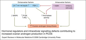
Ovarian hyperandrogenism is mainly attributed to an inherent steroidogenic defect of theca cells in PCOS. Increased luteinising hormone (LH) and increased insulin levels appear to amplify the intrinsic abnormality of theca steroidogenesis. The relatively decreased follicle-stimulating hormone (FSH) levels (in relation to LH levels) and intraovarian factors are also involved (Fig. 1).
Hormonal regulators and intracellular signalling defects contributing to increased ovarian androgen production in PCOS. The intrinsically distorted intracellular signalling stimulates theca cell androgen synthesis in polycystic ovarian syndrome (PCOS). Constitutively decreased MAP2K1 in the intracellular signalling of luteinising hormone (LH) is associated with increased theca androgen biosynthesis. Increased PI3K and IPG activity in the intracellular signaling of insulin also stimulate theca androgen production. Increased insulin interacts with increased LH levels to amplify the inherent steroidogenic defect. Decreased follicle-stimulating hormone (FSH) action reduces aromatase activity in synergy with antiMüllerian hormone (AMH), which may play this role directly, or indirectly through FSH suppression. Reduced aromatase activity hinders the conversion of androgen to oestrogen, contributing to ovarian androgen excess. Increased AMH levels may also act directly on theca cells to stimulate androgen synthesis. In turn, increased androgen levels perpetuate the inhibition of aromatase activity. Green arrows indicate a stimulating effect; red arrows indicate an inhibitory effect. Abbreviations: IPG, phosphoinositoglycan; MAP2K1, mitogen-activated protein kinase kinase 1; PI3K, phosphoinositide 3-kinase.

Abnormalities of the steroidogenic activity of theca cells
Increased androgen production has been demonstrated by genomic and molecular studies to be an intrinsic steroidogenic defect in PCOS theca cells (Refs Reference Legro12, Reference Nelson13). In vitro studies suggest that the enhanced steroidogenic potential of PCOS theca cells resides in the increased enzyme activities of 17α-hydroxylase/17,20-lyase (CYP17α1), 3-beta-hydroxysteroid dehydrogenase type II (HSD3B2) and side-chain cleavage enzyme (CYP11A1); the enzyme activities remain elevated after many passages in culture (Refs Reference Nelson13, Reference Wood14). These three enzymes act at various steps of the pathway of androgen synthesis. CYP11A1 performs the first step of steroid biosynthesis: the conversion of cholesterol to pregnenolone. CYP17α1 (cytochrome P450c17; 17α-hydroxylase/17,20-lyase) has dual functions: the hydroxylase activity catalyses the 17α-hydroxylation of both pregnenolone and progesterone; and the 17,20-lyase activity cleaves the C17–C20 bond of 17α-hydroxypregnenolone and 17α-hydroxyprogesterone to form dehydroepiandrosterone (DHEA) and androstenedione, respectively. HSD3B2 transforms Δ5-steroids (pregnenolone, 17α-hydroxypregnenolone and DHEA) into their Δ4-congenors.
The increased enzyme activity of CYP17A1 is reflected at the transcriptional level. Microarray analyses have shown that CYP17A1 gene expression is altered, with a two- to threefold increase in CYP17A1 promoter function (Ref. Reference Wood14). A 16 bp sequence of the CYP17A1 gene promoter appears to play the key role in regulating promoter function (Ref. Reference Wickenheisser15) through binding of nuclear factors. Nuclear factor 1C (NF-1C), which represses CYP17A1 promoter function, is decreased in PCOS theca cells and could thereby contribute to hyperandrogenaemia by increasing CYP17A1promoter activity (Refs Reference Wickenheisser15, Reference Wickenheisser, Nelson-Degrave and McAllister16). Furthermore, transcription factors that upregulate CYP17A1 expression are overexpressed in the polycystic ovary. For example, the increased mRNA abundance of GATA6 exhibited in PCOS theca cells upregulates the activity of the CYP17A1 promoter (Ref. Reference Wickenheisser, Nelson-Degrave and McAllister16).
Retinoids have recently been shown to play a part in ovarian steroidogenesis. In PCOS, androgen production by theca cells is stimulated by retinoids, which appear to modulate gene expression (Ref. Reference Wickenheisser17). Specifically, 9-cis retinoic acid and retinol enhance CYP17A1 promoter function (Refs Reference Wood14, Reference Wickenheisser17). Additionally, the upregulation of the retinoic acid receptor gene in PCOS ovaries (Ref. Reference Oksjoki18) may further enhance local retinoid activity.
At the post-transcriptional level, molecular studies have revealed alterations in CYP17A1 mRNA: the half-life of CYP17A1 mRNA is elevated twofold in theca cells from PCOS compared with normal cells, leading to increased CYP17A1 mRNA accumulation and increased CYP17A1 enzyme production (Refs Reference Wickenheisser19, Reference Wood20).
Mechanisms acting at the level of enzyme activity have also been intensively investigated. A role for increased serine phosphorylation of the CYP17A1 enzyme, which promotes 17/20-lyase activity via enhanced interaction of serine-phosphorylated CYP17A1 with cytochrome b 5, has been suggested (Refs Reference Pandey and Miller21, Reference Zhang22, Reference Akhtar, Kelly and Kaderbhai23). In parallel, the reduced expression of protein phosphatase 2A observed in PCOS serves to maintain serine phosphorylation of CYP17A1 (Ref. Reference Pandey, Mellon and Miller24).
Other intracellular signalling defects have also been linked with excessive androgen biosynthesis in PCOS theca cells. Inhibition of mitogen-activated protein kinase kinase 1 (MAP2K1) signalling has been demonstrated in cultures of PCOS theca cells. This pathway appears to mediate the action of LH on theca androgen synthesis (Ref. Reference Nelson-Degrave25) (Fig. 1).
Gonadotropins
Impaired gonadotropin dynamics may contribute to excessive androgen production in PCOS (Ref. Reference Jonard and Dewailly26). Increased LH pulse frequency and amplitude leading to persistently increased LH levels may directly enhance theca androgen synthesis. However, it has been suggested that elevated LH levels result from an impaired negative feedback on LH secretion, due to excessive androgen action on the hypothalamic–pituitary axis (Ref. Reference Jonard and Dewailly26).
The relatively reduced FSH levels (in relation to LH) may have an indirect role. The decreased stimulation of aromatase by FSH results in the decreased conversion of androgen to oestrogen and aggravates the ovarian androgen excess (Fig. 1).
Insulin
Insulin appears to be a triggering factor that aggravates the inherent dysregulation of theca steroidogenesis in PCOS. Insulin seems to act in synergy with LH to stimulate androgen synthesis in PCOS ovarian theca cells (Ref. Reference Poretsky11). In lean women with typical PCOS and normal insulin sensitivity, assessed by the euglycaemic clamp, reducing insulin levels by diazoxide was associated with decreased androgen levels, suggesting increased insulin sensitivity of the androgenic pathway in PCOS (Ref. Reference Baillargeon and Carpentier27).
A significant role of insulin in theca cell androgen production is also supported by in vitro studies (Fig. 1). Insulin appears to stimulate ovarian P450c17 (CYP17A1) mRNA expression and enzyme activity through its receptor in theca cells. This action of insulin is mediated via the phosphoinositide 3-kinase (PI3K)/protein kinase B (PKB) pathway, which is activated in PCOS theca cells (Ref. Reference Munir28). Increased insulin levels may thus further amplify androgen synthesis (Ref. Reference Baillargeon and Carpentier27).
Inositoglycan mediators may also accentuate the effects of insulin on theca androgen production (Ref. Reference Nestler29). The generation of inositoglycans occurs at the cell membrane after insulin binding to its receptor, independently of the insulin receptor tyrosine kinase and PI3K activation. The intracellular signals that are activated downstream of inositoglycans are under exploration.
Intraovarian factors
Intraovarian factors of granulosa cell origin, such as antiMüllerian hormone (AMH) and inhibins, may contribute to the steroidogenic activity of theca cells. AMH is a dimeric glycoprotein of the transforming growth factor β (TGF-β) superfamily involved in follicular dynamics (Ref. Reference Knight and Glister30). AMH type II receptors (AMHRII) have recently been detected on theca cell membranes of maturing follicles and could mediate a paracrine effect of AMH on androgen production (Ref. Reference Ingraham31). Additionally, AMH may indirectly contribute to the ovarian androgen excess by inhibiting FSH action or by suppressing aromatase activity (Ref. Reference Jonard and Dewailly26) (Fig. 1). The significant positive relationship between AMH and testosterone levels (Ref. Reference Pigny32) is compatible with the putative role of AMH in perpetuating ovarian androgen excess. Similarly, theca cells have been shown to express inhibin receptors on their membrane, supporting the notion of a paracrine action of inhibins on theca steroidogenesis (Refs Reference Knight and Glister30, Reference McConell33). However, existing data are scant and do not allow for clear conclusions about cause-and-effect associations (Refs Reference Pigny34, Reference Welt35).
Adrenal hyperandrogenism
There is a body of evidence to suggest that adrenal hyperandrogenism by putative dysregulation of CYP17A1 is a genetically determined trait in PCOS (Refs Reference Goodarzi36, Reference Yildiz37). Increased peripheral metabolism of cortisol has also been proposed to contribute to the functional adrenal hyperandrogenism (Ref. Reference Tsilchorozidou, Honour and Conway38). In particular, the enhanced inactivation of cortisol by 5α-reductase or the impaired reactivation of cortisone by 11-beta-hydroxysteroidogenase 1 could lead to decreased feedback suppression of adrenocorticotropic hormone (ACTH) secretion. Notably, insulin resistance may in part account for the enhanced 5α-reduction of cortisol without affecting cortisol production. In this setting, the hypothalamic–pituitary–adrenal axis may be stimulated, leading to increased adrenal androgen production in PCOS (Ref. Reference Yildiz37). Nevertheless, aberrations related to adrenal function appear to contribute to a limited extent to the hyperandrogenism of PCOS (Ref. Reference Rosenfield39).
Ovarian dysfunction: pathophysiology and molecular aspects
PCOS represents the commonest cause of normogonadotropic anovulation, accounting for 55–91% of the entire World Health Organization-II (WHO-II) cohort (Ref. Reference Broekmans40). Oligomenorrhoea/anovulation is a major clinical concern and is present in 70–80% of women with PCOS (Ref. Reference Azziz7).
Anovulation in PCOS is attributed to the disturbances of folliculogenesis that characterise the syndrome. The follicular defect in PCOS consists of accelerated early follicular growth and distortion of the subsequent stages towards the selection of the dominant follicle (follicular arrest) (Ref. Reference Jonard and Dewailly26). Webber et al. (Ref. Reference Webber41) have reported a higher density of small preantral (and specifically primary) follicles in ovarian biopsies from women with PCOS compared with controls. The deceleration of atresia, demonstrated by the same investigators (Ref. Reference Webber42), could compensate for the enhanced recruitment and may explain why the polycystic ovary does not experience premature follicle depletion (Ref. Reference Nikolaou and Gilling-Smith43).
In normal folliculogenesis (Fig. 2a), growth factors [e.g. growth differentiation factor 9 (GDF-9), bone morphogenetic protein 15 (BMP-15)] stimulate the transition from the primordial to the primary stage, while FSH regulates the subsequent stages of folliculogenesis towards the selection of the dominant follicle. During folliculogenesis, androgens and insulin play a synergistic role with LH, which exerts its main action at the mid to late follicular stages. The balance between FSH and AMH might be crucial for aromatase activity at the time of and after the selection of the dominant follicle. The increased oestradiol production by the dominant follicle suppresses FSH levels, leading to the demise of the subordinate follicles and resulting in mono-ovulation. In PCOS, accelerated early follicular growth mainly due to androgen excess leads to small-follicle excess. Reduced levels of oocyte-secreted growth factors, mainly GDF-9 and BMP-15, contribute to amplified early folliculogenesis. Small-follicle excess leads to increased AMH levels, which interferes with follicular FSH responsiveness. Increased insulin induces LH receptor expression and premature luteinisation. The attenuated FSH responsiveness and the premature luteinisation of granulosa cells distort the selection of the dominant follicle, leading to follicular arrest (Fig. 2b).
Luteinising hormone and follicle-stimulating hormone
In polycystic ovaries, overexpression of LH receptor mRNA in granulosa cells from antral follicles of 5 mm diameter may render granulosa cells prematurely overresponsive to LH (Refs Reference Jakimiuk44, Reference Willis45). The untimely and exaggerated effect of LH on PCOS granulosa cells has been implicated in the stagnation of follicular maturation. The LH-induced increase in ovarian androgen levels may also contribute to this effect (Refs Reference Jonard and Dewailly26, Reference Zeleznik, Little-Ihrig and Ramasawamy46, Reference Loumaye47). Additionally, the absence of the intercycle FSH peak, but most importantly the refractoriness of granulosa cells to FSH action, have been linked with anovulation in PCOS (Ref. Reference Jonard and Dewailly26).
Coffler et al. (Ref. Reference Coffler48) reported that the response to recombinant human FSH (rhFSH) occurred at a higher threshold in PCOS subjects compared with controls. Even elevated levels of FSH were shown to be insufficient to sustain the release of oestradiol from PCOS granulosa cells, in contrast to the lasting response of normal cells. The initial vigorous oestradiol release has been suggested to reflect the increased number of responsive follicles, whereas the temporary nature of this response suggests additional defects within the microenvironment of the polycystic ovary (Ref. Reference Coffler48). Increased AMH levels may account for these defects, acting either to inhibit FSH action or to directly suppress aromatase activity (Ref. Reference Jonard and Dewailly26) (see below).
Insulin
Hyperinsulinaemia is considered a ‘second hit’ and not the primary cause of disturbed follicular maturation and anovulation in PCOS (Ref. Reference Jonard and Dewailly26). In particular, excessive insulin levels might be linked to premature luteinisation by amplifying the expression of LH receptors on granulosa cells (Ref. Reference Tao and Yan49).
The paradox of ovarian hypersensitivity in the milieu of peripheral insulin resistance has been challenged (Ref. Reference Poretsky50). The use of different isoforms of the insulin receptor and of different insulin signalling pathways might allow the ovary to remain insulin sensitive in the context of peripheral insulin resistance. The preponderance of insulin receptor A in human granulosa cells offers a mechanism whereby insulin might regulate granulosa cell responsiveness to gonadotropins independently of metabolic effects, which are mediated by insulin receptor B in other insulin-sensitive target tissues (Ref. Reference Phy51). The signalling pathways mediating the steroidogenic activity of insulin in granulosa cells remain unknown, although PI3K (Ref. Reference Poretsky52) and MAP2K1 appear not to be involved (Ref. Reference Seto-Young53).
However, there are data to suggest that granulosa cells demonstrate resistance to both metabolic and steroidogenic effects of insulin. Specifically, insulin-dependent glucose uptake and glycolysis appear to be impaired in granulosa cells (Refs Reference Rice54, Reference Wu55). Although these data lack in vivo confirmation (Ref. Reference Foong56), the concept of energy deficiency due to the impaired glycolysis in PCOS granulosa cells merits exploration. Additionally, an in vivo study in women with PCOS has suggested that granulosa cells harbour a degree of resistance to the stimulatory insulin effect on aromatase activity (Ref. Reference Coffler57). Among women with PCOS, the responsiveness to rhFSH, expressed by the increment of oestradiol levels, was enhanced only after improving insulin sensitivity with pioglitazone. In particular, insulin may interact with FSH to modulate aromatase activity in a concentration-dependent manner, since at increased insulin levels the FSH action is blunted (Ref. Reference Bhatia and Price58).
Overall, increased intrafollicular insulin levels and disturbed insulin action appear to modulate the metabolic and the steroidogenic pathways in granulosa cells in PCOS (Fig. 2b).
Androgens
Intraovarian hyperandrogenism may be causatively linked with anovulation in PCOS. Intraovarian androgen excess could impair folliculogenesis in a dual fashion, in part by stimulating the growth of small follicles (Ref. Reference Jonard and Dewailly26) and in part by hindering follicular maturation towards the dominant stage (Ref. Reference Maciel59). Androgens appear to have diverse actions on ovarian granulosa cells according to the stage of the oocyte developmental program and the concurrent environmental milieu.
Early follicles acquire the androgen receptor before other receptors (FSH receptor, AMH receptor) (Ref. Reference Rice60). Therefore, androgens could affect folliculogenesis at the initial FSH-independent phase (recruitment) and, in concert with other growth factors, contribute to the exaggerated early follicular growth in PCOS (Refs Reference Rice60, Reference Vendola61). During the FSH-dependent period of follicular maturation, androgens may promote FSH receptor expression (Ref. Reference Luo and Wiltbank62), and enhance FSH-induced granulosa cell differentiation, as demonstrated in primate models (Ref. Reference Weil63).
These effects are at variance with evidence showing inhibition of granulosa cell proliferation and maturation by androgens (Ref. Reference Pradeep64). Hickey et al. have suggested that the ability of androgens to modulate FSH-induced granulosa cell proliferation and differentiation may be partly contingent on the stage of folliculogenesis and the concurrent activity of oocyte-secreted factors (Ref. Reference Hickey65). At more-advanced stages of folliculogenesis, androgens may obstruct follicular maturation, in contrast with their promoting effects on earlier follicular growth.
In addition, androgen excess may hinder gonadotropin-induced oestrogen and progesterone synthesis in the PCOS follicle (Refs Reference Zeleznik, Little-Ihrig and Ramasawamy46, Reference Foong56, Reference Greisen, Ledet and Ovesen66). Foong et al. (Ref. Reference Foong56) have shown that the elevated intrafollicular levels of androgens in PCOS patients on gonadotropin therapy for in vitro fertilisation might account for the reduced gonadotropin-induced secretion of progesterone.
Studies on prenatally androgenised animal models provide indirect evidence of androgenic impact on ovarian granulosa cell steroidogenesis. Reduced intrafollicular oestradiol levels in prenatally androgenised female monkeys receiving rhFSH therapy could reflect an arrest in the transition from 5α-reductase to aromatase activity during folliculogenesis (Ref. Reference Dumesic67). This could apply to the pathophysiology of PCOS, since granulosa cells of small PCOS follicles possibly have elevated 5α-reductase activity, which, as a result of increased levels of nonaromatisable androgens, could suppress oestradiol production (Ref. Reference Jakimiuk, Weitsman and Magoffin68).
The androgen-induced inhibition of aromatase activity (Ref. Reference Greisen, Ledet and Ovesen66) may contribute to the distortion of the later stages of folliculogenesis. This abnormality may lead to the failure of the selection of the dominant follicle and the demise of the subordinate cohort of follicles, which are required for mono-ovulation (see above) (Ref. Reference Jonard and Dewailly26).
AMH
The increased intrafollicular AMH levels found in polycystic ovaries may to some extent inhibit the effects of FSH on folliculogenesis and on granulosa cell steroidogenesis (Refs Reference Pellatt32, 69, 70, 71). However, AMH appears not to affect the earlier stages of folliculogenesis, since AMH receptors (AMHRII) appear after the preantral stage of the human growing follicle (Ref. Reference Rice60). The putative role of AMH in inhibition of FSH action may have implications for the pathophysiology of PCOS follicular arrest. In accordance with this notion, inverse negative correlations of AMH with FSH, oestradiol serum levels and follicle count have been demonstrated in women with PCOS (Refs Reference Pellatt71, Reference Pigny72).
Other oocyte-secreted factors
The oocyte-secreted factors GDF-9 and BMP-15 have also been implicated in the complex network of follicular growth and maturation. Their effects could be exerted from the initiation of follicular growth onwards (Fig. 2). Follicle development beyond the primary stage was blocked in the presence of inactivating mutations of the genes encoding GDF-9 and BMP-15, indicating an obligatory role of these factors in folliculogenesis (Refs Reference Knight and Glister30, Reference McNatty73). Accordingly, data showing reduced GDF-9 expression in primary oocytes from polycystic ovaries (Ref. Reference Teixeira Filho74) could have implications for the distorted progression of follicular growth in PCOS.
Normal folliculogenesis and the follicular defect in PCOS. (a) Folliculogenesis is the dynamic process during which a cohort of follicles grows to the dominant stage. After the primary stage, recruited follicles grow to the antral stage; selection of the dominant follicle then culminates with ovulation. The initial recruitment of primordial follicles to enter growth is FSH independent and regulated by an array of growth factors, such as GDF-9 and BMP-15. FSH regulates the subsequent stages of folliculogenesis towards the selection of the dominant follicle. During folliculogenesis, androgens and insulin have minor, enhancing effects, while LH exerts its main action at the mid to late follicular stages, when LH receptors are normally expressed in granulosa cells. The balance between FSH and AMH, regulated by the inverse relationship between the two hormones, might be crucial for aromatase activity at the time of the selection of the dominant follicle. (b) In PCOS, accelerated early follicular growth mainly due to androgen excess leads to small-follicle excess. Reduced levels of oocyte-secreted growth factors, mainly GDF-9 and BMP-15, contribute to amplified early folliculogenesis. Small-follicle excess leads to increased AMH levels, which interfere with follicular FSH responsiveness. Increased insulin induces LH receptor expression and premature luteinisation. The attenuated FSH responsiveness and the premature luteinisation of granulosa cells distort the selection of the dominant follicle, leading to the follicular arrest. Red arrows indicate follicular abnormalities in PCOS. Abbreviations: AMH, antiMüllerian hormone; BMP-15, bone morphogenetic protein 15; FSH, follicle-stimulating hormone, GDF-9, growth-differentiation factor 9; LH, luteinising hormone; LH-R, LH receptor.

Metabolic aberrations: pathophysiology and molecular aspects in PCOS
Obesity is the predominant metabolic aberration in PCOS (Ref. Reference Gambineri75), since more than 50% of women with PCOS are overweight or obese (Ref. Reference Azziz76). Remarkably, among overweight and obese premenopausal women, PCOS has a fourfold higher prevalence rate (Ref. Reference Alvarez-Blasco77) compared with that in the general population (Refs Reference Diamanti-Kandarakis1, Reference Escobar-Morealle2), suggesting that obesity might play a determining role in PCOS.
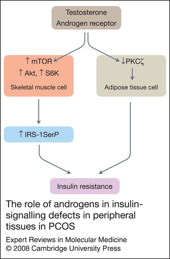
Metabolic derangement in PCOS is reflected at the molecular level. Insulin-signalling defects have been shown in skeletal muscle and in adipose tissue from women with PCOS (Fig. 3). These defects appear to be partly intrinsic and partly conferred by the superimposed effects of the in vivo hormonal and metabolic milieu. Androgens, adipokines and other pro-inflammatory molecules may also affect insulin action in peripheral tissues.
The role of androgens in insulin-signalling defects in peripheral tissues in PCOS. Testosterone, acting through its own receptor, interferes with insulin signalling in peripheral tissues. In adipose tissue, testosterone acts via decreased PKCζ; in skeletal muscle, it acts via increased mTOR and S6K. Abbreviations: IRS, insulin receptor substrate; mTOR, mammalian target of rapamycin; PKC, protein kinase C; SerP, serine phosphorylation; S6K, ribosomal S6-kinase.

Insulin resistance in adipose tissue
Studies in subcutaneous adipocytes isolated from women with PCOS have demonstrated impaired insulin-stimulated glucose transport (Refs Reference Ciaraldi78, Reference Dunaif79). A recent study has suggested that decreased responsiveness rather than decreased sensitivity is the main defect of insulin action on subcutaneous adipocytes (Ref. Reference Lystedt80).
An older study showed diminished insulin-stimulated autophosphorylation of the insulin receptor in PCOS (Ref. Reference Dunaif79). Recently, this defect was demonstrated in erythrocytes only in the insulin-resistant subgroup of PCOS women, probably reflecting a distinct subphenotype with this molecular defect among the entire PCOS cohort (Ref. Reference Mor81). Current research has focused more avidly on post-receptor defects of the insulin-signalling system. Impaired insulin-stimulated tyrosine phosphorylation of insulin receptor substrate 1 (IRS-1) has emerged as an additional signalling defect in PCOS (Ref. Reference Diamanti-Kandarakis and Papavasiliou82). Increased glycogen synthase kinase 3β (GSK-3β) in adipose tissue may also contribute to insulin resistance through increased serine phosphorylation of IRS-1 (Ref. Reference Goodarzi83).
Initial studies showed decreased expression of the insulin-regulated glucose transporter GLUT-4 in PCOS adipocytes (Ref. Reference Rosenbaum, Haber and Dunaif84). This abnormality was also recently confirmed in omental adipocytes from women with PCOS (Ref. Reference Seow85).
Microarray analyses of omental adipose tissue biopsies from women with PCOS revealed abnormal expression of additional specific insulin-signalling pathway genes, which were not found in specimens from non-PCOS obese women. Overexpression of genes for PI3KR1 (the regulatory p85a subunit of PI3K) and ectonucleotide pyrophosphatase/phosphodiesterase 1 (ENPP1; a negative regulator of insulin receptor tyrosine kinase activity) appear to be PCOS-specific defects of insulin signalling in adipose tissue. Most importantly, these gene expression abnormalities were found in the visceral fat depot, which is considered to play a major metabolic and endocrine role (Ref. Reference Corton86).
Insulin resistance in skeletal muscle
Reduced insulin receptor tyrosine kinase activity due to increased serine phosphorylation was suggested as a mechanism for insulin resistance in PCOS skeletal muscle (Ref. Reference Dunaif87). Studies of muscle biopsies from PCOS patients did not reveal alterations of the upstream signalling proteins (e.g. insulin receptor, IRS-1 and the p85 regulatory subunit of PI3K) (Ref. Reference Dunaif88); however, in vivo work showed a significant reduction in insulin-mediated glucose uptake (Refs Reference Diamanti-Kandarakis and Papavasiliou82, Reference Dunaif88) with decreased insulin-mediated IRS-1-associated PI3K activity (Ref. Reference Dunaif88). A compatible finding was increased Ser312 IRS-1 phosphorylation in cultures of PCOS skeletal myocytes (Ref. Reference Corbould89). In a study of freshly isolated muscle tissue from PCOS women, enhanced mitogenic signalling (activation of the MAP2K1 isoforms ERK1/2) was associated with IRS-1 Ser312 phosphorylation and subsequent inhibition of IRS-1 association with the p85 subunit of PI3K. This mechanism could provide a link between altered mitogenic signalling and insulin resistance in skeletal muscle (Ref. Reference Corbould90).
The role of androgens in metabolic aberrations in PCOS
Hyperandrogenaemia may be among the inciting factors of metabolic aberrations in PCOS. Androgen excess during intrauterine life or in the early postnatal period has been shown to precipitate visceral adiposity and insulin resistance (Ref. Reference Abbott, Dumesic and Franks91). Furthermore, girls with premature pubarche have insulin resistance throughout puberty (Ref. Reference Ibanez92), which can be alleviated with combined treatment of insulin sensitisers and antiandrogens (Ref. Reference Ibanez and de Zegher93).
Androgens may aggravate metabolic aberrations via the androgen receptor, which is overly expressed in the visceral fat depot, as compared with the subcutaneous compartment (Refs Reference Rodriguez-Cuenca94, Reference De Pergola95). The preferential upregulation of lipolysis in visceral adipose tissue may reflect an androgen-induced metabolic defect of PCOS (Ref. Reference De Pergola95). Accordingly, laparoscopic ovarian electrocautery in PCOS women reduced androgen levels and attenuated insulin resistance, ameliorating the intracellular insulin-signalling defects in adipose tissue (Ref. Reference Corton86).
Androgens appear to contribute to peripheral insulin resistance in PCOS by acting directly upon the insulin-signalling pathways (Fig. 3). In cultured subcutaneous adipocytes, testosterone selectively induces metabolic insulin resistance acting via the androgen receptor (Ref. Reference Corbould96). This insulin-signalling defect did not involve PI3K, but the impaired phosphorylation of the downstream mediator protein kinase Cζ (PKCζ) (Ref. Reference Corbould96). However, in vivo studies in female-to-male transsexuals have failed to reveal a clear effect of exogenous androgens on insulin (Refs Reference Polderman97, Reference Elbers98).
Skeletal muscle appears to be also affected by androgen-induced insulin resistance. Androgens may interfere with insulin signalling by amplifying phosphorylation of Akt- mammalian target of rapamycin (mTOR) and ribosomal S6-kinase (S6K), and consequently increasing Ser636/639 phosphorylation of IRS-1 (Ref. Reference Allemand99).
Other putative factors
The dynamic balance between pro-inflammatory and anti-inflammatory adipokines seems to be altered in PCOS, favouring a low-grade chronic inflammation (Refs Reference Puder100, Reference Sayin101). Exaggerated tumour necrosis factor (TNF)-α production by mononuclear cells, in response to hyperglycaemia within the physiological range (postprandial state), may further exacerbate metabolic and hormonal abnormalities in PCOS (Ref. Reference González102). TNF-α via its own receptor and via activated NF-κB induces serine phosphorylation of IRS-1, aggravating insulin resistance (Refs Reference González102, Reference Mlinar111).
Recently, advanced glycation end-products (AGEs) and their receptors (receptors for advanced glycated end-products; RAGEs), implicated in the molecular cascades of inflammatory and metabolic stress (Ref. Reference Vlassara103), have been found to be elevated and positively correlated with androgens in women with PCOS (Ref. Reference Diamanti-Kandarakis104). A wealth of data suggests that the interaction of AGEs with RAGEs impairs insulin signalling at the IRS level in two ways: via generation of reactive oxygen species (ROS) (Ref. Reference Vlassara103); and independently of oxidative stress, via stimulation of serine kinases (Ref. Reference Miele105). The role of these hazardous molecules in other insulin-resistant states (Ref. Reference Vlassara103) has been determined and the newly reported findings in women with PCOS (Ref. Reference Diamanti-Kandarakis104) tentatively expand the deleterious spectrum of AGE-related clinical implications. Furthermore, increased AGE levels may be associated with the reproductive/endocrine component of PCOS. Preliminary data have shown increased immunostaining of AGEs and RAGEs in different compartments of ovarian tissue in polycystic ovaries (Ref. Reference Diamanti-Kandarakis106). The source of AGEs in normoglycaemic women with PCOS could be endogenous or exogenous, since a diet rich in glycotoxins has been associated with hyperinsulinaemia, hyperandrogenaemia and increased accumulation of AGEs in rat ovaries (Ref. Reference Diamanti-Kandarakis107).
The growing recognition of the intrinsic linkage of PCOS with metabolic abnormalities has prompted considerations on the long-term sequelae of the syndrome. Among affected women, the potential exacerbation of metabolic aspects along with ageing reinforces the concept of PCOS as a ‘life-long health issue’ (Refs Reference Margolin108, Reference Krentz, von Mühlen and Barrett-Connor109).
The dynamic interplay of androgens, anovulation and metabolic aberrations in PCOS
The major determinants of PCOS – hyperandrogenaemia, ovarian dysfunction and metabolic abnormalities – appear to have interconnecting roles in the pathophysiology of PCOS.
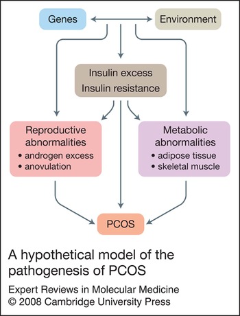
Environmental factors interact with still unknown genetic traits to initiate the pathogenic sequence leading to PCOS (Ref. Reference Diamanti-Kandarakis110). This interaction appears to start before birth and continues throughout the lifespan (Fig. 4). The order of events among the main pathogenetic components of PCOS is not yet clear. However, insulin resistance and the compensatory hyperinsulinaemia may insult ovarian function, contributing to excessive androgen production (Ref. Reference Nestler29) and to anovulation (Ref. Reference Phy51) in PCOS (Fig. 4).
A hypothetical model of the pathogenesis of PCOS. The clinical phenotype of PCOS reflects the contribution of reproductive (androgen excess, anovulation) and metabolic abnormalities (in the adipose tissue and skeletal muscles). The pathogenesis of both reproductive and metabolic components of PCOS involves the dynamic interaction of environmental and genetic factors, which is set in motion during the intrauterine period and lasts throughout life. Insulin resistance and insulin excess at the systemic and the local tissue level serve as the potential link in the bidirectional relationship between the reproductive and the metabolic ‘axes’ of PCOS.

Central adiposity may play a key role in the perpetuation of androgen excess, anovulation and metabolic aberrations. Excess free fatty acids (FFAs) released from visceral fat may contribute to metabolic (Ref. Reference Mlinar111) and to reproductive (Ref. Reference Mai112) abnormalities in PCOS. Furthermore, central obesity is intimately associated with inflammatory stress. Pro-inflammatory cytokines (Refs Reference Kelly113, Reference Diamanti-Kandarakis114) could help explain the links between reproductive and metabolic aberrations of PCOS (Refs Reference Spaczynski, Arici and Duleba115, Reference Kwintkiewicz116).
Altered adipose tissue production of adipokines (local rather than systemic) is also linked with visceral adiposity and androgen excess in PCOS. Serum resistin levels have been shown to be elevated in obese PCOS women (Refs Reference Panidis117, Reference Seow, Juan and Ho120), and recent studies in primary human theca cells suggest that resistin may synergise with insulin to enhance androgen production (Ref. Reference Munir118). Another study has shown no difference in resistin levels between PCOS patients and body mass index (BMI)-matched controls, whereas obesity, independently of PCOS, has been associated with increased resistin levels (Ref. Reference Escobar-Morreale, Villuendas and Botella-Carretero119). However, the direct linkage of resistin to the PCOS pathophysiology remains under investigation.
Clinical implications
The multifactorial clinical spectrum of PCOS requires a multifaceted therapeutic approach. Traditional drugs (oral contraceptives, clomiphene and antiandrogens) have been supplemented by novel pharmaceutical modalities (insulin sensitisers and statins). However, lifestyle modification still remains the therapeutic ‘sine qua non’.
Lifestyle modification
Lifestyle modification is a fundamental treatment option, in cases where adverse lifestyle factors are bound to aggravate the presentation of PCOS. All other interventions should come as adjuvant treatment modalities. Modification aims to decrease weight and sustain reduced body weight in the long-term.
Amelioration of insulin resistance through diet and exercise-induced weight loss appears to contribute to improvement of clinical, metabolic and hormonal parameters of PCOS. Even modest weight loss (approximately 10% of initial body weight) has been shown to increase the frequency of ovulation, improve hormonal indices, and attenuate metabolic aberrations in women with PCOS (Refs Reference Moran121, Reference Hoeger122). Although lifestyle modification is of greater urgency in obese patients, every woman with PCOS can benefit from a balanced diet and regular exercise (Refs Reference Diamanti-Kandarakis110, Reference Marsh and Brand-Miller123). Energy restriction has been in the foreground of most dietary intervention programmes, whereas the quality of diet may also play a role (Refs Reference Diamanti-Kandarakis110, Reference Moran121, Reference Marsh and Brand-Miller123). Of major concern is that long-term weight loss is difficult to achieve and sustain in the majority. On these grounds, additional medical therapy is warranted.
Insulin sensitisers
There is an intriguing pathophysiological framework to support the therapeutic advantages of insulin sensitisers in the management of PCOS, including metabolic aspects, ovulatory function and hyperandrogenaemia. Available literature, as a whole, provides credence to this concept and encourages the integration of insulin sensitisers in therapeutic strategies of PCOS. Metformin and thiazolineodiones are the two more widely used insulin sensitisers.
Metformin
Metformin is a widely studied insulin sensitiser with a reassuring safety profile (Ref. Reference Nestler124). Metformin facilitates weight loss, reduces visceral adiposity and attenuates insulin resistance, mainly at the hepatic and muscle tissue level, contributing to a more favourable metabolic (Ref. Reference Diamanti-Kandarakis and Diamanti-Kandarakis125) and hormonal (Ref. Reference Kolodziejczyk126) profile.
The metabolic effects of metformin are well established in other metabolic disorders and, specifically, in obese and nonobese women with PCOS (reviewed in Ref. Reference Diamanti-Kandarakis and Diamanti-Kandarakis125). In a meta-analysis of 13 controlled studies of women with PCOS from different ethnic populations, it was confirmed that metformin has a significant effect in reducing fasting insulin levels (Ref. Reference Lord, Flight and Norman127). Severe obesity may be a confounding factor, but tentatively is not the sole predictor of resistance to the beneficial effects of metformin (Ref. Reference Diamanti-Kandarakis and Diamanti-Kandarakis125). An intriguing aspect is that metformin treatment appears to be devoid of significant risks when used during pregnancy (Ref. Reference Jakubowicz128). Administering metformin in pregnant women with PCOS may alleviate the risk of early miscarriage (Ref. Reference Jakubowicz128). However, the safety of metformin during pregnancy awaits confirmation by larger studies. Metformin appeared to have lowering effects on several cardiovascular and atherogenic molecules, such as C-reactive protein (Ref. Reference Morin Papunen129), endothelin 1, adhesion molecules (ICAM, VCAM) (Ref. Reference Diamanti-Kandarakis130) and AGEs (Ref. Reference Diamanti-Kandarakis131).
Extending the efficacy of metformin across the entire clinical spectrum of PCOS, several studies have witnessed the attenuation of hyperandrogenaemia with metformin treatment (Ref. Reference Kolodziejczyk126) and this has been also attested by the meta-analysis comparing metformin with either placebo or no treatment (Ref. Reference Lord, Flight and Norman127). The greatest efficacy has been observed in hyperinsulinaemic hyperandrogenic PCOS patients, suggesting that the androgen-lowering effect of metformin resides, at least partly, in the reduction of insulin levels (Ref. Reference Genazzani132). In particular, an advantageous effect on androgen levels has also been been demonstrated in lean patients and has been shown to be independent of BMI (Ref. Reference Genazzani133). Most strikingly, metformin has been shown to act directly on ovarian theca cells to reduce androgen production (Ref. Reference Attia134).
For targeting the PCOS symptoms of menstrual irregularity or hirsutism, metformin treatment is not considered as the first-line medication, although metformin appears to be clearly superior to placebo in regularising menstrual cycles (Ref. Reference Costello135). The Cochrane Library review (Ref. Reference Jakubowicz128) concluded that metformin treatment was effective in increasing ovulation rates in women with PCOS, compared with placebo, but there was no mention with regard to live birth rates. Available data suggest that anovulatory women with PCOS may not always respond to metformin, although the determinants of the response to treatment remain elusive (Ref. Reference Jakubowicz128).
A recent study with the primary outcome chosen as live-birth rate compared the efficacy of metformin with that of clomiphene citrate, an anti-oestrogenic substance traditionally used for ovulation induction (Ref. Reference Legro136). Among a sizeable cohort of PCOS women, metformin treatment resulted in inferior rates of live births and the combination therapy did not achieve a significant advantage over clomiphene monotherapy. These findings may limit the initial enthusiasm from head-to-head trials (Ref. Reference Palomba137). However, complications of pregnancy were more common among subjects in the clomiphene group, as compared with the ones in the metformin group, which should be considered when assigning clomiphene for infertility treatment in PCOS.
Thiazolinediones
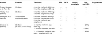
Several studies have shown that thiazolinediones, namely rosiglitazone and pioglitazone, are beneficial in attenuating insulin resistance and hyperandrogenaemia and in restoring ovulation in PCOS (Refs Reference Romualdi138, Reference Brettenthaler139). A recent study among severely obese and insulin-resistant PCOS women has shown a significant decline of free testosterone levels with rosiglitazone treatment, which was comparable that achieved with metformin (Ref. Reference Sepilian and Nagamani140). Interestingly, thiazolinediones may also exert direct effects on CYP17A1 gene expression in theca cells (Ref. Reference Seto-Young141), although evidence is still sparse. Studies comparing thiazolinediones and metformin do not reveal differences in their insulin-sensitising effects (Refs Reference Diamanti-Kandarakis and Diamanti-Kandarakis125, Reference Ortega-Gonzalez142, Reference Glueck143, Reference Kilicdag144, Reference Baillargeon145) (Table 1).
Studies comparing metformin with thiazolinediones in metabolic terms

aAdapted from Table 1 in Ref. Reference Romualdi125 (© Humana Press Inc.), with kind permission of Springer Science and Business Media.
Symbols: →, not significantly altered; ↑, significantly increased; ↓, significantly decreased.
Abbreviations: BMI, body mass index; HDL, high-density lipoprotein; LDL, low-density lipoprotein; non-resp., women non-responders to metformin treatment; resp., women responders to metformin treatment; W/H, waist-to-hip ratio.
Existing data indicate that treatment with thiazolidinediones does not significantly alter the lipidaemic profile [triglycerides, low-density-lipoprotein–cholesterol (LDL-C) and high-density-lipoprotein–cholesterol (HDL-C)] (Refs Reference Romualdi138, Reference Brettenthaler139, Reference Legro146). However, conclusions regarding effects of thiazolinediones on lipid or other parameters in the PCOS should be drawn for each thiazolinedione separately, because their effects are differentiated in various metabolic and reproductive pathways (Ref. Reference Diamanti-Kandarakis and Diamanti-Kandarakis125).
The role of thiazolinediones in infertility therapy in PCOS awaits further exploration. However, thiazolinedione treatment should not be administered to pregnant women, since data on the safety profile during pregnancy are lacking.
Antiandrogen treatment: flutamide
Antiandrogen treatment is the cornerstone in the therapeutic approach to hirsutism and acne. Considering the risk of feminisation of the male fetus, contraception is essential when antiandrogens are administered in women of reproductive age. The potential adverse metabolic impact of antiandrogen therapy has fuelled debate on when and for how long antiandrogen treatment is indicated.
Few clinical studies have addressed the effects of the androgen receptor antagonist flutamide on metabolic features of PCOS. Available studies among lean and obese PCOS patients have shown either neutral or a mild positive effect of flutamide treatment on insulin sensitivity (Refs Reference Diamanti-Kandarakis147, Reference Moghetti148, Reference Sahin149). Notable inconsistency applies to the clinical data on the effects of flutamide on the lipidaemic profile (Refs Reference Diamanti-Kandarakis150, Reference Vrbikova151).
Oral contraceptives
The oral contraceptive pill (OCP) has been for decades the main pharmaceutical modality to regulate menstrual cyclicity and to attenuate the clinical signs of hyperandrogenism in PCOS. Additionally, treatment with OCPs alleviates the risk of endometrial hyperplasia, which is associated with chronic anovulation and oligomenorrhoea.
However, the increasing recognition of the metabolic aspects of PCOS has generated debate on the metabolic impact of treating PCOS women with OCPs or with ethinylestradiol/cyproterone acetate (OCP/CA pill). Trials among PCOS women taking OCPs as monotherapy have given varied results concerning the metabolic effects, and some of them have raised concerns that OCP treatment may result in the deterioration of insulin sensitivity, glucose tolerance and lipid profile (Refs Reference Nader152, Reference Diamanti-Kandarakis153, Reference Mastorakos154, Reference Charitidou155). The metabolic effects of OCPs appear to be significantly dependent on the clinical phenotype of the patient, since obese women appear to be more vulnerable to these metabolic aberrations (Ref. Reference Diamanti-Kandarakis153). The type of progestin used in combination with oestrogen appears to play an additional role (Refs Reference Diamanti-Kandarakis153, Reference Mastorakos154). However, a recent meta-analysis of four randomised controlled trials has not revealed an adverse metabolic risk with the OCP treatment compared with metformin, in terms of clinical (type 2 diabetes, cardiovascular disease) and surrogate (fasting glucose, insulin and total cholesterol levels) metabolic outcomes. Interestingly, though, the paucity of randomised controlled trials addressing this issue has been highlighted (Ref. Reference Costello135).
CA is a progestinoid with potent antiandrogenic effects, which is often used in combination with ethinylestradiol (OCP/CA pill) to improve menstrual regularity and clinical signs of hyperandrogenism in affected women. Treatment with the OCP/CA pill has been associated with increased serum HDL-C (Refs Reference Morin-Papunen156, Reference Luque Ramirez157) and triglyceride concentrations in both nonobese and obese PCOS subjects (Refs Reference Diamanti-Kandarakis and Diamanti-Kandarakis125, Reference Morin-Papunen156). The OCP/CA pill has also been incriminated for a worsening of glucose tolerance (Refs Reference Diamanti-Kandarakis and Diamanti-Kandarakis125, Reference Mastorakos154), although this effect has been reported to be minor, whereas hyperinsulinaemia has been shown in some (Ref. Reference Charitidou155) but not in all (Ref. Reference Luque Ramirez157) of the available studies.
Statins
Statins may prove to be an additional therapeutic tool for the steroidogenic abnormalities in PCOS. However, available data are limited and should be interpreted with caution until further research has been carried out. A recent study among women receiving combined treatment of statin and OCP has shown a significant statin-attributable attenuation of clinical and biochemical hyperandrogenism in concert with amelioration of cardiovascular risk factors (Ref. Reference Banaszewska158). This clinical study has provided support to previous in vitro findings showing that statin inhibits proliferation and steroidogenesis of ovarian theca-interstitial cells from PCOS women (Ref. Reference Kwintkiewicz159). Owing to the potential fetal toxicity of statins, effective contraception is essential when statin treatment is assigned in women of reproductive age (Ref. Reference Edison and Muenke160).
Conclusions
PCOS remains a syndrome of unknown causation. The integration of available data to formulate a concrete aetiological base remains a difficult task. Hyperandrogenaemia and anovulation are recognised by a significant portion of literature to be inextricably linked with PCOS. Insulin resistance is also essentially integrated in the PCOS spectrum, playing a key role in the clinical aspects of PCOS. These entities interweave with each other to confer a variable degree of contribution to the final phenotype of the syndrome. Accumulating evidence suggests that the web of aberrations associated with PCOS extends far beyond the hormonal/reproductive disturbances. This critical dimension, newly added to our view of PCOS, provides momentum for the investigation of alternative presentations of PCOS extending throughout the lifespan of women.
Acknowledgements and funding
The author thanks Dr Charikleia Christakou for her excellent assistance during the preparation of the manuscript, and the peer reviewers for their constructive comments.