Case report
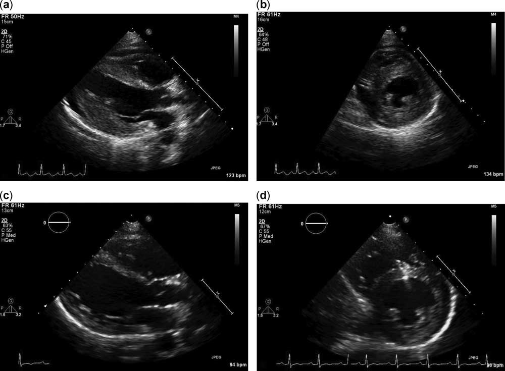
A 14-year-old girl with recent influenza A infection – confirmed by respiratory virus panel 10 days prior and treated with oseltamivir – was referred to our medical centre with a 1-day history of malaise, abdominal pain, and peripheral oedema. She had no significant past medical history or family history to report. Vital signs were significant for tachycardia (150 beats/minute), tachypnoea (28 respirations/minute), and hypotension (blood pressure 84/54 mmHg). Physical examination revealed an obese female (88 kg) with mild increased work of breathing, bilateral crackles at the lung bases, tender hepatomegaly, cool distal extremities, a prolonged capillary refill, and pitting oedema of bilateral lower extremities. Admission arterial blood gas demonstrated a metabolic acidosis with a lactate of 7.8 mmol/L and both serum troponin-I and serum B natriuretic peptide levels were elevated at 2.6 ng/ml (peak 15.8 ng/ml) and 987 pg/ml (peak 2570 pg/ml), respectively. Initial chest x-ray showed mild cardiomegaly with mild pulmonary vascular congestion and initial electrocardiogram showed sinus tachycardia with low-voltage QRS complexes. Admission echocardiogram (Fig 1a, b, Data Supplement Movie 1) demonstrated moderately depressed left ventricular systolic function (shortening fraction 21%, ejection fraction 29%), a moderate sized pericardial effusion, and moderate left ventricular hypertrophy (septal wall thickness=1.6 cm and left ventricular posterior wall thickness=1.6 cm). Viral studies (from serum and respiratory secretions) and a blood culture sent on admission were negative.
Transthoracic echocardiogram images. Parasternal long- ( a ) and short-axis ( b ) views from the admission study demonstrate moderate left ventricular hypertrophy and depressed left ventricular systolic function (septal wall thickness=1.6 cm, left ventricular posterior wall thickness=1.6 cm, shortening fraction 21%, and ejection fraction 29%). A follow-up echocardiogram was performed 2 weeks after hospital discharge. Parasternal long- ( c ) and short-axis ( d ) views demonstrate improved left ventricular wall measurements and normal left ventricular systolic function (septal wall thickness=1.3 cm, left ventricular posterior wall thickness=1.0 cm, shortening fraction 33%, and ejection fraction 68%).
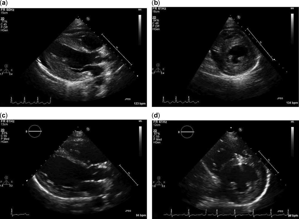
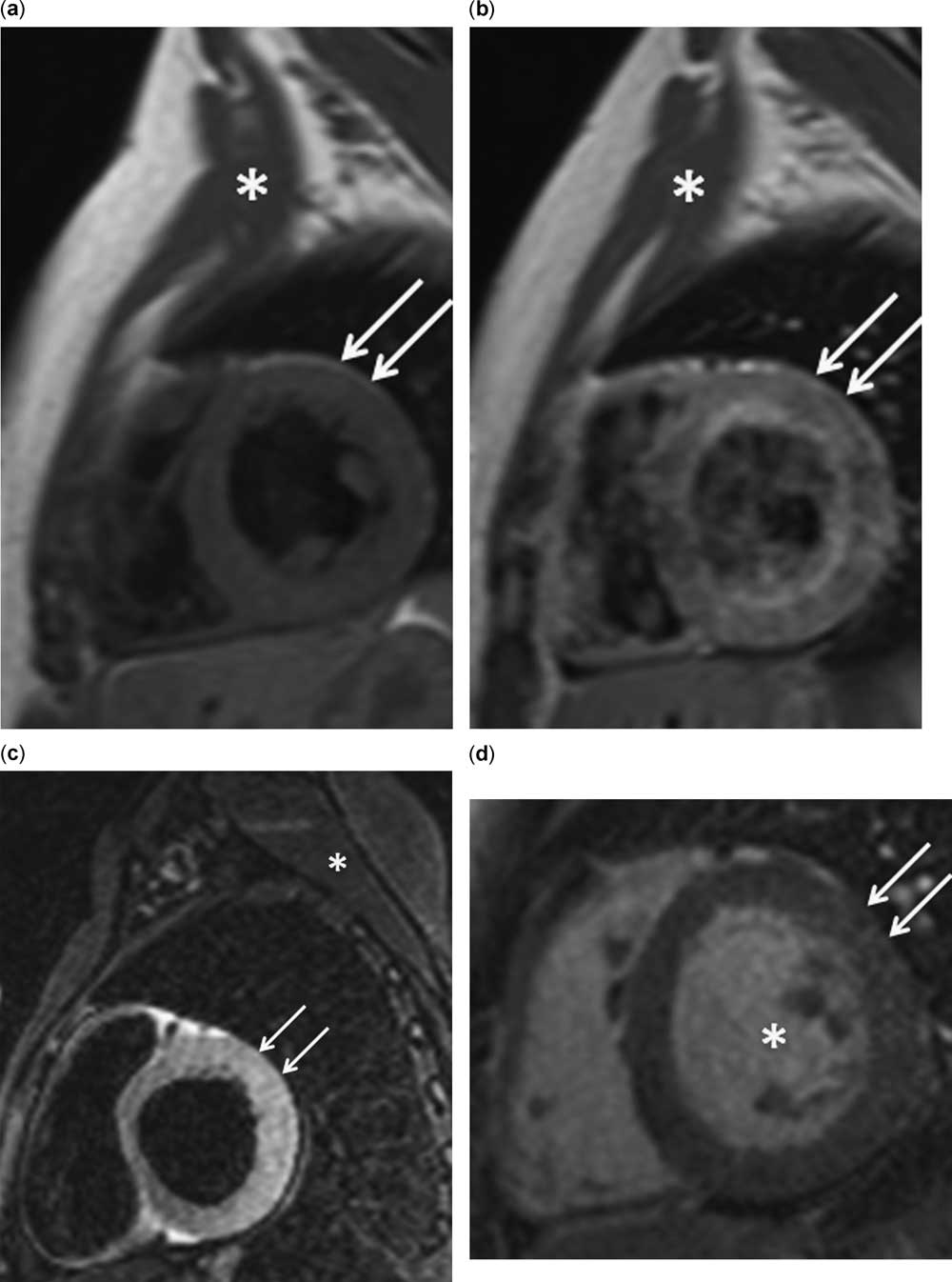
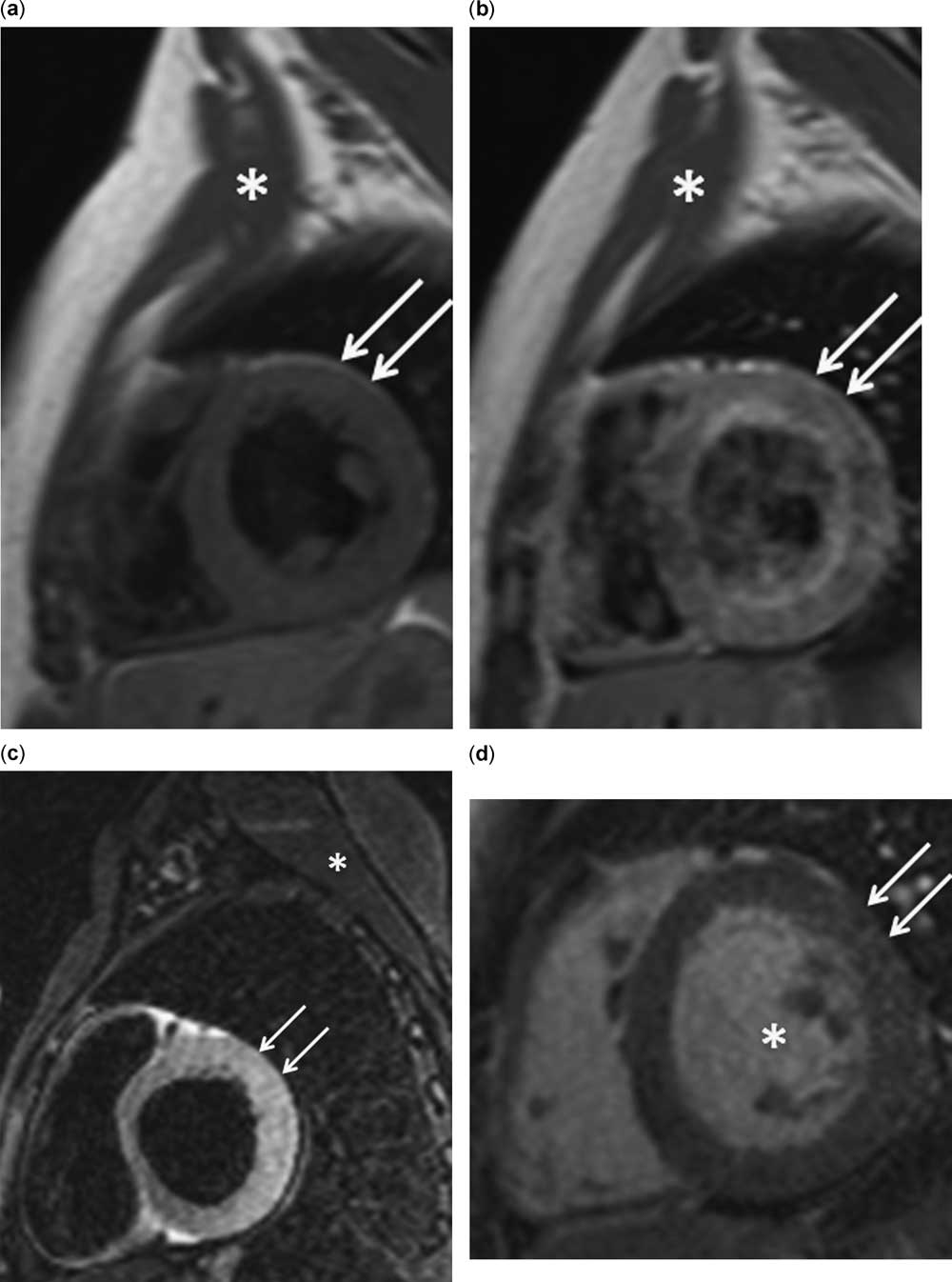
The diagnosis was presumed to be viral myocarditis, in the setting of a recent influenza A infection, managed with dopamine and milrinone for hypotension and poor perfusion, as well as furosemide for pulmonary oedema and volume overload. A single dose of intravenous immunoglobulin (IVIG, dose 2 g/kg) was given on hospital day 1. On hospital day 2, the patient developed worsening pulmonary oedema and an increased oxygen requirement prompting intubation and mechanical ventilation. A cardiac catheterisation was performed on hospital day 3, which documented elevated right atrial pressure (mean 8 mmHg), elevated pulmonary capillary wedge pressure (mean 24 mmHg), and a normal cardiac index (3.9 L/minute/m2). Endomyocardial biopsies were found to be non-specific with a single focus of myocyte damage and surrounding oedema with no lymphocytic infiltrate and negative viral polymerase chain reaction studies – for multiple viruses including influenza A. The patient was extubated on hospital day 5 and gradually weaned off of dopamine and milrinone and transitioned to oral cardiac medications – furosemide, aldactone, and enalapril. She did not develop significant ectopy or dysrrhythmia during her hospital course. A follow-up echocardiogram on hospital day 7 showed marked improvement in left ventricular systolic function – shortening fraction 32%, ejection fraction 66% – with unchanged, moderate left ventricular hypertrophy. Owing to the concern for an underlying cardiomyopathy, cardiac magnetic resonance imaging was performed on hospital day 10 and was notable for significant global myocardial oedema and hyperaemia with no evidence of delayed enhancement (Fig 2) and normal left ventricular systolic function – ejection fraction 72%. The patient was discharged after 11 days of hospitalisation. She was asymptomatic at 2-week follow-up, with a repeat echocardiogram demonstrating improved left ventricular thickness (septal wall thickness=1.3 cm and left ventricular posterior wall thickness=1.0 cm) and continued normal left ventricular systolic function (Fig 1c, d, Data Supplement Movie 2). There is a plan for repeat cardiac magnetic resonance imaging ~6 months from her initial presentation.
Cardiac magnetic resonance imaging on hospital day 10. A T1-weighted turbo spin-echo dark blood image acquired for assessment of myocardial hyperaemia demonstrates that before gadolinium administration ( a ), there is evidence of resolving wall thickness (septal wall thickness=1.3 cm and left ventricular posterior wall thickness=1.0 cm). The signal intensity of the myocardium (arrows) is similar to that of the skeletal muscle (asterisk). Immediately after the administration of gadolinium ( b ), early gadolinium myocardial enhancement is observed with >80% increase in myocardial signal intensity compared with the pre-gadolinium image. Signal intensity⩾45% on post-gadolinium images are required to meet Lake Louise criteria for myocardial hyperaemia. A T2-weighted dark blood image ( c ) acquired for assessment of myocardial oedema – without the use of a contrast agent – shows a four-fold increased signal intensity in the myocardium (arrows), due to water enhancement, relative to the skeletal muscle (asterisk) signifying global myocardial oedema. Lake Louise criteria requires a two-fold increase for consideration of myocardial oedema. An inversion recovery image acquired 10 minutes after the administration of gadolinium ( d ) shows that the myocardium (arrows) appears relatively dark compared with the blood pool (asterisk), confirming normal viability without evidence of delayed enhancement to suggest myocardial necrosis or fibrosis.
Discussion
Transient left ventricular wall thickening in the acute phase of myocarditis is presumed to be caused by myocardial oedema.Reference Hauser, Gordon, Cieszkowski and Timmis 1 – Reference Hiramitsu, Morimoto and Kato 3 Hiramitsu et alReference Hiramitsu, Morimoto and Kato 3 evaluated 25 patients (mean age 45±21.3 years) with myocarditis, documenting left ventricular wall thickening in 14 (56%) of the cases immediately after the onset of disease (septal wall thickness=13.2±3.2 mm and left ventricular posterior wall thickness=12.1±2.5 mm), more commonly in patients with fulminant disease, with near normalisation in wall measurements at 1 week after onset. Hiramitsu et al also performed serial echocardiograms and endomyocardial biopsies on a small cohort of adult patients with myocarditis during the acute and convalescent phases of disease. The thickness of the left ventricle decreased significantly from the acute to the convalescent phase with myocardial oedema found on endomyocardial biopsy in 22 patients (88%) in the acute phase but only in seven patients (28%) in the convalescent phase, supporting the theory that left ventricular wall thickening found with acute myocarditis is due to myocardial oedema.Reference Hiramitsu, Morimoto and Kato 2
Our patient was referred for cardiac magnetic resonance imaging to rule out an underlying cardiomyopathy in the setting of persistent left ventricular hypertrophy on repeat echocardiogram despite normalising ventricular function, which contrasts previous reports of early normalisation in left ventricular wall thickness with acute myocarditis.Reference Hiramitsu, Morimoto and Kato 3 The cardiac magnetic resonance imaging results were helpful in providing non-invasive evidence to support the diagnosis of myocarditis and in demonstrating that the left ventricular thickening seen by echocardiography was related to myocardial oedema and not myocardial hypertrophy. Cardiac magnetic resonance imaging has become the primary tool for non-invasive assessment of myocardial inflammation in patients with suspected myocarditis and current recommendations have been published that include indications for cardiac magnetic resonance imaging, protocol standards, and diagnostic criteria (i.e., Lake Louise Criteria) in this clinical setting.Reference Friedrich, Sechtem and Schulz-Menger 4
This is the first report to non-invasively demonstrate transient ventricular wall thickening related to global myocardial oedema in a child with acute myocarditis, underscoring the utility of cardiac magnetic resonance imaging in these patients. More research is needed to determine whether cardiac magnetic resonance imaging can be used for non-invasive risk stratification of children presenting with acute heart failure in order to assist the clinician in decisions regarding endomyocardial biopsy, immunosuppressive therapy, ventricular assist devices, and transplantation.
Acknowledgment
We would like to acknowledge the contribution of doctors Kimberly Y. Lin, Stephen M. Paridon, and Robert E. Shaddy to the care of this patient and to the writing of this brief report.
Financial Support
This research received no specific grant from any funding agency, commercial or not-for-profit sectors.
Conflicts of Interest
None.
Supplementary material
To view supplementary material for this article, please visit http://dx.doi.org/10.1017/S1047951114000018.