Introduction
South America is characterized by high biogeographic complexity due to the heterogeneity of climates and topographies, its great range in latitude (Ortiz-Jaureguizar and Cladera, Reference Ortiz-Jaureguizar and Cladera2006), and its isolation during a large part of the Cenozoic Era (the so called ‘splendid isolation’ of Simpson, Reference Simpson1980) until the definitive formation of the Isthmus of Panama (Woodburne, Reference Woodburne2010; O'Dea et al., Reference O'Dea, Lessios, Coates, Eytan, Restrepo-Moreno, Cione and Stallard2016). These conditions generated a complex paleobiogeographic and evolutionary scenario that favored the origin and radiation of some clades of endemic mammals, such as Litopterna, Notoungulata, and Xenarthra (Pascual, Reference Pascual2006; Croft, Reference Croft, Patterson and Costa2012). Among them, only Xenarthra have living representatives, although their current diversity is a poor representation compared to several intervals of the Cenozoic (Abba et al., Reference Abba, Tognelli, Seitz, Bender and Vizcaíno2012; Gaudin and Croft, Reference Gaudin and Croft2015; Gibb et al., Reference Gibb, Condamine, Kuch, Enk, Moraes-Barros, Superina, Poinar and Delsuc2015). The monophyly of this group has been repeatedly proved by molecular and morphological analyses (Gaudin, Reference Gaudin1995, Reference Gaudin2004; Delsuc et al., Reference Delsuc, Ctzeflis, Stanhope and Douzery2001, Reference Delsuc, Scally, Madsen, Stanhope, De Jong, Catzeflis, Springer and Douzery2002; Pujos et al., Reference Pujos, De Iuliis, Argot and Werdelin2007; Delsuc and Douzery, Reference Delsuc, Douzery, Hedges and Kumar2009). Recent molecular evidence indicates that Xenarthra is one of the most basal clades of Placentalia, as the sister group of the remaining mammals (Epitheria) (O'Leary et al., Reference O'Leary, Bloch, Flynn, Gaudin, Giallombardo, Giannini, Goldberg, Kraatz, Luo, Meng, Ni, Novacek, Perini, Randall, Rougier, Sargis, Silcox, Simmons, Spaulding, Velazco, Weksler, Wible and Cirranello2013). However, some authors support the interpretation of Xenarthra as the sister group of the Afrotheria (Meredith et al., Reference Meredith, Janecka, Gatesy, Ryder, Fisher, Teeling and Rabosky2011).
Xenarthra includes two large clades, Pilosa (Vermilingua + Tardigrada) and Cingulata (armadillos, pampatheres, peltephilids, and glyptodonts) (Delsuc et al., Reference Delsuc, Scally, Madsen, Stanhope, De Jong, Catzeflis, Springer and Douzery2002; McDonald and Naples, Reference McDonald, Naples, Janis, Gunnell and Uhen2007; Gaudin and Croft, Reference Gaudin and Croft2015). The earliest record of Xenarthra is the Paleocene (or earliest Eocene?; Gelfo et al., Reference Gelfo, Goin, Woodburne and Muizon2009) cingulate dasypodid Riostegotherium yanei Oliveira and Bergqvist, Reference Oliveira and Bergqvist1998 (Bergqvist et al., Reference Bergqvist, Abrantes and Avilla2004). Although the Paleogene record of this clade is quite scarce, the beginning of the large radiations and taxonomic diversifications seem to be closely related to certain climatic-environmental events, particularly since the Oligocene (Zachos et al., Reference Zachos, Pagani, Sloan, Thomas and Billups2001; Ortiz-Jaureguizar and Cladera, Reference Ortiz-Jaureguizar and Cladera2006; Gibb et al., Reference Gibb, Condamine, Kuch, Enk, Moraes-Barros, Superina, Poinar and Delsuc2015). However, the paleontological record is much more complete in the Neogene (Gaudin and Croft, Reference Gaudin and Croft2015).
Most knowledge of the evolutionary history of Xenarthra comes from southern South America (Carlini et al., Reference Carlini, Ciancio and Scillato-Yané2005; Croft et al., Reference Croft, Carlini, Ciancio, Brandoni, Drew, Engelman and Anaya2016), but knowledge of the group from middle and low latitudes has recently increased (Cozzuol, Reference Cozzuol2006; Ciancio et al., Reference Ciancio, Carlini, Campbell and Scillato-Yané2013; Tejada-Lara et al., Reference Tejada-Lara, Salas-Gismondi, Pujos, Baby, Benammi, Brusset, De Franceschi, Espurt, Urbina and Antoine2015; Amson et al., Reference Amson, Carrillo and Jaramillo2016; Antoine et al., Reference Antoine, Abello, Adnet, Altamirano Sierra, Baby, Billet, Boivin, Calderón, Candela, Chabain, Corfu, Croft, Ganerød, Jaramillo, Klaus, Marivaux, Navarrete, Orliac, Parra, Pérez, Pujos, Rage, Ravel, Robinet, Roddaz, Tejada-Lara, Vélez-Juarbe, Wesselingh and Salas-Gismondi2016), including Andean and sub-Andean areas (Croft et al., Reference Croft, Flynn and Wyss2004, Reference Croft, Flynn and Wyss2007, Reference Croft, Anaya, Auerbach, Garzione and MacFadden2009, Reference Croft, Carlini, Ciancio, Brandoni, Drew, Engelman and Anaya2016; Reguero and Candela, Reference Reguero and Candela2008, Reference Reguero, Candela, Salfity and Marquilas2011; Contreras et al., Reference Contreras, Tomassini, Perez and Oliva2013; Bonini et al., Reference Bonini, Schmidt, Reguero, Cerdeño, Candela and Solís2017). Particularly between 30°S and 15°S, several authors have reported numerous Neogene localities bearing abundant paleofaunas (some of them from elevations of > 2500 masl) in which xenarthrans are one of the best-represented groups. Among these localities are Quebrada Honda, Inchasi, and Cerdas in Bolivia (MacFadden et al., Reference MacFadden, Wang, Cerling and Anaya1994; Croft et al., Reference Croft, Anaya, Auerbach, Garzione and MacFadden2009, Reference Croft, Carlini, Ciancio, Brandoni, Drew, Engelman and Anaya2016; Saint-André et al., Reference Saint-André, Pujos, Cartelle, De Iuliis, Gaudin, McDonald and Mamani Quispe2010), Chucal in Chile (Croft et al., Reference Croft, Flynn and Wyss2004, Reference Croft, Flynn and Wyss2007), and northwestern Argentina (e.g., Valle de Santa María, Chiquimil, Andalhuala, and Uquía), with exposures assignable to the early Miocene–Pliocene, ca. 18–2.5 Ma; Marshall et al., Reference Marshall, Berta, Hoffstetter, Pascual, Reig, Bombin and Mones1984; Reguero and Candela, Reference Reguero, Candela, Salfity and Marquilas2011; Esteban et al., Reference Esteban, Nasif and Georgieff2014, Reference Esteban, Nasif, Armella, Madozzo Jaén, Muruaga and Grosse2017).
The Argentine Province of Jujuy has one of the most important continental late Miocene–Pleistocene sequences of South America, in areas currently near 2700 masl (Pingel et al., Reference Pingel, Strecker, Alonso and Schmitt2012). More precisely in the Quebrada de Humahuaca (Eastern Cordillera), the localities of Uquía, Esquina Blanca, Chucalezna, San Roque, and Maimará (Salfity et al., Reference Salfity, Brandan, Monaldi and Gallardo1984; Reguero et al., Reference Reguero, Candela and Alonso2007; Reguero and Candela, Reference Reguero and Candela2008, Reference Reguero, Candela, Salfity and Marquilas2011; Pingel et al., Reference Pingel, Strecker, Alonso and Schmitt2012; Pujos et al., Reference Pujos, Candela, Galli, Coira, Reguero, de los Reyes and Abello2012; Abello et al., Reference Abello, De Los Reyes, Candela, Pujos, Voglino and Quispe2015; Galli et al., Reference Galli, Coira, Alonso, Iglesia Llanos, Prezzi and Mahlburg Kay2016; Bonini et al., Reference Bonini, Schmidt, Reguero, Cerdeño, Candela and Solís2017) have yielded a large diversity of vertebrates in which xenarthrans are well represented (Reguero and Candela, Reference Reguero and Candela2008, Reference Reguero, Candela, Salfity and Marquilas2011; Pujos et al., Reference Pujos, Candela, Galli, Coira, Reguero, de los Reyes and Abello2012).
Recently, Zurita et al. (Reference Zurita, Camacho, Miño-Boilini, Candela, Cuadrelli, Krmpotic and Solís2017) reported the first finds of fossil xenarthrans at a new locality (Calahoyo) at > 3600 masl at the eastern end of the Puna. These remains were exhumed from the Tafna Formation (Turner, Reference Turner1964, but see discussion below) and suggest a late Miocene–Pliocene age for the sequence. New intensive field work yielded a significant number of new remains assigned to this clade from Calahoyo as well as from a new locality (Casira) with precise geographic and stratigraphic provenance.
The aims of this paper are to: (1) report new records of assemblages of xenarthrans with good stratigraphic control; and (2) compare the faunal assemblages of Casira and Calahoyo, and with other Neogene assemblages to assess their potential biochronological significance in the temporal correlation with other stratigraphic units. In addition, we present the first certain record of the genus Simomylodon Saint-André et al., Reference Saint-André, Pujos, Cartelle, De Iuliis, Gaudin, McDonald and Mamani Quispe2010 from Argentina and the oldest record of dermal ossicles for sloths in South America.
Geographic and geological setting
The specimens analyzed in this work come from outcrops exposed by retrograde erosion in the vicinity of the Calahoyo and Casira localities, both in the northern part of the Laguna de Los Pozuelos Basin, Jujuy Province, Argentina (Fig. 1). This area is included within the Central Andes and in the geological Puna Province, a high-elevation tableland at > 3700 masl. with Ordovician basement rock and intramontane basins with N-S orientation and Cenozoic volcano-sedimentary fill (Rubiolo, Reference Rubiolo1999). This unit has well-defined boundaries with the Eastern Cordillera to the east and passes transitionally to the Frontal Cordillera and the Famatina System to the south (Ramos, Reference Ramos1999; Ramos and Coira, Reference Ramos and Coira2008).
(1) Map showing the geographic positions of localities mentioned in the text: (a) Chucal, Chile; (b) Cerdas, (c) Inchasi, and (d) Quebrada Honda, Bolivia; (e) Calahoyo and Casira (analyzed during this study), (f) San Roque, (g) Esquina Blanca, (h) Maimará, (i) Entre Ríos (Chiquimil), and (j) Puerta de Corral Quemado, northwestern Argentina. (2) Geological context of the Calahoyo and Casira study area within the Laguna de los Pozuelos Basin.

The sedimentary sequences from which some of the older remains were analyzed comes from the Calahoyo Basin (sensu Camacho et al., Reference Camacho, Zurita, Miño-Boilini, Rodríguez-Bualo, Canavidez and Scillato-Yané2015a), where two units are exposed. The first presents a basal section (Fig. 2.1) composed of fine sediments with massive intraclasts or with weak lamination, interpreted as gravitational deposits of mud currents. Interspersed among these are some conglomerate levels that show flood events. The presence of some taxa (i.e., Stenotatus Ameghino, Reference Ameghino1891b, see below) indicates a middle Miocene age for this section. In another sector (Fig. 2.2), the upper section of this unit is exposed, which is composed mainly of fine sediments with intercalations of conglomerate deposits, which allows recognition of a greater variety of environments, from alluvial to eolian. These upper sections are characterized by faulting and folding in a north-south direction, with an inclination of 35–41° on their flanks (Camacho et al., Reference Camacho, Zurita, Miño-Boilini, Rodríguez-Bualo, Canavidez and Scillato-Yané2015a). According to the paleontological content, i.e., Macrochorobates scalabrinii (Moreno and Mercerat, Reference Moreno and Mercerat1891), the age of this section is late Miocene.
Stratigraphic provenance of the materials: (1) lower and (2) upper section of Unit A, and (3) upper section of Unit B (Tafna Formation, sensu Zurita et al., Reference Zurita, Camacho, Miño-Boilini, Candela, Cuadrelli, Krmpotic and Solís2017).

Within the context of the Pozuelos Basin, for the middle–late Miocene interval, two formations similar to those present in Calahoyo have been recognized: the Cara-Cara Formation on the western slope of the Sierra de Cochinoca (Rubiolo, Reference Rubiolo1999; Rubiolo et al., Reference Rubiolo, Seggiaro, Gallardo, Disalvo, Sanchez, Turel, Ramallo, Sandruss and Godeas2001), and the Tiomayo Formation toward the southwest of the basin, to which Camacho and Kulenmeyer (Reference Camacho and Kulemeyer2017) also allocate the deposits of the first-mentioned formation. However, the absence of limestones, diatomites, and ignimbrites in the analyzed section does not allow a reliable correlation with the mentioned units. Therefore, this sequence is referred to in this work as Unit A, until knowledge of this sequence is increased by a more detailed facies analysis and radiometric dating.
In apparent concordance with the before-mentioned unit, in the western sector of Calahoyo, is another unit composed predominantly of conglomerate deposits that were interpreted by Zurita et al. (Reference Zurita, Camacho, Miño-Boilini, Candela, Cuadrelli, Krmpotic and Solís2017) as the basal member of the Tafna Formation (Turner, Reference Turner1964). This section is considered late Miocene–Pliocene in age according to the paleontological evidence by those authors.
Finally, in the Casira Basin are the most recent fossiliferous sedimentary sequences analyzed in this work. These correspond to the levels of the upper member of Tafna Formation (Zurita et al., Reference Zurita, Camacho, Miño-Boilini, Candela, Cuadrelli, Krmpotic and Solís2017) and are identified as Unit B in this work (Fig. 2.3). This unit is characterized by a greater predominance of sandstones, with a high degree of bioturbation, some interspersed conglomerate levels, and various levels of tuffs and tuffites indicating abundant volcanic activity compared to the previous periods. The fossiliferous levels are fine sands with silty matrix, interpreted by Camacho et al. (Reference Camacho, Zurita, Miño-Boilini, Rodríguez-Bualo and Canavidez2015b) as mainly eolian deposits. As mentioned above, the sequence ranges from silty deposits to levels of ephemeral conglomerates, which can be interpreted as the most distal sectors of an alluvial system. From a structural point of view, the sequences of the upper member of Unit B are slightly tilted (~12°). According to the paleontological evidence, these levels are late Miocene–Pliocene in age, which temporarily and lithologically agrees with the period of abundant explosive volcanism and regional tectonic stability indicated by some authors (Galli et al., Reference Galli, Coira, Alonso, Iglesia Llanos, Prezzi and Mahlburg Kay2016) for the northwestern region.
Materials and methods
The collected materials were identified through bibliographic references and direct comparison with type specimens of each taxon. The specimens here described belong to the Museo de Geología, Mineralogía y Paleontología del Instituto de Geología y Minería. Universidad Nacional de Jujuy, Argentina (JUY-P). Nomenclature for osteoderms of cingulates follows Scillato-Yané (Reference Scillato-Yané1982), Krmpotic et al. (Reference Krmpotic, Carlini and Scillato-Yané2009), and Ciancio et al. (Reference Ciancio, Krmpotic, Scarano and Epele2017). Descriptions for the tardigrades follow the nomenclature of De Iuliis et al. (Reference De Iuliis, Brandoni and Scillato-Yané2008), Brandoni (Reference Brandoni2009), and Oliva and Brandoni (Reference Oliva and Brandoni2012).
Linear measurements in Tables 1–3 are expressed in millimeters (mm). Measurements were taken with a manual Vernier caliper with an error range of 0.5 mm.
Dimensions of the osteoderms (in mm). Number of specimens measured in parentheses.

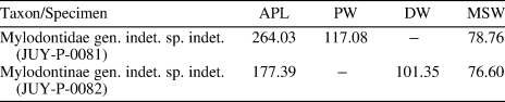
Measurements (in mm) of the femurs of Mylodontidae gen. indet. sp. indet. and Mylodontinae gen. indet. sp. indet. APL = maximum anteroposterior length; DW = distal width; MSW = midshaft width; PW = proximal width.

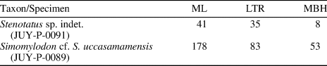
Measurements (in mm) of the mandibles of Stenotatus sp. indet. and Simomylodon cf. S. uccasamamensis. LTR = length of the tooth row; MBH = maximum height of mandibular body; ML = mandible length.

Repositories and institutional abbreviations
JUY-P = Museo de Geología, Mineralogía y Paleontología, Instituto de Geología y Minería, Universidad Nacional de Jujuy, San Salvador de Jujuy, Argentina; MLP = Museo de La Plata, La Plata, Buenos Aires, Argentina; UATF = Universidad Autónoma Tomás Frías, Potosí, Bolivia.
Other abbreviations
C/c = upper and lower caniniform tooth, respectively; masl = meters above sea level; Mf/mf = upper and lower molariform tooth, respectively; MMCO = Middle Miocene Climatic Optimum; SALMA = South American Land Mammal Age. Anatomical abbreviations discf, ectf, fibf, hrm, Mf/mf, odontf, peomc, and symph (see figure captions for definitions) are modified from Pujos et al. (Reference Pujos, De Iuliis, Mamani Quispe, Adnet, Andrade Flores, Billet and Antoine2016).
Systematic paleontology
Class Mammalia Linnaeus, Reference Linnaeus1758
Order Xenarthra Cope, Reference Cope1889
Suborder Cingulata Illiger, Reference Illiger1811
Family Dasypodidae Gray, Reference Gray1821
Subfamily Euphractinae Winge, Reference Winge1923
Tribe Eutatini Bordas, Reference Bordas1933
Genus Stenotatus Ameghino, Reference Ameghino1891b, p. 253
Type species
Stenotatus karaikensis Ameghino, Reference Ameghino1891b, p. 26.
Other species
Stenotatus patagonicus (Ameghino, Reference Ameghino1887), Stenotatus hesternus (Ameghino, Reference Ameghino1889), Stenotatus ornatus (Ameghino, Reference Ameghino1897), Stenotatus centralis (Ameghino, Reference Ameghino1902), and Stenotatus planus Scillato-Yané and Carlini, Reference Scillato-Yané and Carlini1998.
Occurrence
Basal section of the lower unit, Calahoyo Basin, Argentina.
Description
Osteoderms with large piliferous foramina on the posterior margin as in other members of the tribe Eutatini, a character that allows assignment to that tribe (Scillato-Yané, Reference Scillato-Yané1977, Reference Scillato-Yané1982; Carlini and Scillato-Yané, Reference Carlini and Scillato-Yané1999; Soibelzon et al., Reference Soibelzon, Miño-Boilini, Zurita and Krmpotic2010). Each osteoderm (Fig. 3.3) has one central figure on the exposed surface that occupies the entire caudal portion; the piliferous foramina form a single row and vary in number from four to six, as in Stenotatus, Doellotatus Bordas, Reference Bordas1932, and Chasicotatus Scillato-Yané, Reference Scillato-Yané1977. These features separate these remains from Ringueletia Reig, Reference Reig1959, in which the central figure is interrupted on the anterior portion by two lateral figures, and the piliferous foramina form two clearly differentiated rows (Bordas, Reference Bordas1933; Scillato-Yané, Reference Scillato-Yané1982; Scillato-Yané et al., Reference Scillato-Yané, Carlini, Vizcaíno, Ortiz-Jaureguizar, Alberdi, Leone and Tonni1995; Urrutia et al., Reference Urrutia, Montalvo and Scillato Yané2008). The surface of osteoderms JUY-P-0091 is similar to Stenotatus and Chasicotatus (Scillato-Yané et al., Reference Scillato-Yané, Krmpotic and Esteban2010), and differs from Doellotatus, Paraeutatus Scillato-Yané, Reference Scillato-Yané1980, and Proeutatus Ameghino, Reference Ameghino1891c, in which they are characterized by distinctive rugosity (Ameghino, Reference Ameghino1891d; Scillato-Yané, Reference Scillato-Yané1982; Urrutia et al., Reference Urrutia, Montalvo and Scillato Yané2008; González, Reference González2010). The area between the cranial and caudal portions (transverse depression) shows numerous rugosities and irregular furrows, as in Stenotatus, but is different from Chasicotatus in which this region is smooth (Scillato-Yané et al., Reference Scillato-Yané, Krmpotic and Esteban2010). This set of characters suggests that the specimen JUY-P-0091 belongs to the genus Stenotatus. Among the species described for this genus, some characters show similarity with Stenotatus planus, such as the number of piliferous foramina between four and six, unlike in Stenotatus ornatus, Stenotatus centralis, Stenotatus hesternus, and Stenotatus patagonicus, which have two to four large foramina; the longitudinal furrows have small external surface foramina, as in Stenotatus patagonicus and Stenotatus planus, but are not as pronounced as in Stenotatus hesternus. In some osteoderms, the furrows are barely marked as in Stenotatus planus but different from Stenotatus patagonicus in which these furrows are obliquely oriented with respect to the longitudinal axis of the lateral figure, dividing it into two smaller figures (González, Reference González2010). However, the comparative analysis of JUY-P-0091 suggests a strong morphological similarity with those osteoderms described by Croft et al. (Reference Croft, Carlini, Ciancio, Brandoni, Drew, Engelman and Anaya2016) (UATF-V-000869) and recognized as Eutatini indet. from the middle Miocene (ca. 16 Ma) of Cerdas, Bolivia. Both specimens share the following combination of characters: (1) a single row of large piliferous foramina (generally 4–6) along the posterior margin; (2) two longitudinal furrows and two pairs of transverse furrows that define an elongate central figure and two pairs of peripheral figures; and (3) small foramina in the longitudinal furrows (3 or 4 pairs). The material from Cerdas was originally assigned to Stenotatus planus by Croft et al. (Reference Croft, Anaya, Auerbach, Garzione and MacFadden2009). However, Croft et al. (Reference Croft, Carlini, Ciancio, Brandoni, Drew, Engelman and Anaya2016) reassigned the specimen UATF-V-000869 to Eutatini indet. Our observations suggest that both materials belong to Stenotatus and the combination of characters indicates that it corresponds to the same species.
Stenotatus sp. indet. (JUY-P-0091): (1, 2) left hemimandible in occlusal and lateral view, respectively; (3) movable osteoderms. a.mf = upper molariform tooth alveolus; Cp = caudal portion; Crp = cranial portion; esf = external surface foramina; hrm = horizontal ramus; mf = molariform tooth; mfor = mental foramen; pf = piliferous foramina; Td = transverse depression. Scale bars = 20 mm.

Hemimandible
The left hemimandible has a maximum length of 40 mm and its ventral side is slightly convex. The anterior margin of the coronoid process is at the level of the last lower molariform tooth, as in Ringueletia and Doellotatus, different from Proeutatus in which the coronoid process rises behind the mf10. In occlusal view, the horizontal ramus is narrow and thinning even more toward the symphysial region. The dental series is 33 mm long and formed by 10 molariforms with the long axis parallel to the dental series. The mf1–mf4 are small and laterally compressed, becoming larger from mf5–mf8. From mf9, the last two molariforms show gradual reduction. The six posterior molariforms (mf5–mf10) are subelliptical to subrectangular in occlusal view as in Stenotatus, Doellotatus, and Eutatus Gervais, Reference Gervais1867, unlike in Proeutatus, in which they are subtriangular to bilobate.
Materials
JUY-P-0091, left hemimandible and five mobile osteoderms, basal section of the lower unit, Calahoyo Basin, Jujuy Province, northwestern Argentina.
Remarks
Stenotatus has been recorded in Argentina from the Deseado Formation (Deseadan SALMA) in Santa Cruz and Chubut provinces (Ameghino, Reference Ameghino1897); Sarmiento Formation (Colhuehuapian SALMA), Chubut Province (Ameghino, Reference Ameghino1902); Chichinales Formation (Colhuehuapian SALMA), Río Negro Province (Barrio et al., Reference Barrio, Carlini and Goin1989); Santa Cruz Formation (Santacrucian SALMA), Santa Cruz Province (Scott, Reference Scott and Scott1903; Vizcaíno et al., Reference Vizcaíno, Bargo, Kay and Milne2006); Collón-Curá Formation (‘Colloncuran’ SALMA), Neuquén and Río Negro provinces (Roth, Reference Roth1898), and in the Santacrucian SALMA (17.5–16.3 Ma) and ‘Friasian’ (16.3–15.5 Ma) of Patagonia (González, Reference González2010). Outside of Argentina, Stenotatus is known from Cerdas, Bolivia (Miocene, 16.3–15.1 Ma) (Croft et al., Reference Croft, Anaya, Auerbach, Garzione and MacFadden2009, Reference Croft, Carlini, Ciancio, Brandoni, Drew, Engelman and Anaya2016) and the Chucal Formation (Santacrucian SALMA) of Chile (Croft et al., Reference Croft, Flynn and Wyss2004, Reference Croft, Flynn and Wyss2007).

Occurrence
Upper section of the lower unit, Calahoyo Basin, Jujuy Province, northwestern Argentina.
Description
Osteoderms are assigned to the tribe Eutatini because of the large piliferous foramina on the posterior margin (Scillato-Yané, Reference Scillato-Yané1977, Reference Scillato-Yané1982; Carlini and Scillato-Yané, Reference Carlini and Scillato-Yané1999; Soibelzon et al., Reference Soibelzon, Miño-Boilini, Zurita and Krmpotic2010) and the central figure being wider than in Euphractini (Scillato-Yané, Reference Scillato-Yané1982). Each osteoderm has a central figure on the exposed surface that occupies the whole caudal portion as in Stenotatus, Doellotatus, and Chasicotatus, but differs from Ringueletia in which the central figure is interrupted on the anterior part by two lateral figures (Bordas, Reference Bordas1933; Scillato-Yané, Reference Scillato-Yané1982; Scillato-Yané et al., Reference Scillato-Yané, Carlini, Vizcaíno, Ortiz-Jaureguizar, Alberdi, Leone and Tonni1995; Urrutia et al., Reference Urrutia, Montalvo and Scillato Yané2008). Three or four furrows, oblique to the longitudinal axis of the lateral figure, divide it into four or five smaller figures. The presence of furrows and lateral figures are characters that distinguish JUY-P-0171 from Stenotatus, Doellotatus, Ringueletia, Chasicotatus, Paraeutatus, and Proeutatus. The piliferous foramina (two to four) are arranged in a single row on the posterior margin, different from those of Doellotatus, Ringueletia, Chasicotatus, Paraeutatus, and Proeutatus. The osteoderms of JUY-P-0171 have a smooth surface as in Stenotatus and Chasicotatus (Scillato-Yané et al., Reference Scillato-Yané, Krmpotic and Esteban2010), different from those of Doellotatus, Paraeutatus, Proeutatus, and Ringueletia in which the surface is very rugose (Ameghino, Reference Ameghino1891d; Scillato-Yané, Reference Scillato-Yané1982; Urrutia et al., Reference Urrutia, Montalvo and Scillato Yané2008; González, Reference González2010). The transverse depression is very reduced and has numerous rugosities, as in Stenotatus, but differs from that of Chasicotatus, in which this region is smooth (Scillato-Yané et al., Reference Scillato-Yané, Krmpotic and Esteban2010).
Materials
JUY-P-0171, fragment of dorsal carapace, three rows of mobile osteoderms, upper section of the lower unit, Calahoyo Basin, Jujuy Province, northwestern Argentina.
Remarks
This specimen presents the following combination of characters: (1) a single row of large piliferous foramina (generally 2–4) along the posterior margin, and (2) two longitudinal furrows and three or four pairs of transverse furrows that define an elongate central figure and four or five pairs of peripheral figures. Therefore, the specimen JUY-P-0171 differs from other genera already recognized in the Eutatini. The combination of observed characters indicates that this material could correspond to a new taxon within the Eutatini. Further studies will corroborate this taxonomic hypothesis.
Tribe Euphractini Winge, Reference Winge1923
Genus Macrochorobates Scillato-Yané, Reference Scillato-Yané1980
Type species
Proeuphractus scalabrinii Moreno and Mercerat, Reference Moreno and Mercerat1891.
Other species
Macrochorobates chapalmalensis (Ameghino, Reference Ameghino1908).
Macrochorobates scalabrinii (Moreno and Mercerat, Reference Moreno and Mercerat1891)
Figure 5; Table 1
- Reference Moreno and Mercerat1891
Proeuphractus scalabrinii Moreno and Mercerat, p. 226.
- Reference Scillato-Yané1980
Macrochorobates Scalabrinii, Scillato-Yané, p. 26.
Macrochorobates scalabrinii (JUY-P-0090): (1) movable osteoderms of the dorsal shield; (2) pelvic buckler osteoderms of the dorsal shield; (3) fixed osteoderm from the dorsal shield; (4) cephalic shield osteoderms; and (5) caudal sheath osteoderms. Cp = caudal portion; Crp = cranial portion; esf = external surface foramina; pf = piliferous foramina; Td = transverse depression. Scale bars = 20 mm.

Holotype
MLP? lost (Mones, Reference Mones1986), osteoderms of the dorsal carapace, Valle de Santa María, Catamarca Province, Argentina; ‘Araucanian’ (late Miocene–early Pliocene).
Occurrence
Upper section of the lower unit, Calahoyo Basin, Argentina.
Description
Osteoderms of the cephalic shield are rectangular to rhomboidal; their exposed surfaces are convex and smooth. Mobile osteoderms of the dorsal carapace are rectangular and anteroposteriorly long. Each osteoderm has a cranial portion in the form of a key. The transverse depression is evidently rugose. On the caudal portion, a central elevated figure is distinguished, with a lateral deviation toward the external margin on the distal portion, as in Macrochorobates and Chorobates Reig, Reference Reig1959, but different from the flat, posteriorly elevated central figure of Paraeuphractus Scillato-Yané, Reference Scillato-Yané1975, and the completely flat one in Euphractus Wagler, Reference Wagler1830 (Scillato-Yané, Reference Scillato-Yané1982). This central figure is delimited by two wide furrows and has numerous small external surface foramina as in Macrochorobates and Chorobates (Scillato-Yané, Reference Scillato-Yané1982; Carlini and Scillato-Yané, Reference Carlini and Scillato-Yané1995; Urrutia et al., Reference Urrutia, Montalvo and Scillato Yané2008). The lateral figures are less elevated than the central one, and are slightly flat as in Macrochorobates (Castellanos, Reference Castellanos1947), Euphractus, and Paraeuphractus, unlike those of Chorobates and Proeuphractus in which the lateral figures are convex and higher than the central figure (Scillato-Yané, Reference Scillato-Yané1982). Some osteoderms display shallow furrows oblique to the longitudinal axis of the lateral figure, which divide it into two smaller figures, anterior and posterior, as in Macrochorobates (Moreno and Mercerat, Reference Moreno and Mercerat1891; Contreras et al., Reference Contreras, Tomassini, Perez and Oliva2013), Chorobates (Carlini and Scillato-Yané, Reference Carlini and Scillato-Yané1995), Proeuphractus Ameghino, Reference Ameghino1886 (Esteban et al., Reference Esteban, Nasif, Montalvo and Visconti2003; Urrutia et al., Reference Urrutia, Montalvo and Scillato Yané2008), and Macroeuphractus Ameghino, Reference Ameghino1887 (Scillato-Yané, Reference Scillato-Yané1982; Urrutia et al., Reference Urrutia, Montalvo and Scillato Yané2008). The marginal foramina form a single row on the posterior and both lateral margins, as in Proeuphractus (Scillato-Yané, Reference Scillato-Yané1982; Esteban et al., Reference Esteban, Nasif and Montalvo2001; Tauber, Reference Tauber2005; Urrutia et al., Reference Urrutia, Montalvo and Scillato Yané2008) and Macroeuphractus (Mones and Mehl, Reference Mones and Mehl1990). In Chorobates, the posterior and external lateral margins each bear a single row of piliferous foramina (Scillato-Yané, Reference Scillato-Yané1982; Carlini and Scillato-Yané, Reference Carlini and Scillato-Yané1995), whereas in Paraeuphractus, there is a double row of perforations exclusively on the posterior margin (Scillato-Yané, Reference Scillato-Yané1982). Fixed osteoderms are subhexagonal. On the exposed surface, there is a central elongated figure, slightly convex, with deviation toward the external lateral margin, as in Chorobates, Macrochorobates, and Proeuphractus, but different from Euphractus in which the figure is flat without deviation (Scillato-Yané, Reference Scillato-Yané1982). The central figure is surrounded by slightly convex peripheral figures as in Macrochorobates, but different from the completely flat figures of Euphractus and Paraeuphractus, and the very convex and globose figures of Proeuphractus and Chorobates (Scillato-Yané, Reference Scillato-Yané1982). The peripheral figures are limited by furrows with numerous foramina as in Macrochorobates, Chorobates, and Proeuphractus (Scillato-Yané, Reference Scillato-Yané1982; Carlini and Scillato-Yané, Reference Carlini and Scillato-Yané1995; Urrutia et al., Reference Urrutia, Montalvo and Scillato Yané2008). The marginal foramina are numerous on a single row on the posterior and both lateral margins, as in Macrochorobates (Contreras et al., Reference Contreras, Tomassini, Perez and Oliva2013) and Proeuphractus (Scillato-Yané, Reference Scillato-Yané1982; Esteban et al., Reference Esteban, Nasif and Montalvo2001; Urrutia et al., Reference Urrutia, Montalvo and Scillato Yané2008), but differ from Chorobates in which the foramina are on the posterior and external lateral margins (Scillato-Yané, Reference Scillato-Yané1982; Carlini and Scillato-Yané, Reference Carlini and Scillato-Yané1995) and from Paraeuphractus in which the foramina are numerous, form a double row, and are present on the posterior margin (Scillato-Yané, Reference Scillato-Yané1982). Semimobile osteoderms of the pelvic shield are similar in morphology to mobile osteoderms; numerous piliferous foramina in a single row are present on the posterior and both lateral margins. Finally, osteoderms of the caudal sheath have smooth exposed surfaces and vary from rhomboidal to pentagonal; each has one or two foramina on its posterior region.
This set of characters allows the assignment of JUY-P-0090 to the genus Macrochorobates. Concerning the specific assignment, the ornamentation of the osteoderms of Macrochorobates scalabrinii and Macrochorobates chapalmalensis is similar, however, some differences justifying assignment to the former can be recognized. Osteoderms of JUY-P-0090 and JUY-P-0033 (previously described by Zurita et al., Reference Zurita, Camacho, Miño-Boilini, Candela, Cuadrelli, Krmpotic and Solís2017) differ from Macrochorobates chapalmalensis in: (1) larger size; (2) the central figure of mobile osteoderms have a less acute posterior angle; and (3) the more convex peripheral figures of the fixed osteoderms. As reported by Moreno and Mercerat (Reference Moreno and Mercerat1891) and Contreras et al. (Reference Contreras, Tomassini, Perez and Oliva2013), two other characteristics of Macrochorobates scalabrinii observed in this material are: (1) in some mobile osteoderms, very shallow, barely distinguishable furrows divide the lateral figures into two parts; and (2) piliferous foramina are present on the posterior and both lateral margins, are variable in number, and form a single row.
Materials
JUY-P-0090, three osteoderms from the cephalic shield, eight from mobile bands, two fixed osteoderms, two osteoderms from the pelvic shield, and two osteoderms from the caudal sheath.
Remarks
Macrochorobates scalabrinii has been recorded in the Andalhuala Formation, Catamarca Province (Marshall and Patterson, Reference Marshall and Patterson1981; Bossi et al., Reference Bossi, Ovejero and Strecker1987; Esteban and Nasif, Reference Esteban and Nasif1996; Esteban et al., Reference Esteban, Nasif and Montalvo2001). Radiometric dating for this unit estimated the age to be between 7.14 and 5.59 Ma (Marshall and Patterson, Reference Marshall and Patterson1981; Latorre et al., Reference Latorre, Quade and McIntosh1997; Reguero and Candela, Reference Reguero, Candela, Salfity and Marquilas2011; Bonini et al., Reference Bonini, Schmidt, Reguero, Cerdeño, Candela and Solís2017). This species has also been cited for the Epecuén Formation (Huayquerian SALMA), Buenos Aires Province (Scillato-Yané, Reference Scillato-Yané1980, Reference Scillato-Yané1982); Cerro Azul Formation (Huayquerian SALMA), La Pampa Province (Esteban et al., Reference Esteban, Nasif and Montalvo2001; Urrutia et al., Reference Urrutia, Montalvo and Scillato Yané2008); Salicas Formation, La Rioja Province (Tauber, Reference Tauber2005; Brandoni et al., Reference Brandoni, Schmidt, Candela, Noriega, Brunetto and Fiorelli2012); and Las Flores Formation, San Juan Province (Contreras, Reference Contreras2007; Contreras et al., Reference Contreras, Tomassini, Perez and Oliva2013) in which the fossil-bearing level is between 6.9 and 6.3 Ma (Ré et al., Reference Ré, Jordan and Kelley2003). According to Tonni et al. (Reference Tonni, Scillato-Yané, Cione and Carlini1998) and Cione and Tonni (Reference Cione, Tonni, de Barrio, Etcheverry and Caballé y E. Llambias2005), the occurrence of Macrochorobates scalabrinii in the sediments overlying the Arroyo Chasicó Formation, Buenos Aires Province, allowed assignment of these levels to the Macrochorobates scalabrinii Biozone as the biostratigraphic base for the early Huayquerian Age/Stage. However, it should be noted that there are no known remains of this species in any other unit containing a Chasicoan fauna, the absence of which others have interpreted as a real pattern and not bias in the record (Verzi et al., Reference Verzi, Montalvo and Deschamps2008; Contreras et al., Reference Contreras, Tomassini, Perez and Oliva2013).
Suborder Tardigrada Latham and Davies in Forster, Reference Latham, Davies and Forster1795
Superfamily Mylodontoidea Gill, Reference Gill1872
Family Mylodontidae Gill, Reference Gill1872
Mylodontidae gen. indet. sp. indet.
Figure 6; Table 2
Mylodontidae gen. indet. sp. indet., distal portion of right femur (JUY-P-0081): (1) anterior view; (2) posterior view. lc = lateral condyle; le = lateral epicondyle; mc = medial condyle; me = medial epicondyle; pt = patellar trochlea. Scale bar = 20 mm.

Occurrence
Upper member of the upper unit (Tafna Formation sensu Zurita et al., Reference Zurita, Camacho, Miño-Boilini, Candela, Cuadrelli, Krmpotic and Solís2017), Casira Basin, Argentina.
Description
Femur. Only the distal half, anteroposteriorly compressed, is preserved. The anterior side is flat, except for a shallow depression limited distally by the patellar facet, which, together with the tibial facet, forms a continuous surface. The patellar facet is trochlea-shaped, formed by two, slightly convex articular facets that are connected through a slightly concave region to the medial portion. The posterior side is mainly flat, characterized by a higher region related to the popliteal fascia, near the intercondylar fossa. Both margins of the femur (medial and lateral) are almost parallel to slightly concave, as in Mylodontidae. On the distal extremity, the articular surfaces of the condyles and the patellar facet are connected by the articular isthmus, as in most Mylodontinae, Scelidotheriinae, and the megatheriine Megathericulus patagonicus Ameghino, Reference Ameghino1904 (Esteban, Reference Esteban1996; De Iuliis et al., Reference De Iuliis, Brandoni and Scillato-Yané2008; Pitana, Reference Pitana2011; Miño-Boilini, Reference Miño-Boilini2012). This anatomy differs from other Megatheriinae in which the trochlea is continuous with the lateral articular facet (De Iuliis et al., Reference De Iuliis, Brandoni and Scillato-Yané2008) and Nothrotheriidae and some Megalonychidae (separated by nonarticular bone; Brandoni, Reference Brandoni2009; De Iuliis et al., Reference De Iuliis, Gaudin and Vicars2011; Brandoni and McDonald, Reference Brandoni and McDonald2015). The intercondylar fossa is deep; the medial condyle is pyriform in shape, whereas the lateral condyle is quadrangular and smaller than the medial condyle.
Materials
JUY-P-0081, distal fragment of a right femur.
Remarks
The morphological evidence clearly suggests that the Casira material belongs to the family Mylodontidae. However, the poor preservation of the fossil precludes a more precise identification.

Occurrence
Upper section of the upper unit (Tafna Formation, sensu Zurita et al., Reference Zurita, Camacho, Miño-Boilini, Candela, Cuadrelli, Krmpotic and Solís2017), Casira Basin, Argentina.
Description
Femur. Only the posterior face of the proximal epiphysis and part of the diaphysis are preserved. Proximally, the head of the femur is poorly preserved, and it is not possible to determine whether the ‘fovea capitis’ was present. In posterior view, the greater trochanter is subtriangular in contour, as in some Mylodontinae (e.g., Glossotherium Owen, Reference Owen1840, Paraglossotherium Esteban, Reference Esteban1993, and Paramylodon Brown, Reference Brown1903), whereas the trochanteric fossa is well developed as in Glossotherium and Paramylodon. Distally, the base of the greater trochanter is continuous with the diaphysis, without developing a third trochanter (Esteban, Reference Esteban1996). The lesser trochanter is 23 mm from the distal margin of the femoral head; it is elliptical with the longest axis vertical, with a convex surface. Distal to the lesser trochanter, the medial margin of the diaphysis becomes concave, whereas the lateral margin is almost straight, with a smooth convexity at the middle-distal sector of the diaphysis, a condition observed in some Mylodontinae, such as Mylodon Owen, Reference Owen1840, Paraglossotherium, and Paramylodon. In summary, the presence of: (1) a well-developed and subtriangular major trochanter; (2) the development of the intertrochanteric fossa; and (3) the absence of third trochanter are characters found in almost all genera of Mylodontinae previously mentioned (Esteban, Reference Esteban1996). The poor preservation of the femur hampers generic and specific assignment of this material.
Materials
JUY-P-0082, proximal two-thirds of a right femur.
Remarks
The morphological evidence clearly suggests that the Casira material belongs to the family Mylodontidae. However, the poor preservation of the fossil precludes a more precise identification.
Genus Simomylodon Saint-André et al., Reference Saint-André, Pujos, Cartelle, De Iuliis, Gaudin, McDonald and Mamani Quispe2010
Type species
Simomylodon uccasamamensis Saint-André et al., Reference Saint-André, Pujos, Cartelle, De Iuliis, Gaudin, McDonald and Mamani Quispe2010
cf. Simomylodon sp. indet.
Figure 8
cf. Simomylodon sp. indet. (JUY-P-0173): (1, 2) posterior portion of skull, in lateral and dorsal view, respectively; (3) palate in ventral view. a.C = upper caniniform tooth alveolus; a.Mf = upper molariform tooth alveolus; fr = frontal; fss = frontosquamosal suture; par = parietal; pss = parietosupraoccipital suture; sq = squamosal; zyg.sq = zygomatic process of squamosal. Arrow indicates posteroanterior orientation. Scale bars = 20 mm.

Occurrence
Upper section of the upper unit (Tafna Formation, sensu Zurita et al., Reference Zurita, Camacho, Miño-Boilini, Candela, Cuadrelli, Krmpotic and Solís2017), Casira Basin, Argentina.
Description
Skull. The incomplete posterior fragment of a skull belongs to a juvenile based on the unfused sutures. In dorsal view, the parietal-supraoccipital and frontal-squamosal sutures are evident; the latter is at the level of the anterior margin of the squamosal, as observed by Esteban (Reference Esteban1996) in Mylodontinae. The right squamosal is preserved, with a fragment of the zygomatic apophysis. In lateral view, the suture between squamosal and parietal is horizontal.
The preserved portion of the palate extends from the posterior margin of the C alveolus to the anterior margin of the Mf4 alveolus. The palatine surface shows numerous vascular foramina. The general morphology of this region, the portion of the alveoli Mf2 and Mf3, and the absence of diastema between C and Mf1 are similar to those of Simomylodon.
Materials
JUY-P-0173, posterior portion of the skull.
Remarks
The Casira material almost certainly is referrable to the genus Simomylodon. Nevertheless, a definitive identification cannot be made confidently at the present time because of the limited material available for study.
Simomylodon cf. S. uccasamamensis Saint-André et al., Reference Saint-André, Pujos, Cartelle, De Iuliis, Gaudin, McDonald and Mamani Quispe2010
Figures 9, 10; Table 3
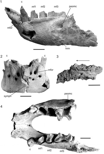
Simomylodon cf. S. uccasamamensis (JUY-P-0089): (1, 2, 4) dentary in lateral, anterior, and occlusal views, respectively; (3) palate in ventral view. c = caniniform; hrm = horizontal ramus; mf = lower molariform tooth; mfor = mental foramen; peomc = posteroexternal opening of mandibular canal; symph = symphysis. Arrow indicates posteroanterior orientation. Scale bars = 20 mm.

Simomylodon cf. S. uccasamamensis (JUY-P-0089): (1–3) left astragalus: (1) medial view (dorsal at top, anterior at left); (2) lateral view (posterior upward); (3) plantar view (posterior to the right); (4) osteoderm in inner and external view, respectively. discf = discoid facet; ectf = ectal facet; fibf = fibular facet; odontf = odontoid facet; sulct = sulcus tali; tib = tibial. Scale bars = 20 mm.

Occurrence
Upper section of the upper unit (Tafna Formation, sensu Zurita et al., Reference Zurita, Camacho, Miño-Boilini, Candela, Cuadrelli, Krmpotic and Solís2017), Casira Basin, Argentina.
Description
Palate. The preserved portion of palate extends from the posterior margin of the Mf1 alveolus to the anterior margin of the Mf4 alveolus. The palate is subtriangular with the tooth rows diverging anteriorly. The palatine surface is rugose with numerous vascular foramina, as in Pleurolestodon Rovereto, Reference Rovereto1914 (Kraglievich, Reference Kraglievich1923) and Simomylodon (Saint-André et al., Reference Saint-André, Pujos, Cartelle, De Iuliis, Gaudin, McDonald and Mamani Quispe2010; Boscaini et al., Reference Boscaini, Gaudin, Mamani Quispe, Münch, Antoine and Pujos2019).
Mandible
In lateral view, the anterior portion of the horizontal ramus of the specimen JUY-P-0089 is preserved. The ventral margin is approximately straight behind the level of the mf2. Although the coronoid process is not preserved, it can be seen that the anterior margin of this process begins behind the level of the posterior margin of the mf3, as in all Mylodontidae except for Mylodon (Esteban, Reference Esteban1996). Only one posterior aperture of the mandibular canal is preserved; it is below the occlusal plane, near the anterior margin of the coronoid process and perpendicular to the axis of the posterior margin of the mf3 (as in Mylodontidae, except Mylodon and Ranculcus Ameghino, Reference Ameghino1891a). In lateral view, the symphysis is sigmoid, convex at the level of the mental foramina, and concave between this level and the symphyseal margin as in Simomylodon (Boscaini et al., Reference Boscaini, Gaudin, Mamani Quispe, Münch, Antoine and Pujos2019). In occlusal view, the posterior margin of the mandibular symphysis is U-shaped.
Dentition
The complete dentition is preserved for the left mandibular ramus and the first and last element for the right. As in Mylodontinae, the dental formula is 1c and 3mf. The dentition is heterodont, with a pair of caniniforms (except Mylodon) and three pairs of molariforms mf1 to mf3. There is a marked anterior divergence of the dental series as in Pleurolestodon acutidens (Kraglievich, Reference Kraglievich1921) and Simomylodon uccasamamensis (see Boscaini et al., Reference Boscaini, Gaudin, Mamani Quispe, Münch, Antoine and Pujos2019). The caniniform is proportionally smaller than the molariforms, as in Glossotheridium, Pleurolestodon, and Simomylodon. It is subtriangular in cross section, with a more concave lingual surface (as in Pleurolestodon acutidens and in some of the Simomylodon specimens described by Boscaini et al., Reference Boscaini, Gaudin, Mamani Quispe, Münch, Antoine and Pujos2019) than in other Mylodontinae (Kraglievich, Reference Kraglievich1921). The caniniform is not separated from mf1 by a diastema, as in Pleurolestodon acutidens (Kraglievich, Reference Kraglievich1921; Oliva and Brandoni, Reference Oliva and Brandoni2012) and Simomylodon uccasamamensis (see Boscaini et al., Reference Boscaini, Gaudin, Mamani Quispe, Münch, Antoine and Pujos2019). The mf1 is elliptical in cross section, with a lingual inclination on its posterior half. Its anterolingual face is slightly concave, whereas the posterior one is convex as in Glossotheridium, Pleurolestodon, and Simomylodon (Saint-André et al., Reference Saint-André, Pujos, Cartelle, De Iuliis, Gaudin, McDonald and Mamani Quispe2010; Boscaini et al., Reference Boscaini, Gaudin, Mamani Quispe, Münch, Antoine and Pujos2019). In occlusal view, the mf2 is subrectangular to rhomboidal in section, anterolabially and posterolingually oriented as in Glossotherium, Pleurolestodon, Promylodon Ameghino, Reference Ameghino1883, Ranculcus, and Simomylodon (Saint-André et al., Reference Saint-André, Pujos, Cartelle, De Iuliis, Gaudin, McDonald and Mamani Quispe2010). The mf3 is bilobed, typical of Mylodontoidea, anteroposteriorly longer than transversally, typical of Mylodontinae as in Pleurolestodon, Simomylodon, Glossotherium, Glossotheridium Kraglievich, Reference Kraglievich1934, Kiyumylodon Rinderknecht, Perea and McDonald, Reference Rinderknecht, Perea and McDonald2007, and Mylodon (Webb, Reference Webb, Redford and Eisenberg1989). On the external and internal faces there are well-defined furrows; the labial face is wide and shallow, whereas the lingual face is shorter and very deep. Both furrows limit a marked constriction in the middle of the tooth, defining the anterior and posterior lobes. The anterior lobe is subtriangular in cross section, with the anterior face flat or concave, as in Pleurolestodon, Ranculcus, Simomylodon, Glossotherium, Glossotheridium, Mylodon, and Kiyumylodon. The posterior lobe is also subtriangular but has a greater projection lingually, as in Pleurolestodon, Simomylodon, and in Mylodontinae in general (Esteban, Reference Esteban1996; Boscaini et al., Reference Boscaini, Gaudin, Mamani Quispe, Münch, Antoine and Pujos2019).
Astragalus
In dorsal view, the discoid facet and the basal margin of the odontoid facet are preserved. In lateral view, the fibular facet extends from the anterolateral margin of the discoid facet up to the ectal facet, as in Simomylodon, forming the articular surface of the fibula. In plantar view, the ‘sulcus tali’ separates the ectal facet and the sustentacular facet for the calcaneus, as in Scelidotheriinae, Pseudoprepotherium Hoffstetter, Reference Hoffstetter1961, Thinobadistes Hay, Reference Hay1919, Lestodon Gervais, Reference Gervais1855, Pleurolestodon, and Simomylodon. This feature has been considered by several authors as the primitive condition for Mylodontidae (Ameghino, Reference Ameghino1907; Kraglievich, Reference Kraglievich1923; Saint-André et al., Reference Saint-André, Pujos, Cartelle, De Iuliis, Gaudin, McDonald and Mamani Quispe2010).
Dermal ossicles
A single, small, subtriangular ossicle with rounded angles is preserved. It is 17 mm long (maximum) and 14 mm wide. The external surface is rugose, with irregularly arranged foramina, whereas the internal surface, recognized by its interwoven texture according to the criteria of Ricqlès et al. (Reference Ricqlès, Suberbiola, Gasparini and Olivero2001), is more even. In general, these structures are very similar to those described for other Mylodontidae, e.g., Glossotherium chapadmalense Kraglievich, Reference Kraglievich1925, Mylodon darwinii Owen, Reference Owen1840, and Paramylodon harlani (Owen, Reference Owen1840) (Sinclair, Reference Sinclair1910; Hill, Reference Hill2006; Haro et al., Reference Haro, Tauber and Krapovickas2016; McDonald, Reference McDonald2017).
Materials
JUY-P-0089, portions of the palate, mandible, and left astragalus, seven fragmentary dorsal vertebrae, and one dermal ossicle.
Remarks
Simomylodon is monospecific and is known from the late Miocene (or early Pliocene) to the late Pliocene from the Bolivian Altiplano, central South America (Saint-André et al., Reference Saint-André, Pujos, Cartelle, De Iuliis, Gaudin, McDonald and Mamani Quispe2010; Boscaini et al., Reference Boscaini, Gaudin, Mamani Quispe, Münch, Antoine and Pujos2019). This represents the southernmost record of the genus and the first record of this taxon in Argentina.
Discussion
The new records presented in this contribution from Neogene sediments of the Argentine Puna significantly increase the diversity of the xenarthra in a poorly known region. The previously known xenarthran diversity of the Puna came from Calahoyo (Zurita et al., Reference Zurita, Camacho, Miño-Boilini, Candela, Cuadrelli, Krmpotic and Solís2017), which included Pyramiodontherium bergi (Tardigrada, Megatheriinae) and Macrochorobates scalabrinii (Cingulata, Dasypodidae) and Eosclerocalyptus Ameghino, Reference Ameghino1919 (Cingulata, Glyptodontidae). On this basis, the age proposed by the authors for the fossil-bearing levels was estimated as late Miocene–Pliocene.
The additional taxa reported in this contribution increase the taxonomic diversity and extend the temporal range of the fossil-bearing levels to the middle Miocene. From a biochronologic and paleobiogeographic standpoint, the presence of the dasypodid Stenotatus in the most basal levels cropping out in Calahoyo is very significant. The biochron of this genus extends from the Santacrucian SALMA (17.5–16.3 Ma) to the ‘Friasian’ SALMA (16.3–15.5 Ma; González, Reference González2010). There is a close morphological similarity with the material assigned to Eutatini n. gen. n. sp. by Croft et al. (Reference Croft, Carlini, Ciancio, Brandoni, Drew, Engelman and Anaya2016) from Cerdas, southern Bolivia, which has radiometric dates of 16.3 and 15.1 Ma (early–middle Miocene; MacFadden et al., Reference MacFadden, Anaya and Swisher1995; Madden et al., Reference Madden, Guerrero, Kay, Flynn, Swisher, Walton, Kay, Madden, Cifelli and Flynn1997; Croft et al., Reference Croft, Anaya, Auerbach, Garzione and MacFadden2009). Our observations reveal, however, that materials from both Cerdas and Calahoyo must be interpreted as Stenotatus sp. indet. In Calahoyo, the shared presence of Stenotatus along with the rodents (currently under study) collected in the same lower levels and tentatively assigned to Prolagostomus Ameghino, Reference Ameghino1887 or Pliolagostomus Ameghino, Reference Ameghino1887, indicate paleofaunistic similarity to that recorded in Cerdas. This evidence suggests a middle Miocene age for the lower levels of Calahoyo. Croft et al. (Reference Croft, Carlini, Ciancio, Brandoni, Drew, Engelman and Anaya2016) interpreted that the paleofauna of Cerdas would have developed in humid subtropical environments coeval with the end of the MMCO (Zachos et al., Reference Zachos, Pagani, Sloan, Thomas and Billups2001, Reference Zachos, Dickens and Zeebe2008). In agreement with these observations, phytolith analyses for the basal levels of Calahoyo support this interpretation based on the presence of phytoliths related to tropical/subtropical plants (unpublished data, S. Contreras, 2019). The MMCO occurred at ca. 15 Ma and represents a midlatitude warming event with ~ 6°C relative temperature higher than present (You et al., Reference You, Huber, Müller, Poulsen and Ribbe2009). Farther south, in other early–middle Miocene localities (Anta and San José formations) of northwestern Argentina (Tucumán and Salta provinces), Anzótegui et al. (Reference Anzótegui, Mautino, Garralla, Herbst, Robledo, Horn, Muruaga and Grosse2017) described similar paleoclimatic conditions, represented by heterogeneous vegetation paleocommunities that indicate warm and humid, but seasonally dry, environments. This vegetation extended to the south, with tropical elements found in Patagonia (Barreda et al., Reference Barreda, Anzótegui, Prieto, Aceñolaza, Bianchi, Borromei, Brea, Caccavari, Cuadrado, Garralla, Grill, Guerstein, Lutz, Mancini, Mautino, Ottone, Quattrocchio, Romero, Zamaloa and Zucol2007).
Thus, the evidence suggests that the basal section of the lower unit exposed in the Calahoyo Basin would have been deposited under very different environmental conditions than the current ones, which is a cold and dry climate (with an average of 10°C and with annual rainfall between 100 and 800 mm; Matteucci, Reference Matteucci, Morello, Matteucci, Rodriguez and Silva2012). This implies tropical/subtropical humid environments.
The record of the Euphractini Macrochorobates scalabrinii in levels overlying those bearing Stenotatus sp. indet. suggests that the sequences of the lower sedimentary unit present at Calahoyo is middle–late Miocene in age. Our observations indicate that the materials assigned by Zurita et al. (Reference Zurita, Camacho, Miño-Boilini, Candela, Cuadrelli, Krmpotic and Solís2017) to Macrochorobates chapadmalensis (Euphractini) belong instead to Macrochorobates scalabrinii. This reassignment, in agreement with the records of Pyramiodontherium bergi (Tardigrada, Megatheriinae), allows us to assign a late Miocene age with certainty to the lower member of the unit interpreted as Tafna Formation by these authors. From a tectonic and paleoenvironmental standpoint, during this period the uplift of the Andes Cordillera contributed to decrease of temperatures and more arid conditions (Barreda et al., Reference Barreda, Anzótegui, Prieto, Aceñolaza, Bianchi, Borromei, Brea, Caccavari, Cuadrado, Garralla, Grill, Guerstein, Lutz, Mancini, Mautino, Ottone, Quattrocchio, Romero, Zamaloa and Zucol2007). In this interval, northwestern Argentina had neotropical plant taxa, and vegetation was characterized by a balance between xeric herbaceous-shrubby communities and hydrophilous forests (Barreda et al., Reference Barreda, Anzótegui, Prieto, Aceñolaza, Bianchi, Borromei, Brea, Caccavari, Cuadrado, Garralla, Grill, Guerstein, Lutz, Mancini, Mautino, Ottone, Quattrocchio, Romero, Zamaloa and Zucol2007). This variety of paleocommunities would have developed under a warm climate with areas of dry season (Anzótegui et al., Reference Anzótegui, Mautino, Garralla, Herbst, Robledo, Horn, Muruaga and Grosse2017). In this scenario, it has been interpreted that the dasypodid Macrochorobates scalabrinii was adapted to temperate-warm conditions and open, semiarid environments with the development of grasslands or partially forested areas (Contreras et al., Reference Contreras, Tomassini, Perez and Oliva2013). The observed combination of characters in the material interpreted as Eutatini indet. from the upper levels of Calahoyo suggests that this could belong to a new taxon, which could be an endemic taxon to this part of the Puna of South America. Although there are no palaeoecological studies related to the tardigrade Pyramiodontherium, the record of this Megatheriinae suggests the development of forested areas (Scillato-Yané et al., Reference Scillato-Yané, Carlini, Vizcaíno, Ortiz-Jaureguizar, Alberdi, Leone and Tonni1995). Both taxa found in the upper levels of Calahoyo (Macrochorobates scalabrinii and Pyramiodontherium bergi) suggest climatic and environmental conditions different from those of the basal levels, with the development of more xeric conditions and with temperatures a bit colder than the lower levels of Calahoyo. It is noteworthy that these are the northernmost records of both species. In summary, the sedimentary sequences of Calahoyo suggest a middle–late Miocene age, probably involving the end of the MMCO.
In Casira, the xenarthran diversity was previously considered completely different from that of Calahoyo. They are restricted to the tardigrade mylodontid Mylodontinae gen. indet. sp. indet., and the mylodontines cf. Simomylodon and Simomylodon cf. S. uccasamamensis. The latter taxon shows similarities with Simomylodon uccasamamensis, a species whose geographic and stratigraphic distribution was previously restricted to localities in Bolivia (Boscaini et al., Reference Boscaini, Gaudin, Mamani Quispe, Münch, Antoine and Pujos2019) and known from the late Miocene to late Pliocene, with a temporal extension from 5.3–2.6 Ma (Boscaini et al., Reference Boscaini, Gaudin, Mamani Quispe, Münch, Antoine and Pujos2019). Associated with these last remains, octodontid rodents related to dryer climates (currently under study) were found.
An interesting subject is the first certain record of a dermal ossicle associated with the materials assigned to Simomylodon (JUY-P-0089); this represents the oldest record of this structure in sloths. The previous record was presented by Nasif et al. (Reference Nasif, Esteban and Georfieff2008) from ‘Araucanian’ levels of northwestern Argentina. The material was assigned to Mylodontinae gen. indet. sp. indet. because they were found isolated.
Conclusions
The Xenarthra exhumed from Calahoyo and Casira suggest that the sedimentation of the fossil-bearing units occurred during the interval from the middle–late Miocene/Pliocene. The lower levels of Calahoyo are similar from a paleofaunal standpoint to Cerdas (southern Bolivia), which probably lived at the end of the MMCO. In support of this hypothesis, preliminary analyses of phytolith assemblages indicate a humid, warm environment. In turn, the presence of Macrochorobates scalabrinii in the upper levels of Calahoyo indicates the presence of faunal elements common to the ‘Araucanian’ (late Miocene) communities of northwestern Argentina. At Casira, the only known xenarthran is Simomylodon cf. S. uccasamamensis. What is remarkable is the absence of Cingulata, despite the strong sampling efforts, and the high diversity of this group observed in Calahoyo. This difference in paleofaunal composition suggests environmental conditions that were not very favorable for Cingulata but highly favorable for Tardigrada, given the remarkable frequency of this clade in Casira.
In summary, this contribution is particularly significant because it not only increases the knowledge of the diversity of Puna xenarthrans but also documents a new locality for the middle Miocene of South America, previously recognized mainly in Argentine Patagonia and in a few other localities from South America.
Acknowledgments
The authors want to express gratitude to the authorities and people of Cieneguillas, Calahoyo, Casira, and Santa Catalina for their hospitality during fieldwork; E. Silva de Cruz of the Instituto de Geología y Minera de la Universidad Nacional de Jujuy for providing us with access to the geological bibliography of the province; and P. Cuaranta and M. Claros for participating in field work. We also thank the Secretaría de Cultura of Jujuy Province for the authorization. Both reviewers (Gillette and Tambuso) and the Editor (McDonald) are also thanked for their helpful suggestions that greatly improved this contribution. This contribution was partially supported by grants PI Q003/14 (SGCyT-UNNE), PI Q002/17 (SGCyT-UNNE) and PICT 2017-0765 (FONCYT).