Case report
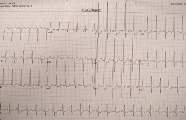
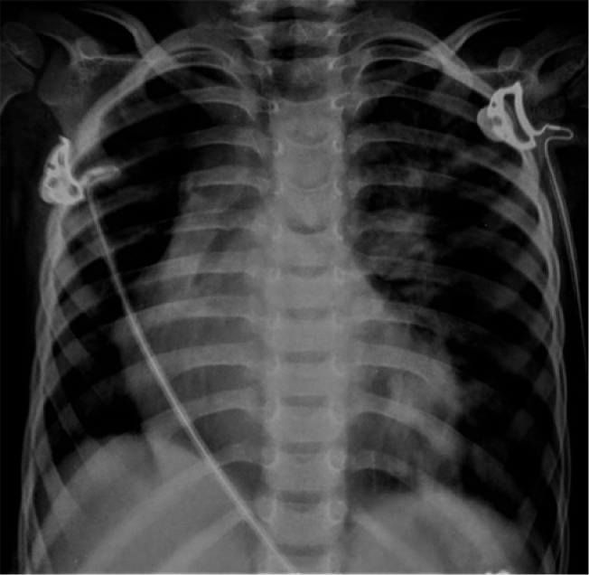
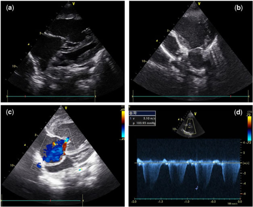
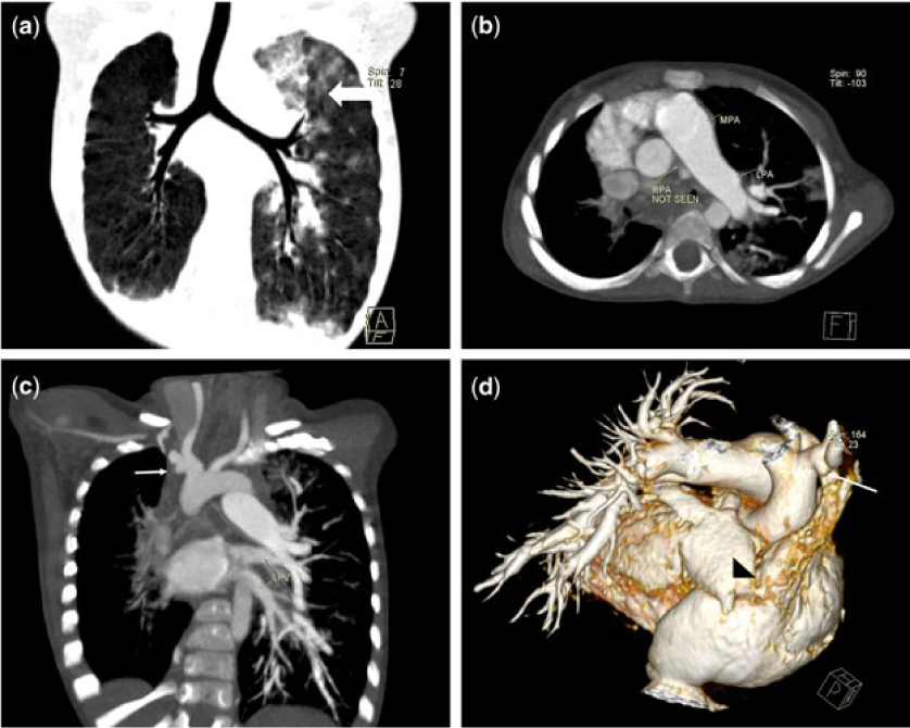
A 2-year and 10-month-old boy was referred to us for generalised oedema who had responded to diuretics given by his family doctor. He had past history of recurrent upper respiratory tract infection, none of which required hospitalisation. Birth, developmental, and maternal history were non-contributory. Cardiovascular examination was unremarkable except for positive hepato-jugular reflux. Mild hepatomegaly was present. His electrocardiogram (Fig 1) revealed right axis deviation with right ventricular pressure overload with strain. His chest X-ray (Fig 2) showed tracheal and mediastinal shift to right, and decreased right lung volume with compensatory hyperinflation of left lung with left upper lobe herniating into right mediastinum. Two-dimensional trans-thoracic echocardiography revealed grossly dilated right atrium (RA) and right ventricle (RV) with severe RV dysfunction. Colour Doppler examination revealed severe tricuspid regurgitation (TR) with TR maximum velocity of 5.1 m/s, severe pulmonary artery hypertension (PAH). Main pulmonary artery (MPA) was dilated and continued as left pulmonary artery (LPA), confluence and the proximal right pulmonary artery (RPA) could not be visualised in otherwise anatomically normal heart (Fig 3). CT pulmonary angiography (Fig 4) was done, which corroborated previous findings and showed dilated MPA continuing as LPA with RPA proximal interruption which was reformed at hilum at collaterals where it measured 3.5 mm. Nodular infiltrates with surrounding ground glass opacities seen in the left upper lobe of the lung were likely to be of infective aetiology or its sequelae. A diagnosis of isolated proximal RPA interruption with severe PAH and RV dysfunction was accepted. Patient was started on diuretics, sildenafil, and digoxin. Parents were counselled for cardiac catheterisation and heart lung transplant, for which they did not consent. At 3 months follow-up, patient had better functional capacity and no signs of overt right heart failure. Follow-up echo revealed persistent PAH. RV function though better compared to baseline had not normalised.
12 lead electrocardiogram showing sinus rhythm with right axis deviation (+150°) with tall R waves in lead V1 and V2 suggestive of RV hypertrophy with associated ST segment depression in V1 to V4 suggestive of RV strain.

Chest X-ray showing mediastinal widening with trachea shift to right; decreased right lung volume with compensatory hyperinflation of left lung with left upper lobe herniating into right mediastinum. The right lobe of diaphragm is higher than usual.

Two-dimensional trans-thoracic echocardiography. ( a ) Subcostal long axis view demonstrating enlarged RA and RV with septum bulging into LV cavity. ( b ) RV outflow view showing dilated MPA with the pulmonary valve; MPA is seen to continue as LPA and RPA is not visualised. ( c ) & ( d ) Colour Doppler examination showing severe tricuspid regurgitation with severe PAH, RV systolic pressure of 104 mmHg.

CT pulmonary angiography ( a ) coronal plane images showing tracheal shift, mildly hypoplastic right lung. Fluffy ground glass opacities throughout the left lung more prominent in left upper lobe. ( b ) Transverse section image at level of MPA bifurcation showing MPA continuing as LPA with absent RPA. ( c ) Coronal section image showing left aortic arch with diverticulum seen from innominate artery (white arrow) supporting the maldevelopment of sixth arch as embryological basis. Also note hypoplastic right pulmonary veins. ( d ) Three-dimensional volume rendered images showing the absence of RPA, Dilated RA, innominate artery diverticulum (white arrow), and hypoplastic right pulmonary veins (black arrow head).

Discussion
Unilateral absence of the pulmonary artery is a rare congenital malformation frequently associated with congenital heart disease especially when LPA is absent. Congenital anomalies associated are Tetralogy of Fallot, right-sided aortic arch, atrial or ventricular septal defect, and PDA. In about one-third of cases of absence of RPA, it is found in an otherwise normal heart. The condition was first described in 1868 by Fraentzel.Reference Fraentzel 1 Many times the distal segment is reformed at hilum from collaterals or PDA, making interruption of pulmonary artery a more appropriate term.
Two theories explaining embryogenesis deserve a mention. First theory, which is more accepted and popular, deals with maldevelopment of aortic arches.Reference Shakibi, Rastan, Nazarian, Paydar, Aryanpour and Siassi 2 , Reference Turner, Vincent and Epstein 3 Abnormal early resorption of the proximal right sixth arch obliterates the proximal RPA. Persistence of the distal right sixth arch links the distal RPA to the right dorsal aorta, the primitive innominate artery. Late closure of the distal right sixth arch, a vessel histologically identical to the contralateral ductus arteriosus, creates RPA discontinuity and the innominate artery diverticulum. This theory is supported by the fact that in most cases the affected pulmonary artery is on the side opposite to the aortic arch, and a diverticulum of the innominate artery is frequently demonstrated angiographically.Reference Pfefferkorn, Löser, Pech, Toussaint and Hilgenberg 4
Cucci et al,Reference Cucci, Doyle and Lewis 5 in contrast to the previous authors favouring resorption of the proximal sixth aortic arch, believe that the absence of a primary division of the pulmonary trunk is due to defective septation of the truncus arteriosus. Cucci’s hypothesis states that the posterodorsal shift of the right conotruncal ridge is necessary for the development of absent RPA, whereas posterodorsal shift of the left conotruncal ridge causes absence of LPA, and by necessity, tetralogy of Fallot with large aorta and small pulmonary artery.
There are three major case reviews of unilateral interruption of pulmonary artery. These reviews highlight the variable presentation and clinical outcomes.Reference Shakibi, Rastan, Nazarian, Paydar, Aryanpour and Siassi 2 , Reference Ten Harkel, Blom and Ottenkamp 6 , Reference Pool, Vogel and Blount 7 Isolated interruption of pulmonary artery has variable age of presentation, cases ranging from 6 months to 6th decade have been reported. Most of the adult patients are asymptomatic and incidentally detected by decreased lung volume in one of the hemithorax on chest X-ray. Hemoptysis will be present in about 20%. Such episodes of hemoptysis are usually minor and self-limited, but massive hemoptysis may rarely occur, requiring emergency treatment by means of pneumonectomy or embolisation of systemic collateral vessels. Pulmonary hypertension is noted in about 20% cases of isolated interruption of pulmonary artery with otherwise structurally normal heart. Why some patients develop pulmonary hypertension in the absence of shunt lesion is yet unknown and warrants further studies. Entire cardiac output passing through remaining lung is said to result in pulmonary hypertension. Haworth in his experimental animal model ligated LPA in new born pigs, and he found that the perfused RPA had persistent muscularity in vessels less than 75 µm.Reference Haworth, de Leval and Macartney 8 Pool et alReference Pool, Vogel and Blount 7 also in their series documented medial hypertrophy in patients with PAH. However, the theory fails to explain how many cases of congenital lung agenesis and following pneumonectomy do not develop pulmonary hypertension.Reference Malcon, Malcon, Cavada, Caruso and Real 9 , Reference Singh, Jhim and Kumar 10 Timing of pulmonary artery maldevelopment may influence development of systemic pulmonary collaterals and hence eventual PAH.Reference DeRuiter, Gittenberger-de Groot, Poelmann, VanIperen and Mentink 11 Those who develop pulmonary hypertension present at much younger age and with features of right heart failure. Differential diagnoses for isolated interruption of pulmonary artery are simple pulmonary hypoplasia, hypogenetic lung syndrome (scimitar syndrome), Swyer–James syndrome, and tubercular sequelae.Reference Turner, Vincent and Epstein 3
There is no consensus regarding the treatment for isolated interruption of pulmonary artery. Most cases especially without pulmonary hypertension or hemoptysis are managed conservatively. Pulmonary vasodilators like sildenafil should be given in patients with PAH. Diuretics may provide symptomatic improvement in cases with right heart failure. Treatment options include partial or total pneumonectomy, closure of selected collateral arteries not solely responsible for pulmonary blood flow, or a primary versus staged pulmonary artery anastomosis. Some advocate an early repair to promote distal pulmonary artery growth and lung development. Shakibi et alReference Shakibi, Rastan, Nazarian, Paydar, Aryanpour and Siassi 2 reported the use of Dacron graft for primary repair in a child with pulmonary hypertension; the child later succumbed and autopsy finding revealed a haemorrhagic lung with multiple intra-alveolar and intra-bronchial haemorrhages. They in their review inferred the unsuitability of patients with pulmonary hypertension for primary repair. Invasive pulmonary pressure determination and evaluation of any shunt lesions should be done in cases with borderline PA pressure. In selected patients with pulmonary hypertension, depending on RV function and invasive hemodynamic studies, heart lung or isolated lung transplantation may be advised.Reference Franke, Wiebe and Harringer 12 – Reference Goldstein, Sweet, Mao, Huddleston and Grady 14 Our case, which to the best of our knowledge is the only reported case from Indian subcontinent with isolated interruption of pulmonary artery with pulmonary hypertension and RV dysfunction, highlights the disease pathophysiology and merits the discussion of heart lung transplant in treatment of such cases.
Conclusion
Isolated interruption of pulmonary artery constitutes important congenital anomaly with wide clinical spectrum and implications. Early diagnosis of associated anomalies or pulmonary hypertension is must and will dictate the treatment plan. When diagnosed as right heart failure due to the pulmonary hypertension as seen in our case, patient should be considered for heart lung transplantation.
Acknowledgements
None
Financial Support
This research received no specific grant from any funding agency, commercial or not-for-profit sectors.
Conflicts of Interest
None
Ethical Standards
The authors assert that all procedures contributing to this work comply with the ethical standards of the Helsinki Declaration of 1975, as revised in 2008, and written informed parental consent was obtained before reporting the case.