Basic taxonomy of Pasteurella multocida
The taxonomy and nomenclature of Pasteurella have undergone many changes since first isolated by Pasteur as the causative agent of fowl cholera in 1880 (Fajfar-Whetstone et al., Reference Fajfar-Whetstone, Coleman, Biggs and Fox1995). Nearly a half century later (in 1929), bacteria with common biochemical and morphological features were grouped together as Pasteurella septica and thereafter (in 1939) as Pasteurella multocida (Weber et al., Reference Weber, Wolfson, Swartz and Hooper1984; Fajfar-Whetstone et al., Reference Fajfar-Whetstone, Coleman, Biggs and Fox1995). Later, Mutters et al. (Reference Mutters, Ihm, Pohl, Frederiksen and Mannheim1985) reclassified the members of the genus Pasteurella into 11 species including P. multocida within which three subspecies (subsp.) multocida, septica and gallicida were defined based on sugar (d-sorbitol and dulcitol) fermentation. Molecular techniques based on M13 core fingerprinting and α-glucosidase activity were also developed to better differentiate between P. multocida subsp. multocida and subsp. septica (Hunt Gerardo et al., Reference Hunt, Citron, Claros, Fernandez and Goldstein2001). P. multocida subsp. multocida, the most important pathogenic member of the genus Pasteurella sensu stricto, has a broad host spectrum that includes many wild and domestic animal species. P. multocida subsp. gallicida and septica are specifically isolated from fowl and domestic pets (cats and dogs), respectively (Mutters et al., Reference Mutters, Ihm, Pohl, Frederiksen and Mannheim1985). To define isolates associated with tiger bites, a fourth subspecies (tigris) was recently proposed by Capitini et al. (Reference Capitini, Herrero, Patel, Ishitani and Boyce2002) on the basis of phenotype and 16S rRNA gene sequence comparison.
The capsule structure and lipopolysaccharide (LPS) composition of P. multocida are the basis for the classification of the bacterium into serogroups and serotypes, respectively (Heddleston et al., Reference Heddleston, Gallagher and Rebers1972; Rimler and Rhoades, Reference Rimler, Rhoades, Adlam and Rutter1989). Passive hemagglutination and gel diffusion precipitin tests (reviewed in Rimler and Rhoades, Reference Rimler, Rhoades, Adlam and Rutter1989; Confer, Reference Confer1993) have been traditionally used to classify P. multocida into five capsular groups (A, B, D, E and F) and 16 somatic serotypes (1–16), respectively (Carter, Reference Carter1955; Heddleston and Rebers, Reference Heddleston and Rebers1972). Serogroups A and, to a lesser extent, D cause fowl cholera in birds (Rhoades and Rimler, Reference Rhoades and Rimler1989). Serogroup F strains are predominantly isolated from diseased poultry, in particular, turkeys and more recently from a fatal case of fibrinous peritonitis in calves (Shewen and Conlon, Reference Shewen, Conlon, Gyles and Thoen1993; Catry et al., Reference Catry, Chiers, Schwarz, Kehrenberg, Decostere and de Kruif2005). In pigs, atrophic rhinitis and pneumonia are associated primarily with toxigenic strains of serogroup D and serogroup A, respectively (Dungworth, Reference Dungworth, Jubb, Kennedy and Palmer1985), while P. multocida serogroups B and E are associated with hemorrhagic septicemia in cattle and water buffaloes in tropical regions of Africa and Asia (Carter and de Alwis, Reference Carter, de Alwis, Adlam and Rutter1989; Rimler and Rhoades, Reference Rimler, Rhoades, Adlam and Rutter1989; Shewen and Conlon, Reference Shewen, Conlon, Gyles and Thoen1993). In North America, serogroups B and E are rarely isolated from cattle (Confer, Reference Confer1993). Worldwide, P. multocida serogroup A isolates are one of the major causes of bovine respiratory diseases (BRDs) (Frank, Reference Frank, Adlam and Rutter1989; Rimler and Rhoades, Reference Rimler, Rhoades, Adlam and Rutter1989).
P. multocida association with BRD
It is clear that P. multocida is one of the primary bacterial pathogens associated with the clinical syndromes defining BRD complex including neonatal calf (enzootic) pneumonia and beef cattle pneumonia (shipping fever) (Lillie, Reference Lillie1974; Watts et al., Reference Watts, Yancey, Salmon and Case1994; Fulton et al., Reference Fulton, Purdy, Confer, Saliki, Loan, Briggs and Burge2000; Welsh et al., Reference Welsh, Dye, Payton and Confer2004). Both syndromes are considered multifactorial; that is, multiple infectious and non-infectious factors are involved in development of disease (Ames, Reference Ames1997; Mosier, Reference Mosier1997). Unfortunately, many epidemiological studies of these conditions failed to differentiate among the variety of pathogens involved.
Strong evidence exists to link P. multocida to respiratory disease in dairy calves. This includes culture from lung tissue (Van Donkersgoed et al., Reference Van Donkersgoed, Ribble, Boyer and Townsend1993; Sivula et al., Reference Sivula, Ames and Marsh1996a; Singer et al., Reference Singer, Case, Carpenter, Walker and Hirsh1998; Hirose et al., Reference Hirose, Kobayashi, Ito, Kawasaki, Zako, Kotani, Ogawa and Sato2003), from nasopharyngeal swabs (NPS) (Catry et al., Reference Catry, Decostere, Schwarz, Kehrenberg, de Kruif and Haesebrouck2006) and from transtracheal washes (TTW) (Singer et al., Reference Singer, Case, Carpenter, Walker and Hirsh1998; Virtala et al., Reference Virtala, Gröhn, Mechor, Erb and Dubovi2000). The significance of P. multocida in calf pneumonia is more apparent than with shipping fever. Multiple studies involving dairy calves have identified P. multocida in much higher prevalence than Mannheimia haemolytica (Bryson et al., Reference Bryson, McFerran, Ball and Neill1978; Virtala et al., Reference Virtala, Gröhn, Mechor, Erb and Dubovi2000; Nikunen et al., Reference Nikunen, Hartel, Orro, Neuvonen, Tanskanen, Kivela, Sankari, Aho, Pyrala, Saloniemi and Soveri2007). Studies conducted by Nikunen et al. (Reference Nikunen, Hartel, Orro, Neuvonen, Tanskanen, Kivela, Sankari, Aho, Pyrala, Saloniemi and Soveri2007) on 84 calves with BRD in Finland found a significant relationship between the presence of P. multocida in calves with clinical signs of respiratory disease and the elevated concentrations of serum acute phase proteins (i.e. fibrinogen, haptoglobin, serum amyloid-A, LPS-binding protein and α1-acid glycoprotein), suggesting a strong pathogenic role for P. multocida in respiratory disease of calves. No significant relationship was observed for other pathogens including Mycoplasma sp., which were isolated in over 90% of calves as compared to 14% for P. multocida. Recent examination of 396 calves from 280 different dairy farms for occurrence of bacterial, mycoplasmal and viral pathogens found that P. multocida was the most commonly isolated bacterial pathogen and was detected in 26.4, 32.6 and 42.3% of tracheobronchial lavage samples from healthy, suspected or diseased calves, respectively (Autio et al., Reference Autio, Pohjanvirta, Holopainen, Rikula, Pentikäinen, Huovilainen, Rusanen, Soveri, Sihvonen and Pelkonen2007). Seventy-five percent of the P. multocida isolates were indole-negative phenotypes, underlining the need to investigate the role of indole-negative P. multocida isolates in calf respiratory disease. Nonetheless, the presence of the bacterium does not equal disease, as seroconversion to P. multocida occurred in control calves, confirming the ubiquitous nature of the organism and the multifactorial nature of the disease (Virtala et al., Reference Virtala, Mechor, Grohn, Erb and Dubovi1996).
Evidence for involvement of P. multocida in shipping fever has been obtained by several methods. The bacterium was isolated from lungs of fatal cases in feedlot cattle by numerous investigators (Watts et al., Reference Watts, Yancey, Salmon and Case1994; Fulton et al., Reference Fulton, Purdy, Confer, Saliki, Loan, Briggs and Burge2000; Storz et al., Reference Storz, Lin, Purdy, Chouljenko, Kousoulas, Enright, Gilmore, Briggs and Loan2000a; Welsh et al., Reference Welsh, Dye, Payton and Confer2004; Gagea et al., Reference Gagea, Bateman, van Dreumel, McEwen, Carman, Archambault, Shanahan and Caswell2006). Indeed, M. haemolytica is the most common isolated bacterium from shipping fever, but the proportion of fatal cases of respiratory disease in feedlot cattle attributable to P. multocida appears to be increasing (Welsh et al., Reference Welsh, Dye, Payton and Confer2004). Potential causes for this shift include changes in virulence among the pathogens, efficacy of antimicrobial agents, and changes in identification and/or management of sick cattle (Welsh et al., Reference Welsh, Dye, Payton and Confer2004). P. multocida was cultured from nasal or NPS, TTW and/or bronchoalveolar lavage (BAL) of sick animals (Allen et al., Reference Allen, Viel, Bateman, Rosendal, Shewen and Physick-Sheard1991; DeRosa et al., Reference DeRosa, Mechor, Staats, Chengappa and Shryock2000; Fulton et al., Reference Fulton, Cook, Step, Confer, Saliki, Payton, Burge, Welsh and Blood2002). A positive correlation was found between the species of bacteria isolated from multiple sites (e.g. NPS and TTW) (DeRosa et al., Reference DeRosa, Mechor, Staats, Chengappa and Shryock2000). The presence of P. multocida in BAL fluid accounted for ∼40% of respiratory disease (Allen et al., Reference Allen, Viel, Bateman, Nagy, Rosendal and Shewen1992a). However, the presence or absence of P. multocida in the nasal pharynx did predict disease (Allen et al., Reference Allen, Viel, Bateman, Rosendal, Shewen and Physick-Sheard1991; Fulton et al., Reference Fulton, Cook, Step, Confer, Saliki, Payton, Burge, Welsh and Blood2002). Similarly, seroconversion occurred in a high percentage of calves and thus was not predictive of disease (Virtala et al., Reference Virtala, Gröhn, Mechor, Erb and Dubovi2000). Nonetheless, low antibody concentration to P. multocida at feedlot entry correlated with decreased value to the owner (carcass value−total feedlot expenses) (Fulton et al., Reference Fulton, Cook, Step, Confer, Saliki, Payton, Burge, Welsh and Blood2002). Therefore, these various findings suggest that P. multocida is ubiquitous and can be found in both healthy and sick cattle; and not all calves harboring or exposed to the bacterium will develop pneumonia. However, its presence (particularly in large numbers) in the nares, trachea or bronchioles of a sick calf strongly suggests its involvement in the disease process.
Although knowledge of P. multocida serogroup and serotype specificity from BRD cases is limited, the reported data clearly singled out P. multocida serogroup A strains as the most frequently isolated from BRD cases (Blanco-Viera et al., Reference Blanco-Viera, Trigo, Jaramillo-Meza and Aguilar-Romero1995; Ewers et al., Reference Ewers, Lubke-Becker, Bethe, Kiebling, Filter and Wieler2006; Autio et al., Reference Autio, Pohjanvirta, Holopainen, Rikula, Pentikäinen, Huovilainen, Rusanen, Soveri, Sihvonen and Pelkonen2007; Nikunen et al., Reference Nikunen, Hartel, Orro, Neuvonen, Tanskanen, Kivela, Sankari, Aho, Pyrala, Saloniemi and Soveri2007). Studies by Blanco-Viera et al. (Reference Blanco-Viera, Trigo, Jaramillo-Meza and Aguilar-Romero1995) showed that 61 and 25% of P. multocida isolated from pneumonic lung lesions belonged to capsular groups A and D, respectively, with serotype 3 (77%) being the most frequent. Kumar et al. (Reference Kumar, Shivachandra, Biswas, Singh and Srivastava2004) investigated the prevalence of P. multocida serotypes in samples from different animal species in India. Cattle samples found to be positive for P. multocida were predominantly serotype A:3, in agreement with reports by Frank (Reference Frank, Adlam and Rutter1989). Studies in our laboratories found that 80% of P. multocida isolates from cattle with pneumonia were serotype A:3 (Dabo et al., Reference Dabo, Confer and Murphy1997), and recently, more than 92% of P. multocida isolated from cattle were found to be serogroup A (Ewers et al., Reference Ewers, Lubke-Becker, Bethe, Kiebling, Filter and Wieler2006).
Clinical disease: shipping fever
The term ‘undifferentiated bovine respiratory disease’ is often considered synonymous with ‘shipping fever’, although BRD may also be considered to include other disease entities, including enzootic calf pneumonia. For this discussion, respiratory disease in beef cattle will be referred to as shipping fever, which affects post-weaning beef calves, most commonly after transport to a stocker or feedlot operation. Diagnostic criteria are often vague, but typically include depression, inappetence, cough, nasal discharge and fever (or some combination of these). Most epidemiologic investigations relied upon producer treatment as a proxy for diagnosis. This frequently is no more exclusive than ‘animals that are pulled for treatment and found to have no signs referable to a system other than the respiratory tract’ (Bagley et al., Reference Bagley, Bingham and Hall2003; Cusack, Reference Cusack2004). Studies have shown limited correlation between treatment and more objective measures such as mortality, BAL cytology and lung lesions at slaughter (Allen et al., Reference Allen, Viel, Bateman, Nagy, Rosendal and Shewen1992a, Reference Allen, Viel, Bateman, Rosendal and Shewenb; Griffin et al., Reference Griffin, Perino and Wittum1994; Ribble et al., Reference Ribble, Meek, Janzen, Guichon and Jim1995a; Wittum et al., Reference Wittum, Woollen, Perino and Littledike1996; Bryant et al., Reference Bryant, Perino, Griffin, Doster and Wittum1999). Clearly the limitations of field-based diagnosis contribute significantly to the difficulty in studying shipping fever (Kelly and Janzen, Reference Kelly and Janzen1986).
Morbidity and mortality due to shipping fever average approximately 14 and 1%, respectively (USDA National Animal Health Monitoring System (NAHMS)). However, both vary widely, with reported ranges for morbidity from 4.6% (Snowder et al., Reference Snowder, Van Vleck, Cundiff and Bennett2006) to 89% (Storz et al., Reference Storz, Purdy, Lin, Burrell, Truax, Briggs, Frank and Loan2000b) and for mortality from 1% (Snowder et al., Reference Snowder, Van Vleck, Cundiff and Bennett2006) to 13% (Storz et al., Reference Storz, Purdy, Lin, Burrell, Truax, Briggs, Frank and Loan2000b). Outbreaks are common, with initial cases appearing 7–10 days after arrival and typically peaking prior to 20 days (Jensen et al., Reference Jensen, Pierson, Braddy, Saari, Lauerman, England, Horton and McChesney1976; Alexander et al., Reference Alexander, MacVean and Salman1989). Mortality appears shortly after initiation of the outbreak, peaking at 16 days (Ribble et al., Reference Ribble, Meek, Jim and Guichon1995b). However, this timeframe can be lengthened by continued introduction of new animals into the herd or pens (Alexander et al., Reference Alexander, MacVean and Salman1989). Although disease occurs throughout the year, shipping fever has seasonal variation, peaking in fall to winter (Jensen et al., Reference Jensen, Pierson, Braddy, Saari, Lauerman, England, Horton and McChesney1976; Ribble et al., Reference Ribble, Meek, Janzen, Guichon and Jim1995a; Loneragan et al., Reference Loneragan, Dargatz, Morley and Smith2001).
Numerous predisposing factors are frequently cited for shipping fever. The most well documented of these are viral infections, which will be discussed in regard to their specific association with P. multocida. Other proposed factors are generally termed ‘stressors’ and include transportation, co-mingling with other cattle, dust, cold, sudden and extreme changes of temperature and humidity, dehydration, hypoxia, endotoxin, cold coupled with wetness, and acute metabolic disturbances (Lillie, Reference Lillie1974; Irwin et al., Reference Irwin, McConnell, Coleman and Wilcox1979). These are thought to alter the respiratory mucosa or hinder cattle's immune system, either directly or through the effects of endogenous agents such as cortisol, making the animals more susceptible to opportunistic infections. The intuitive nature of these suggestions is appealing, and they have become widely accepted and repeated throughout the literature. However, limited epidemiologic investigations have been done to confirm or refute the significance of these factors, and studies done thus far have been inconclusive. The most consistent finding in epidemiologic studies of shipping fever is the immense variability in occurrence, from operation to operation and year to year. This makes interpretation of interventions difficult, particularly in multi-year studies.
Several predisposing factors have been conclusively demonstrated. Newly received cattle are at greater risk for disease (Jensen et al., Reference Jensen, Pierson, Braddy, Saari, Lauerman, England, Horton and McChesney1976; Kelly and Janzen, Reference Kelly and Janzen1986; Martin and Meek, Reference Martin and Meek1986; MacVean et al., Reference MacVean, Franzen, Keefe and Bennett1986; Alexander et al., Reference Alexander, MacVean and Salman1989; Ribble et al., Reference Ribble, Meek, Jim and Guichon1995b). Calves purchased through a salebarn are more at risk than those arriving directly from farm sources (Wilson et al., Reference Wilson, Church and Acres1985; Gummow and Mapham, Reference Gummow and Mapham2000). However, it is possible that other factors commonly associated with salebarn calves are responsible for the increased risk, rather than the salebarn process itself. These may include co-mingling of cattle from multiple sources, as well as the stress of repeated unloading, loading, handling, etc. Marketing through a salebarn may also reflect different management practices (e.g. better managers market direct, while poorer managers rely upon the salebarn). When examining what strategies can prevent disease, metaphylaxis (administration of parenteral antimicrobials to calves that are at high risk for respiratory disease) has consistently reduced morbidity (Van Donkersgoed, Reference Van Donkersgoed1992; Frank et al., Reference Frank, Briggs, Loan, Purdy and Zehr2000, Reference Frank, Briggs, Duff, Loan and Purdy2002; Macartney et al., Reference Macartney, Bateman and Ribble2003; Cusack, Reference Cusack2004), while results of vaccination have been equivocal at best (Mosier et al., Reference Mosier, Confer and Panciera1989; Perino and Hunsaker, Reference Perino and Hunsaker1997).
Clinical disease: enzootic calf pneumonia (ECP)
Similar to shipping fever, the term ‘enzootic calf pneumonia’ (ECP) is more a description than a diagnosis. In some ways, the distinction between ECP and shipping fever is arbitrary, with ECP occurring prior to shipping fever and after weaning. However, pneumonia is infrequent in pre-weaned beef calves on pasture. Thus, ECP usually refers to respiratory disease in dairy and veal calves raised in confinement. Diagnostic criteria for ECP are similar to those for shipping fever, both in content and vague nature. Studies have shown relatively poor correlation between producer-diagnosed ECP and that detected by a veterinarian (Van Donkersgoed et al., Reference Van Donkersgoed, Ribble, Boyer and Townsend1993; Virtala et al., Reference Virtala, Grohn, Mechor and Erb1999; Svensson et al., Reference Svensson, Hultgren and Oltenacu2006). Nevertheless, most field studies relied upon treatment by the producer as a proxy for disease, greatly complicating the conclusions that can be reached from such research.
ECP has traditionally been defined as developing between 2 and 6 months of age (Ames, Reference Ames1997). Studies looking at younger calves found peak occurrence from nearly 4 weeks to 6 weeks of age (Waltner-Toews et al., Reference Waltner-Toews, Martin and Meek1986; Curtis et al., Reference Curtis, Erb and White1988a; Van Donkersgoed et al., Reference Van Donkersgoed, Ribble, Boyer and Townsend1993; Virtala et al., Reference Virtala, Mechor, Grohn, Erb and Dubovi1996). Reported morbidity varies significantly. Two studies found 6–8% of calves affected (Curtis et al., Reference Curtis, Erb and White1988a;Sivula et al., Reference Sivula, Ames, Marsh and Werdin1996b), whereas two other studies found morbidity rates of 15% (Waltner-Toews et al., Reference Waltner-Toews, Martin and Meek1986) to 29% (Van Donkersgoed et al., Reference Van Donkersgoed, Ribble, Boyer and Townsend1993). Mortality ranged from approximately 5% (Waltner-Toews et al., Reference Waltner-Toews, Martin and Meek1986) to nearly 10% (Sivula et al., Reference Sivula, Ames, Marsh and Werdin1996b).
Fewer studies have been done on the epidemiology of ECP compared to shipping fever. Waltner-Toews et al. (Reference Waltner-Toews, Martin and Meek1986) and Van Donkersgoed et al. (Reference Van Donkersgoed, Ribble, Boyer and Townsend1993) found larger herds had higher incidence of ECP than smaller herds. This relationship, however, was not identified by Curtis et al. (Reference Curtis, Scarlett, Erb and White1988b). None of these studies included farms that would be considered large by today's standards (Waltner-Toews et al., Reference Waltner-Toews, Martin and Meek1986). Waltner-Toews et al. (Reference Waltner-Toews, Martin and Meek1986) and others (Bryson et al., Reference Bryson, McFerran, Ball and Neill1978; Svensson et al., Reference Svensson, Hultgren and Oltenacu2006) found a higher incidence in fall and winter but Curtis et al. (Reference Curtis, Scarlett, Erb and White1988b) did not. Calves that had diarrheal disease may be at an increased risk for ECP (Curtis et al., Reference Curtis, Scarlett, Erb and White1988b; Van Donkersgoed et al., Reference Van Donkersgoed, Ribble, Boyer and Townsend1993; Svensson et al., Reference Svensson, Hultgren and Oltenacu2006). However, Van Donkersgoed et al. (Reference Van Donkersgoed, Ribble, Boyer and Townsend1993) found that relationship was no longer significant after adjusting for clustering, and other studies did not identify a relationship between scours and respiratory disease (Virtala et al., Reference Virtala, Grohn, Mechor and Erb1999). Failure of passive transfer (FPT) is an intuitive potential predisposing factor, but investigations of a possible relationship between FPT and ECP yielded conflicting results (Van Donkersgoed et al., Reference Van Donkersgoed, Ribble, Boyer and Townsend1993; Virtala et al., Reference Virtala, Mechor, Grohn, Erb and Dubovi1996, Reference Virtala, Grohn, Mechor and Erb1999). The diagnostic criteria for FPT may account for part of the discrepancies. Virtala et al. (Reference Virtala, Grohn, Mechor and Erb1999) determined that immunoglobulin of less than 1200 mg dl−1 provides the best prediction of disease and thus the best break point for FPT. In contrast, Van Donkersgoed et al. (Reference Van Donkersgoed, Ribble, Boyer and Townsend1993), Sivula et al. (Reference Sivula, Ames and Marsh1996a) and Virtala et al., (Reference Virtala, Mechor, Grohn, Erb and Dubovi1996) used values less than 800 mg dl−1 for diagnosing FPT. Another potential confounder is the diagnosis of respiratory disease. Van Donkersgoed et al. (Reference Van Donkersgoed, Ribble, Boyer and Townsend1993) found no correlation between FPT and owner-diagnosed respiratory disease but found a positive association with that diagnosed by a veterinarian. Prado et al. (Reference Prado, Prado, Payton and Confer2006) found colostral antibodies to P. multocida to be short-lived, largely being undetectable by 2 months of age. Thus, their impact on ECP would likely be minimal.
Housing of calves was a significant influence on ECP in several studies. Several studies (Martin and Meek, Reference Martin and Meek1986; Waltner-Toews et al., Reference Waltner-Toews, Martin and Meek1986; Virtala et al., Reference Virtala, Grohn, Mechor and Erb1999) found hutches to be better than inside pens, whereas Svensson et al. (Reference Svensson, Hultgren and Oltenacu2006) found housing calves individually better than group housing. Sivula et al. (Reference Sivula, Ames and Marsh1996a) were not able to demonstrate a reduction in ECP attributable to housing in hutches. Using the same data set, the authors applied objective criteria to define ‘adequate ventilation of calf housing’ and assessed its relationship with ECP (Sivula et al., Reference Sivula, Ames, Marsh and Werdin1996b). There was a positive correlation between adequate ventilation of housing and average daily gain, but no relationship between either of these and ECP. These findings again could reflect the inadequacy of producer-diagnosed respiratory disease. Such an assertion is bolstered by the finding that producer-diagnosed death due to pneumonia only had a 58.3% sensitivity (Sivula et al., Reference Sivula, Ames, Marsh and Werdin1996b). Numerous other factors have been potentially implicated in ECP. These include sire, vaccination of dam prior to calving, calves born in loose housing, time of day when born, how and when colostrum is provided, navel treatment, weaning based on size rather than age, use of non-medicated milk replacer, and provision of water and mineral.
Pathology of P. multocida pneumonia in cattle
Bovine pneumonia associated with P. multocida infections whether in young dairy calves, in weaned and shipped beef cattle or in calves experimentally challenged is often difficult to discern from pneumonia associated with other bovine bacterial pathogens such as M. haemolytica and Histophilus somni. Dungworth (Dungworth, Reference Dungworth, Jubb, Kennedy and Palmer1985) indicated that ‘less fulminating … bronchopneumonias tend to be more often caused by P. multocida than by P. [Mannheimia] haemolytica’. In fact, isolation of more than one of these pathogens from pneumonic lungs in conjunction with respiratory viruses and/or Mycoplasma spp. occurs frequently (Gagea et al., Reference Gagea, Bateman, van Dreumel, McEwen, Carman, Archambault, Shanahan and Caswell2006). This has led authors to question if P. multocida is a primary pathogen in bovine pneumonia (Lopez, Reference Lopez, McGarvin and Zachary2007). Experimental reproduction of bovine pneumonia with P. multocida alone and its isolation from cases of naturally acquired bovine pneumonia without evidence of other infectious agents indicate that it can likely occur as a primary pathogen in young or stressed cattle.
The gross pulmonary pathological changes have been described differently by various authors, and those designations probably reflect the age of the lesion, whether or not other infectious agents were involved but not identified, and descriptive preferences of the various authors. The lesion is a typical cranioventral bronchopneumonia and has been characterized simply as bronchopneumonia by some authors (Haritani et al., Reference Haritani, Narita, Murata, Hashimoto and Takizawa1989; Mathy et al., Reference Mathy, Mathy, Lee, Walker, Lofthouse and Meeusen2002; Ewers et al., Reference Ewers, Lubke-Becker, Bethe, Kiebling, Filter and Wieler2006; Lopez, Reference Lopez, McGarvin and Zachary2007) or as a bronchopneumonia with various descriptive modifiers denoting lesion age and type of exudate. These include acute fibrinosuppurative (Gagea et al., Reference Gagea, Bateman, van Dreumel, McEwen, Carman, Archambault, Shanahan and Caswell2006), subacute to chronic fibrinopurulent (Mosier, Reference Mosier1997), fibrinous to fibrinopurulent (Dungworth, Reference Dungworth, Jubb, Kennedy and Palmer1985), suppurative (Tegtmeier et al., Reference Tegtmeier, Uttenthal, Friis, Jensen and Jensen1999) and fibrino-necrotizing (Tegtmeier et al., Reference Tegtmeier, Uttenthal, Friis, Jensen and Jensen1999). The presence of fibrinous to fibrinopurulent pleuritis, distended interlobular septa with edema or fibrin and/or abscesses is variable with P. multocida infection. Lopez (Reference Lopez, McGarvin and Zachary2007) designated the P. multocida-associated lesion as bronchointerstitial to bronchopneumonia depending on whether or not there was concurrent viral infection.
Histologic changes typically consist of bronchi and bronchioles filled with neutrophils, macrophages and necrotic epithelium interspersed with a small amount of fibrin (Dungworth, Reference Dungworth, Jubb, Kennedy and Palmer1985; Jericho and Carter, Reference Jericho and Carter1985; Haritani et al., Reference Haritani, Narita, Murata, Hashimoto and Takizawa1989; Dowling et al., Reference Dowling, Hodgson, Dagleish, Eckersall and Sales2004; Lopez, Reference Lopez, McGarvin and Zachary2007). Neutrophil morphology ranges from intact to necrotic. Similar exudate is present in alveoli with peribronchiolar alveoli most severely affected particularly in the early phases of infection. The character of the exudate varies with time with acute lesions having a higher percentage of neutrophils and lower percentage of macrophages compared to chronic lesions. There is usually a variable amount of intra-alveolar hemorrhage, and P. multocida cells and antigen can be detected within alveolar lumens and within neutrophils and macrophages. Both intact and degenerating bacteria are seen within phagosomes of phagocytes (Haritani et al., Reference Haritani, Narita, Murata, Hashimoto and Takizawa1989), and intracellular survival of P. multocida within phagocytes has been demonstrated (Dowling et al., Reference Dowling, Hodgson, Dagleish, Eckersall and Sales2004). Interlobular lymphatics can be dilated with fibrinous to fibrinopurulent exudate. Large foci of necrosis and abscesses have been described occasionally after experimental challenge (Dowling et al., Reference Dowling, Hodgson, Schock, Donachie, Eckersall and McKendrick2002; Mathy et al., Reference Mathy, Mathy, Lee, Walker, Lofthouse and Meeusen2002; Ishiguro et al., Reference Ishiguro, Kitajima, Kubota, Amimoto, Oda, Fukuyama and Shimizu2005). Immunohistochemical studies of natural cases of bovine pneumonia demonstrated, however, that abscesses were more often associated with M. haemolytica, Arcanobacterium pyogenes or Mycoplasma bovis infections and not with P. multocida (Haritani et al., Reference Haritani, Nakazawa, Hashimoto, Narita, Tagawa and Nakagawa1990; Adegboye et al., Reference Adegboye, Hallbur, Cavanaugh, Werdin, Chase, Miskimins and Rosenbusch1995; Gagea et al., Reference Gagea, Bateman, van Dreumel, McEwen, Carman, Archambault, Shanahan and Caswell2006).
Epidemiology of P. multocida pneumonia
True epidemiologic investigation of P. multocida-associated respiratory disease is scant. One reason for this is the ubiquitous nature of the organism. Its mere presence in the upper respiratory tract is not diagnostic of disease; whereas confirmation of its involvement in pneumonia requires post-mortem culture or invasive measures (TTW or BAL), which are not practical for large numbers of cattle. It appears that P. multocida has a synergistic relationship with Mycoplasma species. This was particularly clear for dairy calf pneumonia, as isolation of both from clinical cases is more common than isolating either alone (Virtala et al., Reference Virtala, Gröhn, Mechor, Erb and Dubovi2000; Hirose et al., Reference Hirose, Kobayashi, Ito, Kawasaki, Zako, Kotani, Ogawa and Sato2003). Co-infection with other pathogens has also been noted, including H. somni (Gagea et al., Reference Gagea, Bateman, van Dreumel, McEwen, Carman, Archambault, Shanahan and Caswell2006), M. haemolytica (Van Donkersgoed et al., Reference Van Donkersgoed, Ribble, Boyer and Townsend1993; Fulton et al., Reference Fulton, Purdy, Confer, Saliki, Loan, Briggs and Burge2000), coronavirus (Storz et al., Reference Storz, Lin, Purdy, Chouljenko, Kousoulas, Enright, Gilmore, Briggs and Loan2000a; Autio et al., Reference Autio, Pohjanvirta, Holopainen, Rikula, Pentikäinen, Huovilainen, Rusanen, Soveri, Sihvonen and Pelkonen2007), adenovirus (Autio et al., Reference Autio, Pohjanvirta, Holopainen, Rikula, Pentikäinen, Huovilainen, Rusanen, Soveri, Sihvonen and Pelkonen2007), parainfluenza-3 virus and bovine respiratory syncytial virus (Van Donkersgoed et al., Reference Van Donkersgoed, Ribble, Boyer and Townsend1993; Autio et al., Reference Autio, Pohjanvirta, Holopainen, Rikula, Pentikäinen, Huovilainen, Rusanen, Soveri, Sihvonen and Pelkonen2007). The ability of P. multocida to cause disease in the absence of other pathogens has been debated (Bryson et al., Reference Bryson, McFerran, Ball and Neill1978). In a Finnish study, P. multocida was one of only three pathogens to be isolated more commonly from BAL of diseased than from healthy and suspect cattle (Autio et al., Reference Autio, Pohjanvirta, Holopainen, Rikula, Pentikäinen, Huovilainen, Rusanen, Soveri, Sihvonen and Pelkonen2007). However, no association was found between the isolation of P. multocida and clinical disease when no other pathogen was present (Autio et al., Reference Autio, Pohjanvirta, Holopainen, Rikula, Pentikäinen, Huovilainen, Rusanen, Soveri, Sihvonen and Pelkonen2007). This led them to accept the conclusion of Maheswaran et al. (Reference Maheswaran, Thumbikat and Dileepan2002) that P. multocida is an opportunistic pathogen and not a primary causative agent. In contrast, another Finnish study found P. multocida to be the only agent positively associated with clinical signs and elevated serum acute phase proteins (Nikunen et al., Reference Nikunen, Hartel, Orro, Neuvonen, Tanskanen, Kivela, Sankari, Aho, Pyrala, Saloniemi and Soveri2007). Those authors concluded that there is ‘a strong pathogenic role for P. multocida in respiratory disease in calves in Finland, at least in situations where other known pathogens are absent’. Finland is free of bovine viral diarrhea virus and M. bovis; therefore, findings there may not reflect disease where interaction between P. multocida and those agents may occur.
A major epidemiologic question is whether there is an inherent difference in pathogenic versus commensal populations of P. multocida or whether P. multocida-associated disease is strictly a change in host–bacteria relationship. Bacteriological characterizations have not identified an inherent difference between pathogen and commensal isolates, but the ability to detect such differences may be the limiting factor. Davies et al. (Reference Davies, MacCorquodale, Baillie and Caffrey2003a) found that different subpopulations are responsible for progressive atrophic rhinitis and pneumonia in swine. Similarly, P. multocida isolates from ovine respiratory and reproductive tracts were often distinguishable (Davies et al., Reference Davies, MacCorquodale and Caffrey2003b). Therefore, differences (perhaps quite subtle) may exist between commensals and pathogens.
The entire P. multocida genome has now been sequenced, albeit from a single avian isolate, Pm70 (May et al., Reference May, Zhang, Li, Paustian, Whittam and Kapur2001). This should enable relatively rapid characterization of genetic differences among various isolates and has in fact already yielded valuable information. Several virulence factors have been identified and are discussed elsewhere in this article. This section will focus only on the efforts to use bacterial products or genes encoding these products as markers for epidemiologic investigation. Hunt et al. (Reference Hunt, Citron, Claros, Fernandez and Goldstein2001) identified several genes whose expression is up-regulated in vivo, including membrane proteins and metabolic and biosynthetic pathways. However, those were derived from bacteria inoculated into the bloodstream of mice and may not reflect events occurring in the upper or lower respiratory tract of healthy or sick cattle. Additionally, only one isolate of P. multocida was used; thus no comparative analysis was possible to determine what genes may promote virulence. Boyce and Adler (Reference Boyce and Adler2006) concluded that ‘true virulence genes might be constitutively expressed, upregulated only during initial stages of infection’. Thus, assaying for expression of virulence factors may be confounded by the progression of disease and interaction of host and pathogen. A more relevant comparative study examined the genotypes of a wide range of isolates from multiple host species (cattle, sheep and goats, bison, swine, rabbits, poultry, cats, dogs and humans), with some of the isolates being from diseased animals and others from clinically normal ones (Ewers et al., Reference Ewers, Lubke-Becker, Bethe, Kiebling, Filter and Wieler2006). They identified several genes that were more common in bovine isolates than from isolates obtained from other species. They also had modest success in identifying genes more commonly associated with isolates from diseased animals.
Another question relates to transmission, i.e. is P. multocida pneumonia contagious? The ubiquitous presence and commensal nature of P. multocida means that finding the organism in multiple animals does not demonstrate transmission. Instead, to assess transmission requires a way of discriminating between bacterial isolates. Classic means of distinction, including serotyping and serogrouping, are inadequate (Wilson et al., Reference Wilson, Rimler and Hoffman1992). Purdy et al. (Reference Purdy, Raleigh, Collins, Watts and Straus1997) employed an extensive assessment of enzymatic activity to compare 61 P. multocida isolates collected from multiple locations over the course of 8 years. The profiles that resulted showed no evidence of clustering, suggesting that this practice offered little discriminatory power (Purdy et al., Reference Purdy, Raleigh, Collins, Watts and Straus1997).
Another phenotypic approach to the question of transmission is assessment of antimicrobial resistance. Catry et al. found tetracycline-resistant P. multocida in the nasopharynx of 5 calves on 1 farm, while no resistant P. multocida were found in the other 56 calves from 15 farms (Catry et al., Reference Catry, Decostere, Schwarz, Kehrenberg, de Kruif and Haesebrouck2006). No tetracycline had been used on any farms. The authors suggested that the five isolates were clonal, indicating horizontal transmission. A large retrospective study found evidence of regional differences in antimicrobial resistance among P. multocida isolates, with clustering in specific regions (Singer et al., Reference Singer, Case, Carpenter, Walker and Hirsh1998). No further examination was done to assess whether isolates were clonal or even if the mechanism of antimicrobial resistance was consistent. A smaller study examined the genotypic basis for tetracycline resistance (Kehrenberg et al., Reference Kehrenberg, Catry, Haesebrouck, de Kruif and Schwarz2005). Six Pasteurella and Mannheimia isolates were obtained from a single farm where oxytetracycline had been given prophylactically. The same gene encoding resistance was found in multiple bacterial species, with little or no mutation. The authors concluded that Pasteurella and Mannheimia likely had been transmitted between calves. However, they acknowledged that horizontal transmission of resistance genes could also account for the findings. Because the same genetic material was found in multiple species of bacteria, it is possible that only the genes were passed from animal to animal (within another microbial species) and were subsequently taken up by the resident bacteria.
Genotypic analysis should be more insightful than the phenotypic methods described above for understanding transmissibility of P. multocida. Several methods have been used to examine isolates from several host species reviewed in Hunt et al. (Reference Hunt, Adler and Townsend2000). Molecular techniques that have been successfully used include restriction endonuclease analysis (REA), ribotyping, sequencing of rRNA, random amplification of polymorphic DNA (PCR fingerprinting), pulse field gel electrophoresis (PFGE) and multilocus sequence analysis as well as using PCR for capsular typing and analysis for the presence of the gene coding for dermonecrotoxin. Each of these techniques has both positive attributes and limitations: PFGE is considered the ‘gold standard’ of molecular epidemiology (Olive and Bean, Reference Olive and Bean1999); however, few researchers have employed it in examining P. multocida, perhaps due to its time-consuming nature and relatively high equipment costs. Ribotyping and rRNA sequencing are less sensitive than other approaches in identifying differences among P. multocida isolates (Davies, Reference Davies2004; Dziva et al., Reference Dziva, Christensen, van Leengoed, Mohan and Olsen2004), whereas numerous researchers have employed PCR fingerprinting for epidemiologic investigation of P. multocida in several animal species (Chaslus-Dancla et al., Reference Chaslus-Dancla, Lesage-Decauses, Leroy-Setrin, Martel, Coudert and Lafont1996; Zucker et al., Reference Zucker, Kruger and Horsch1996; Dabo et al., Reference Dabo, Debey, Montelongo and Confer1999a, Reference Dabo, Confer, Montelongo and Lub, Reference Dabo, Confer and Lu2000; Dziva et al., Reference Dziva, Christensen, Olsen and Mohan2001, Reference Dziva, Christensen, van Leengoed, Mohan and Olsen2004). Due to the relative low cost and ease of conducting PCR fingerprinting, this is arguably the technique of choice.
Most studies have used a combination of the above approaches and/or cellular characterization methods (whole cell protein profiles, outer membrane protein (OMP) profile, etc.). Davies et al. (Reference Davies, MacCorquodale, Baillie and Caffrey2003a) used OMP profiles, capsular typing and the presence of dermonecrotoxin to compare P. multocida isolates from porcine pneumonia and found limited diversity among isolates. They offered two potential explanations: either there is little diversity among the commensal population of P. multocida with little potential for diversity in disease state or there are a few virulent clones that account for disease under most circumstances. Similar techniques (capsular typing and OMP profiles) using avian isolates, however, demonstrated wide diversity of strains, suggesting that avian disease more likely represents opportunistic infection (Davies et al., Reference Davies, MacCorquodale and Caffrey2003b).
Davies et al. (Reference Davies, MacCorquodale, Baillie and Caffrey2003a) also examined bovine isolates, using a combination of multilocus sequence analysis, OMP profiling and rRNA sequencing. They found only a limited number of OMP types predominated among bovine isolates, even from isolates obtained from diverse geographical regions. They, therefore, speculated that few clones have a marked capacity to cause disease (similar to the proposed situation in swine). Using ribotyping, we (Dabo et al., Reference Dabo, Debey, Montelongo and Confer1999a) found little heterogeneity among isolates from diverse geographical areas, but the significance of this is uncertain considering the limitations of ribotyping. All of Davies isolates were from diseased animals, whereas we used isolates from healthy and diseased cattle. While we (Dabo et al., Reference Dabo, Debey, Montelongo and Confer1999a) did not identify which isolates were obtained from healthy versus sick animals, some variation was found in ribotypes of isolates obtained from lung which were from diseased animals compared to nasal passage, which were potentially from healthy or diseased animals. This could be consistent with differences existing between commensal and pathogenic isolates. DeRosa et al. (Reference DeRosa, Mechor, Staats, Chengappa and Shryock2000) found only one ribotype among P. multocida isolates obtained from both TTW and NPS of sick calves. They concluded that this method is not sensitive enough to discern relatedness. However, given the findings of others (Dabo et al., Reference Dabo, Debey, Montelongo and Confer1999a; Davies et al., Reference Davies, MacCorquodale, Baillie and Caffrey2003a), it is possible that all isolates in DeRosa's population were clonal in origin. DeRosa also performed antibiograms to compare isolates obtained from the two sources (TTW or NPS). The authors state that ‘although a few paired isolates exhibited identical ribotypes, their antibiotic susceptibility profiles were different’. However, the study included both P. multocida and M. haemolytica, and there is no clarification of which species demonstrated the divergent ribotyping and antibiogram results. When taken together, these findings indicate that an inherent difference exists between commensals and pathogens. It is clear, though, that additional work is required to confirm this, preferably using a consistent methodology with a more sensitive molecular approach. This final point is reinforced by several studies that found different grouping patterns among the same isolates when using multiple molecular or cellular techniques (Dabo et al., Reference Dabo, Debey, Montelongo and Confer1999a; Blackall et al., Reference Blackall, Fegan, Pahoff, Storie, McIntosh, Cameron, O'Boyle, Frost, Bara, Marr and Holder2000; Davies, Reference Davies2004; Davies et al., Reference Davies, MacCorquodale and Reilly2004).
The primary involvement of P. multocida in BRD is well established. Nonetheless, the understanding of the disease entities and the bacterial mechanisms in causing the diseases is still rudimentary. Improved methods of diagnosis of shipping fever and ECP will likely be needed before progress can be made in clarifying the host and environmental factors that contribute to the diseases. In contrast, it is possible that the tools needed for understanding the pathogen's role already exist. Hopefully there will soon be an answer as to what (if anything) distinguishes the commensal P. multocida from that which causes disease.
P. multocida virulence factors
The potential P. multocida virulence factors and features of the bacterium important in BRD infection are limited. The review below will focus on the virulence factors associated with P. multocida serotype A isolates.
Adherence and colonization factors
Attachment is a primary prerequisite for bacterial infections of a host, and ligands that are involved in such adherence are potential virulence factors. Several studies examined the adherence of different P. multocida isolates to different cell types, tissues or organs and reflected serogroup-specific host preference and pathogenicity (Glorioso et al., Reference Glorioso, Jones, Rush, Pentler, Darif and Coward1982; Jacques et al., Reference Jacques, Parent and Foiry1988; Ackermann et al., Reference Ackermann, Cheville and Gallagher1991; Letellier et al., Reference Letellier, Dubreuil, Roy, Fairbrother and Jacques1991; Dugal et al., Reference Dugal, Belanger and Jacques1992; Esslinger et al., Reference Esslinger, Seleim, Herrmann and Blobel1994; Isaacson and Trigo, Reference Isaacson and Trigo1995; Al-Haddawi et al., Reference Al-Haddawi, Jasni, Zamri-Saad, Mutalib, Zulkifli, Son and Sheikh-Omar2000; Borrathybay et al., Reference Borrathybay, Sawada, Kataoka, Ohtsu, Takagi, Nakamura and Kawamoto2003; Ali et al., Reference Ali, Sawada, Hatakeyama, Ohtsuki and Itoh2004a). Fimbriae, as adherence mediators in P. multocida serotype A, have been reported (Glorioso et al., Reference Glorioso, Jones, Rush, Pentler, Darif and Coward1982; Rebers et al., Reference Rebers, Jensen and Laird1988; Ruffolo et al., Reference Ruffolo, Tennent, Michalski and Adler1997). P. multocida serotype A specifically bound HeLa and rabbit pharyngeal cells in vivo and in vitro with fimbriae as putative adhesins (Glorioso et al., Reference Glorioso, Jones, Rush, Pentler, Darif and Coward1982). P. multocida serogroup A, B, D and B:2 strains are known to possess type IV fimbriae (designated PtfA) (Ruffolo et al., Reference Ruffolo, Tennent, Michalski and Adler1997; Doughty et al., Reference Doughty, Ruffolo and Adler2000; Siju et al., Reference Siju, Kumar, Shivachandra, Chaudhuri, Srivastava and Singh2007), and sequence comparison revealed variation within the gene among isolates (Doughty et al., Reference Doughty, Ruffolo and Adler2000). However, the role of type IV fimbriae in P. multocida serotype A adherence to host cells has not been reported. A 39 kDa OMP, lipoprotein B (plpB) from P. multocida serotype A isolates was shown to be an adherence factor and stimulated cross-protective immunity in mice and poultry (Borrathybay et al., Reference Borrathybay, Sawada, Kataoka, Ohtsu, Takagi, Nakamura and Kawamoto2003; Ali et al., Reference Ali, Sawada, Hatakeyama, Ohtsuki and Itoh2004a, Reference Ali, Sawada and Nodab; Tabatabai and Zehr, Reference Tabatabai and Zehr2004; Harper et al., Reference Harper, Boyce and Adler2006).
The P. multocida genome contains several adhesin-related genes including homologs of Bordetella pertussis filamentous hemagglutinin (pfhaB1 and pfhaB2) and Heamophilus influenzae surface fibril (Hsf1 and Hsf2) proteins (May et al., Reference May, Zhang, Li, Paustian, Whittam and Kapur2001). Application of SPAAN (Sachdeva et al., Reference Sachdeva, Kumar, Jain and Ramachandran2005) to P. multocida whole genome revealed several uncharacterized novel adhesions. B. pertussis FHA family of proteins are known or suspected to play a crucial role in bacterial adherence to and colonization of host cells (Barenkamp and St Geme, Reference Barenkamp and St Geme1994; Barenkamp, Reference Barenkamp1996; Barenkamp and St Geme, Reference Barenkamp and St Geme1996a, Reference Barenkamp and St Gemeb; Ward et al., Reference Ward, Lumbley, Latimer, Cope and Hansen1998). We previously cloned and sequenced in bovine P. multocida strains a gene encoding a protein related to B. pertussis FHA proteins (Dabo and Confer, 2001; PmFHA, GenBank accession no. AY035342). H. influenzae type b Hsf is a major vitronectin-binding protein that contributes to serum resistance of that bacterium (Hallstrom et al., Reference Hallstrom, Trajkovska, Forsgren and Riesbeck2006). Both P. multocida FHA and Hsf proteins were found to be transcriptionally activated under nutrient-deficient conditions (Paustian et al., Reference Paustian, May and Kapur2002). Additionally, signature-tagged mutagenesis (SMT) identified four adhesin-related genes in bovine P. multocida strains that attenuate virulence in a murine septicemia model (Fuller et al., Reference Fuller, Kennedy and Lowery2000); these include the two pfhaB1 and pfhaB2 genes, one gene (pfhaC) that is similar to hemolysin accessory gene of B. pertussis FhaC, and one gene (tadD), a homolog of Actinobacillus actinomycetemcomitans tadD protein (Fuller et al., Reference Fuller, Kennedy and Lowery2000). TadB is part of flp (fimbria-like protein) operon, a widespread locus in bacteria known to be essential for virulence in A. actinomycetemcomitans and Haemophilus ducreyi (Kachlany et al., Reference Kachlany, Planet, Bhattacharjee, Kollia, DeSalle, Fine and Figurski2000, Reference Kachlany, Planet, DeSalle, Fine and Figurski2001). The expression of P. multocida tad genes was also up-regulated under starvation conditions (Paustian et al., Reference Paustian, May and Kapur2001). Studies by Tatum et al. (Reference Tatum, Yersin and Briggs2005) showed that P. multocida pfhaB2 mutant was highly attenuated in turkeys following intranasal challenge, suggesting a role in initial colonization by P. multocida; however, pfhaB2 mutant and parent strain were both resistant to complement killing. Recently, Ewers et al. (Reference Ewers, Lubke-Becker, Bethe, Kiebling, Filter and Wieler2006) showed that only 46% of the cattle isolates investigated harbored the fha gene, and a significant association was reported between disease status and bovine P. multocida strains harboring pfhA gene. Hypothetically, P. multocida FHA may also function as a serum resistance factor based on sequence homology to H. somni immunoglobulin-binding p76 protein (Cole et al., Reference Cole, Guiney and Corbeil1993). A related immunoglobulin-binding protein (designated Pm76) was previously identified and sequenced in bovine P. multocida strains (Dabo and Confer, 2001–PmFHA, GenBank accession no. AAK61596).
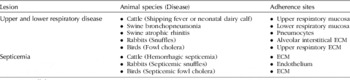
The mechanisms of P. multocida adherence (not colonization) to bovine cells or the bacterial factors involved in such adherence in BRD infections have been poorly addressed. Many pathogens including members of the family Pasteurellaceae exploit host extracellular matrix (ECM) molecules for virulence through adherence and colonization (Duensing and van Putten, Reference Duensing and van Putten1997, Reference Duensing and van Putten1998; Molinari et al., Reference Molinari, Talay, Valentin-Weigand, Rohde and Chhatwal1997; Duensing et al., Reference Duensing, Wing and van Putten1999; Bresser et al., Reference Bresser, Virkola, Jonsson-Vihanne, Jansen, Korhonen and van Alphen2000). The broad host range characteristic of P. multocida suggests recognition of host cell surface components (ECM molecules) common to multiple animal species and tissue types (Table 1). Recent studies in our laboratories investigated the binding of P. multocida to ECM molecules and demonstrated that bovine P. multocida isolates bind to several ECM molecules including fibronectin (Fn) (Dabo et al., Reference Dabo, Confer and Hartson2005). Bacteria bound both soluble and immobilized Fn and preferentially bound the N-terminal heparin-binding fragment (Hep-1) of Fn, similar to that described for other bacterial pathogens (Westerlund and Korhonen, Reference Westerlund and Korhonen1993; Patti et al., Reference Patti, Allen, McGavin and Hook1994; Joh et al., Reference Joh, Wann, Kreikemeyer, Speziale and Hook1999; Dabo et al., Reference Dabo, Confer and Hartson2005). The P. multocida proteins identified as putative Fn-binding proteins include P. multocida OmpA, the TonB-dependent receptor HgbA (hemoglobin-binding protein A), the transferrin-binding protein A (TbpA) and Pm10769 (Dabo et al., Reference Dabo, Confer and Quijano-Blas2003, Reference Dabo, Confer and Hartson2005). P. multocida OmpA bound Madin–Darby bovine kidney (MDBK) via heparin and/or Fn bridging (Dabo et al., Reference Dabo, Confer and Quijano-Blas2003), and monoclonal antibodies against OmpA significantly reduced or abolished bacterial binding to and invasion of MDBK cells (S. M. Dabo and A. W. Confer, unpublished data). The OmpA gene from bovine P. multocida strains exists as an eight-stranded transmembrane anti-parallel β-barrel that displays four surface-exposed loops with putative roles in adherence and serum resistance (Dabo et al., Reference Dabo, Confer and Quijano-Blas2003). In Escherichia coli, OmpA has been associated with virulence, functions as a multifunctional protein displaying phage receptor and porin activities (Datta et al., Reference Datta, Arden and Henning1977; Schweizer and Henning, Reference Schweizer and Henning1997), is involved in the bacterial invasion of host cells (Prasadarao, Reference Prasadarao2002; Prasadarao et al., Reference Prasadarao, Wass, Weiser, Stins, Huang and Kim1996) and contributes to serum resistance (Weiser and Gotschlich, Reference Weiser and Gotschlich1991).
Potential sites for Pasteurella multocida adherence in various diseases and lesions

ECM, extracellular matrix.
Two P. multocida regulatory genes have also been reported as potential virulence factors: DNA adenine methylase (Dam) and the Rci recombinase (rci) gene (Fuller et al., Reference Fuller, Kennedy and Lowery2000; Chen et al., Reference Chen, Paulsen, Scruggs, Banes, Reeks and Lawrence2003). In Salmonella Typhi, Rci protein inverted the DNA in the C-terminal region of the type IV pili, resulting in the inhibition of the minor pilus protein expression and the promotion of bacterial self-association (Morris et al., Reference Morris, Yip, Tsui, Wong and Hackett2003; Tam et al., Reference Tam, Hackett and Morris2004). Whether Rci of P. multocida has a similar action on the bacterium type IV pili is unknown. The dam gene regulated the expression of virulence genes in several Gram-negative bacteria including P. multocida (Blyn et al., Reference Blyn, Braaten and Low1990; Chen et al., Reference Chen, Paulsen, Scruggs, Banes, Reeks and Lawrence2003; Heusipp et al., Reference Heusipp, Falker and Schmidt2007). Overexpression or deletion of dam gene in several organisms resulted in attenuation for mice (Heithoff et al., Reference Heithoff, Sinsheimer, Low and Mahan1999; Chen et al., Reference Chen, Paulsen, Scruggs, Banes, Reeks and Lawrence2003; Badie et al., Reference Badie, Heithoff, Sinsheimer and Mahan2007). P. multocida serotype A harboring a plasmid-mediated alteration of dam expression was highly attenuated in mice (Chen et al., Reference Chen, Paulsen, Scruggs, Banes, Reeks and Lawrence2003). However, a Klebsiella pneumoniae dam mutant showed only partial attenuation in mice (Mehling et al., Reference Mehling, Lavender and Clegg2007).
Neuraminidase (sialidase) is prevalent among P. multocida strains and is expressed in vivo by P. multocida A:3 strains associated with bovine pneumonia (White et al., Reference White, Jolley, Purdy and Straus1995; Straus et al., Reference Straus, Cooley and Purdy1996; Ewers et al., Reference Ewers, Lubke-Becker, Bethe, Kiebling, Filter and Wieler2006); both cell-bound and extracellular sialidases were reported (White et al., Reference White, Jolley, Purdy and Straus1995). Its role in BRD, however, remains largely unknown. Evidence of neuraminidase function in P. multocida disease suggested that the protein is protective in mice (Ifeanyi and Bailie, Reference Ifeanyi and Bailie1992), is associated with virulence (Muller and Krasemann, Reference Muller and Krasemann1974) and may contribute to bacterial adherence, colonization and persistence (Mizan et al., Reference Mizan, Henk, Stallings, Maier and Lee2000). However, the relationship between the ability of P. multocida to produce the enzyme and to cause pneumonia in cattle has not been established. Two sialidase genes (nanB and nanH) were reported in 100 and 88.5% of P. multocida bovine strains investigated, respectively (Mizan et al., Reference Mizan, Henk, Stallings, Maier and Lee2000). Mutation in nanH resulted in a bacterium that was not deficient in sialidase production but had reduced enzyme activity (Mizan et al., Reference Mizan, Henk, Stallings, Maier and Lee2000). As reported by Steenbergen et al. (Reference Steenbergen, Lichtensteiger, Caughlan, Garfinkle, Fuller and Vimr2005) and reviewed by Harper et al. (Reference Harper, Boyce and Adler2006), Pm70 genome database contains several putative sialometabolic gene products, and the uptake, not catabolism, of sialic acid appeared essential for virulence in mice by protecting bacteria from innate host defense mechanisms that may promote persistence (Steenbergen et al., Reference Steenbergen, Lichtensteiger, Caughlan, Garfinkle, Fuller and Vimr2005; Harper et al., Reference Harper, Boyce and Adler2006).
In summary, determining the P. multocida genes involved in bacterial adherence, primarily during the early stage of infection, can assist in our understanding of the molecular pathogenesis of P. multocida. Changes in host–pathogen interaction resulting from changes in environmental signals are most likely the cause of differences between commensal and pathogenic P. multocida. The molecular differences between commensal and pathogenic P. multocida are not at present understood, but the mechanisms clearly involve the ability of the latter to induce inflammatory responses most likely through toll-like receptor (TLR) signaling. TLR signaling is crucial to innate host defense, which provides the first line of defense against infection through bacterial identification and killing and activation of adaptive immunity. Because a growing number of bacterial adhesins are reported to depend on TLR for induction of proinflammatory cytokines (Abramson et al., Reference Abramson, Kedem and Relman2001; Hajishengallis et al., Reference Hajishengallis, Martin, Sojar, Sharma, Schifferle, DeNardin, Russell and Genco2002; Ogawa et al., Reference Ogawa, Asai, Hashimoto and Uchida2002; Lorenz et al., Reference Lorenz, Chemotti, Vandal and Tessier2004; Inatsuka et al., Reference Inatsuka, Julio and Cotter2005; Fischer et al., Reference Fischer, Yamamoto, Akira, Beutler and Svanborg2006), future studies should focus on the identification of proinflammatory P. multocida adhesins as potential vaccine candidates.
LPS
P. multocida LPSs have similar properties (biological and chemical) and structure to R-type LPS (with no polymeric O-antigen) found in many Gram-negative bacteria (Lugtenberg et al., Reference Lugtenberg, van Boxtel and de Jong1984; Rimler, Reference Rimler1990; Confer, Reference Confer1993; Harper et al., Reference Harper, Boyce and Adler2006). These molecules induce classic signs of endotoxic shock (Rimler and Rhoades, Reference Rimler, Rhoades, Adlam and Rutter1989). The LPS structure has been determined for three P. multocida serotype A strains (VP161, X73 and Pm70) (St Michael et al., Reference St Michael, Li and Cox2005a, Reference St Michael, Li, Vinogradov, Larocque, Harper and Coxb, Reference St Michael, Vinogradov, Li and Coxc) and revealed a highly conserved inner but variable outer core (Harper et al., Reference Harper, Boyce and Adler2006). The investigation of the P. multocida Pm70 genome also identified gene clusters that may be responsible for LPS biosynthesis (St Michael et al., Reference St Michael, Vinogradov, Li and Cox2005c). Pathogenic bacteria expressing similar LPS include H. influenzae, Campylobacter jejuni, and Neisseria spp., and changes in the outer core oligosaccharides of these bacteria are often the result of phase-variation strategies in key LPS genes (Raetz and Whitfield, Reference Raetz and Whitfield2002). No such evidence was found in LPS genes in the P. multocida genome (Harper et al., Reference Harper, Boyce, Cox, St Michael, Wilkie, Blackall and Adler2007). Clusters of several putative glycosyltransferase genes, not always found in this genus, were identified in P. multocida Pm70 genome (May et al., Reference May, Zhang, Li, Paustian, Whittam and Kapur2001; St Michael et al., Reference St Michael, Vinogradov, Li and Cox2005c).
A novel finding of P. multocida LPS is the identification of two predicted heptosyl-1-transferases (designated HptA and HptB) expressing a single or two Kdo residue(s), respectively (St Michael et al., Reference St Michael, Li and Cox2005a). hptA is annotated as opsX in P. multocida Pm70 genome for its homology to the same gene in H. influenzae, and hptB is a homolog to waaC of E. coli and K. pneumoniae (St Michael et al., Reference St Michael, Li and Cox2005a; Harper et al., Reference Harper, Boyce, Cox, St Michael, Wilkie, Blackall and Adler2007). Virulence studies show that the hptB mutant was fully virulent (Harper et al., Reference Harper, Boyce, Cox, St Michael, Wilkie, Blackall and Adler2007), whereas the hptA mutant was fully attenuated (Harper et al., Reference Harper, Cox, St Michael, Wilkie, Boyce and Adler2004, Reference Harper, Boyce, Cox, St Michael, Wilkie, Blackall and Adler2007). Therefore, hptA may be critical for the survival of the organism in the host.
Purified P. multocida LPS is highly antigenic and capable of producing widespread vascular alterations and death, but it is poorly immunogenic (Heddleston and Rebers, Reference Heddleston and Rebers1975; Tsuji and Matsumoto, Reference Tsuji and Matsumoto1988; Ryu and Kim, Reference Ryu and Kim2000). Generally, P. multocida LPS is immunogenic or protective only when complexed with proteins (Ganfield et al., Reference Ganfield, Rebers and Heddleston1976; Tsuji and Matsumoto, Reference Tsuji and Matsumoto1988; Ryu and Kim, Reference Ryu and Kim2000) or ribosomes (Phillips and Rimler, Reference Phillips and Rimler1984). Ryu and Kim (Reference Ryu and Kim2000) demonstrated complete protection in mice vaccinated with P. multocida type A:3 LPS–protein complex; however, neither the LPS or protein alone nor the LPS and protein as a mixture provided protection. The authors also showed that the LPS–protein complex induced both humoral and cell-mediated immunity (Ryu and Kim, Reference Ryu and Kim2000). Previously, a bactericidal monoclonal antibody against LPS from a P. multocida serotype A isolate completely protected mice against homologous challenge (Wijewardana et al., Reference Wijewardana, Wilson, Gilmour and Poxton1990; Confer, Reference Confer1993). Sutherland et al. (Reference Sutherland, Davies and Murray1993) also found that polyclonal anti-idiotype antibodies that mimic a linear P. multocida LPS molecule protected mice against homologous challenge (Sutherland et al., Reference Sutherland, Davies and Murray1993). However, studies by Lu et al. (Reference Lu, Aguila, Lai and Pakes1991) failed to show protection in mice against P. multocida infection following passive immunization with affinity-purified rabbit anti-LPS serum. An opsonic but not bactericidal anti-LPS monoclonal antibody only partially protected against P. multocida infection in mice (Ramdani and Adler, Reference Ramdani and Adler1991; Confer, Reference Confer1993).
LPS is also known as a stimulator of host defense against P. multocida infection (Iovane et al., Reference Iovane, Pagnini, Galdiero, Cipollaro de l'Ero, Vitiello, D'Isanto and Marcatili1998; Galdiero et al., Reference Galdiero, Folgore, Nuzzo and Galdiero2000). Purified P. multocida LPS stimulated the expression and release of several cytokines in mice including the inflammatory (IL-1α, IL-6 and TNF-α) and immunoregulatory cytokines (IFN-γ and IL-12), which are known to play an important role in host defense (Iovane et al., Reference Iovane, Pagnini, Galdiero, Cipollaro de l'Ero, Vitiello, D'Isanto and Marcatili1998). Galdiero et al. (Reference Galdiero, Folgore, Nuzzo and Galdiero2000) also demonstrated a role for P. multocida LPS in neutrophil adhesion and migration through bovine endothelial cell monolayers. In another study, LPS from a P. multocida serotype A strain enhanced humoral and cell-mediated immune responses in chickens (Maslog et al., Reference Maslog, Motobu, Hayashida, Yoshihara, Morozumi, Matsumura and Hirota1999).
Capsule and the capsule biosynthetic genes
The genetic organization of the capsule biosynthetic loci from the five P. multocida serogroups was determined and reviewed (Chung et al., Reference Chung, Zhang and Adler1998; Boyce et al., Reference Boyce, Chung and Adler2000a; Townsend et al., Reference Townsend, Boyce, Chung, Frost and Adler2001; Harper et al., Reference Harper, Boyce and Adler2006). Comparative analysis of the five capsular biosynthetic regions identified serogroup-specific regions confirming a genetic basis for the serological differences between the observed strains (Carter, Reference Carter1952; Rimler and Rhoades, Reference Rimler, Rhoades, Adlam and Rutter1989; Townsend et al., Reference Townsend, Boyce, Chung, Frost and Adler2001).
The importance of capsule as virulence determinants in the pathogenesis of P. multocida has been clearly established; however, the focus to date has been primarily on P. multocida capsular types A and B from fowl cholera and hemorrhagic septicemia infections, respectively (reviewed in Harper et al., Reference Harper, Boyce and Adler2006) (Boyce and Adler, Reference Boyce and Adler2000; Chung et al., Reference Chung, Wilkie, Boyce, Townsend, Frost, Ghoddusi and Adler2001; Harper et al., Reference Harper, Boyce and Adler2006). Studies on the specific role of bovine P. multocida capsule in BRD infections are non-existent. Virulence studies in mice indicated that isogenic serotype A mutants were attenuated as compared to their encapsulated wild types (Jacques et al., Reference Jacques, Kobisch, Belanger and Dugal1993; Boyce and Adler, Reference Boyce and Adler2000; Chung et al., Reference Chung, Wilkie, Boyce, Townsend, Frost, Ghoddusi and Adler2001). Transposon insertion into a hyaluronan synthase gene of bovine P. multocida strain attenuated virulence in a septicemic mouse model (Fuller et al., Reference Fuller, Kennedy and Lowery2000). Watt et al. (Reference Watt, Swiatlo, Wade and Champlin2003) reported the loss of virulence of unencapsulated variants of a P. multocida serotype A strain and the restoration of virulence with the capsulated phenotype in mice. The capsular dissociation was not due to alteration in capsular gene nor was it regulated by the two-component (histidine kinase/response) regulator of capsule synthesis which was absent in P. multocida isolates under investigation as well as the Pm70 genome database.
Although P. multocida capsules are implicated in the virulence of P. multocida, vaccination of mice with capsule did not stimulate protection (Esslinger et al., Reference Esslinger, Seleim, Blobel, Pattern, Spencer, Johnson, Hoffmann and Lehane1993, Reference Esslinger, Seleim, Herrmann and Blobel1994; Gutierrez-Pabello et al., Reference Gutierrez-Pabello, Smith, Suarez-Guemes, Donachie, Lainson and Hodgson1995; Pruimboom et al., Reference Pruimboom, Rimler, Ackermann and Brogden1996). In general, many virulent strains of P. multocida express capsule consisting of hyaluronic acid (HA) (types A and B), chondroitin (type F) or heparin (type D) (Carter and Chengappa, Reference Carter and Chengappa1980; Muniandy and Mukkur, Reference Muniandy and Mukkur1993; Boyce et al., Reference Boyce, Chung and Adler2000b; DeAngelis and Padgett-McCue, Reference DeAngelis and Padgett-McCue2000; DeAngelis and White, Reference DeAngelis and White2002; DeAngelis et al., Reference DeAngelis, Jing, Drake and Achyuthan1998, Reference DeAngelis, Gunay, Toida, Mao and Linhardt2002) that are similar to molecules found naturally in the various hosts (Snipes et al., Reference Snipes, Ghazikhanian and Hirsh1987; Rimler and Rhoades, Reference Rimler, Rhoades, Adlam and Rutter1989; Tsuji and Matsumoto, Reference Tsuji and Matsumoto1989; DeAngelis, Reference DeAngelis1996). This ‘molecular mimicry’ prevents the mounting of a strong antibody response to capsular materials, impairs phagocytosis, reduces the action of complement killing and increases adherence to and survival in the host (Harmon et al., Reference Harmon, Glisson, Latimer, Steffens and Nunnally1991). Paradoxically, only P. multocida capsular B strains, and more specifically B:2 strains, produced hyaluronidase, an enzyme that specifically cleaves HA between its repeating disaccharides (N-acetylglucosamine and d-glucuronic acid) (Carter and Chengappa, Reference Carter and Chengappa1980; Rimler and Rhoades, Reference Rimler and Rhoades1994; Harper et al., Reference Harper, Boyce and Adler2006).
Antiphagocytic activity of P. multocida was reported and associated with the presence and thickness of capsule (Truscott and Hirsh, Reference Truscott and Hirsh1988; Harmon et al., Reference Harmon, Glisson, Latimer, Steffens and Nunnally1991; Pruimboom et al., Reference Pruimboom, Rimler, Ackermann and Brogden1996). Decapsulation of P. multocida serotype A:3 with hyaluronidase increased phagocytosis by macrophages, whereas an unencapsulated variant of the bacteria was not internalized (Pruimboom et al., Reference Pruimboom, Rimler, Ackermann and Brogden1996). Moreover, isogenic non-encapsulated P. multocida serotype A strains had significantly increased sensitivity to phagocytosis than the encapsulated wild-type strain (Boyce and Adler, Reference Boyce and Adler2000). Serum resistance correlated with encapsulation in P. multocida serotype A, because spontaneous, isogenic acapsular mutants or decapsulated strains were generally sensitive to complement-mediated killing compared to wild-type strains (Snipes and Hirsh, Reference Snipes and Hirsh1986; Hansen and Hirsh, Reference Hansen and Hirsh1989; Chung et al., Reference Chung, Wilkie, Boyce, Townsend, Frost, Ghoddusi and Adler2001). In contrast, acapsular P. multocida B2 mutants were resistant to complement-mediated killing (Boyce and Adler, Reference Boyce and Adler2000).
Conflicting reports exist with regard to the role of P. multocida capsule in adherence, which may be dependent both on the bacterial strain and on the type of host cells used in the studies. Pruimboom found that P. multocida adhesion to macrophages was mediated by capsular HA (Pruimboom et al., Reference Pruimboom, Rimler, Ackermann and Brogden1996). Enzymatic depolymerization of the capsule decreased P. multocida serotype A adherence to HeLa cells and macrophages (Esslinger et al., Reference Esslinger, Seleim, Herrmann and Blobel1994). Reduced adherence to macrophages was reported for a spontaneous P. multocida serotype A mutant (Pruimboom et al., Reference Pruimboom, Rimler, Ackermann and Brogden1996). P. multocida serotype A strains also had reduced adherence to peripheral blood monocytes as compared to air sac macrophages, and the exposure of monocytes to anti-HA-binding cell surface proteoglycan (CD44) monoclonal antibody decreased bacterial binding (Pruimboom et al., Reference Pruimboom, Rimler and Ackermann1999). In contrast, Glorioso et al. (Reference Glorioso, Jones, Rush, Pentler, Darif and Coward1982) showed increased adherence of P. multocida serogroup A to HeLa cells and rabbit pharyngeal cells following enzymatic depolymerization of the capsule. Others demonstrated that the absence of capsule increases P. multocida adherence to and colonization of lung and tracheal by cells (Jacques et al., Reference Jacques, Belanger, Diarra, Dargis and Malouin1994).
OMPs
Several P. multocida OMPs possess virulence attributes including surface exposure, in vivo expression, immunogenic and bactericidal properties (Confer, Reference Confer1993; Confer et al., Reference Confer, Nutt, Dabo, Panciera and Murphy1996; Marandi and Mittal, Reference Marandi and Mittal1996; Dabo et al., Reference Dabo, Confer and Murphy1997; Vasfi Marandi and Mittal, Reference Vasfi Marandi and Mittal1997; Gatto et al., Reference Gatto, Dabo, Hancock and Confer2002; Borrathybay et al., Reference Borrathybay, Sawada, Kataoka, Ohtsu, Takagi, Nakamura and Kawamoto2003; Davies et al., Reference Davies, MacCorquodale and Reilly2004). Our studies of bovine P. multocida serogroup A isolates identified several surface-exposed and in vivo-expressed OMPs, including an E. coli OmpA and a H. influenzae type b (Hib) P1 homolog proteins (Dabo et al., Reference Dabo, Confer and Murphy1997). P. multocida OmpA is a major immunogenic and antigenic OMP that is conserved, surface-exposed and expressed in vivo (Dabo et al., Reference Dabo, Confer and Murphy1997, Reference Dabo, Confer and Quijano-Blas2003). High antibody responses to this and several other OMPs consistently correlated with resistance of cattle to challenge with virulent P. multocida (Dabo et al., Reference Dabo, Confer and Murphy1997, Reference Dabo, Confer and Quijano-Blas2003).
OmpH is another major antigenic, surface-exposed and conserved OMP porin that is detected in 100% of bovine isolates investigated (Lugtenberg et al., Reference Lugtenberg, van Boxtel and de Jong1984, Reference Lugtenberg, van Boxtel, Evenberg, de Jong, Storm and Frik1986; Chevalier et al., Reference Chevalier, Duclohier, Thomas, Shechter and Wroblewski1993; Ewers et al., Reference Ewers, Lubke-Becker, Bethe, Kiebling, Filter and Wieler2006) and has potential as a vaccine candidate (Luo et al., Reference Luo, Glisson, Jackwood, Hancock, Bains, Cheng and Wang1997, Reference Luo, Zeng, Glisson, Jackwood, Cheng and Wang1999; Vasfi Marandi and Mittal, Reference Vasfi Marandi and Mittal1997). The protein provided protection against P. multocida challenge in mice (Vasfi Marandi and Mittal, Reference Vasfi Marandi and Mittal1997) and chickens (Luo et al., Reference Luo, Glisson, Jackwood, Hancock, Bains, Cheng and Wang1997, Reference Luo, Zeng, Glisson, Jackwood, Cheng and Wang1999), and its expression was shown to be negatively regulated by iron and glucose. Electrophoretic analysis showed that OmpH levels increase in P. multocida fur mutant compared to wild-type strain (Bosch et al., Reference Bosch, Tarrago, Garrido, Campoy, Fernandez de Henestrosa, Perez de Rozas, Badiola and Barbe2001). The expression of OmpH–lacZ fusion in the wild-type strain increases in the presence of chelating agent 2,2′-dipyridyl (DPD), and there was a 5-fold decline in the expression of the gene in the wild type as compared to the fur mutant when grown in the presence of glucose. Given the importance of OmpH as a potential immunogen and the observed inhibitory effect on OmpH expression, those authors recommended growing bacteria in the absence of glucose for bacterin production (Bosch et al., Reference Bosch, Tarrago, Garrido, Campoy, Fernandez de Henestrosa, Perez de Rozas, Badiola and Barbe2001).
Several in vivo studies identified potential P. multocida virulence genes and OMP-associated cross-protection factors (Rimler, Reference Rimler1994; Wang and Glisson, Reference Wang and Glisson1994a, Reference Wang and Glissonb; Hunt et al., Reference Hunt, Citron, Claros, Fernandez and Goldstein2001). When using antisera produced against live P. multocida isolates, Wang and Glisson (Reference Wang and Glisson1994a, Reference Wang and Glissonb) found four high-molecular-weight proteins (ranging from 204 to 153 kDa) that stimulate cross-protection. Hunt et al. (Reference Hunt, Citron, Claros, Fernandez and Goldstein2001) used in vivo expression technology (IVET) to identify two lipoproteins (PM0554 and PM14440) with homology to conserved and widely distributed PCP (peptidoglycan-associated lipoprotein (Pal) cross-reaction protein), and protein D of non-typeable H. influenzae, respectively. The H. influenzae protein D mediates the binding to IgD, a form of immune evasion, and stimulates cross-protection against heterologous challenge in rats (Akkoyunlu et al., Reference Akkoyunlu, Janson, Ruan and Forsgren1996). Anti-PCP serum was bactericidal against H. influenzae clinical isolates (Deich et al., Reference Deich, Anilionis, Fulginiti, Metcalf, Quataert, Quinn-Dey, Zlotnick and Green1990). P. multocida PCP was found to be surface-exposed, while protein D (designated GlpQ) was not (Lo et al., Reference Lo, Boyce, Wilkie and Adler2004); however, neither protein stimulated protection against challenge with live P. multocida in mice and chickens. Boyce et al. (Reference Boyce, Cullen, Nguyen, Wilkie and Adler2006) found significant up-regulation of Pm0803 and the ef-Tu (elongation factor-Tu) during growth in vivo and significant down-regulation of OmpW. ef-Tu (tufA and tufB) is cytoplasmic in location but may be targeted to the outer membrane since its homologs bind host fibronectin in Mycoplasma pneumoniae and host mucin in Lactobacillus johnsonii (Boyce et al., Reference Boyce, Cullen, Nguyen, Wilkie and Adler2006). While the major P. multocida surface proteins have been studied, little is known about the less abundant proteins and their roles in the pathogenesis of P. multocida infections in general.
Iron-regulated and acquisition proteins – IROMPs
Iron acquisition and uptake are essential for bacterial survival and pathogenic bacteria have developed different strategies for that uptake. P. multocida produces both iron-chelating siderophores and outer membrane receptors for the iron-binding host molecules (transferrin, hemoglobin and hemin) (Choi-Kim et al., Reference Choi-Kim, Maheswaran, Felice and Molitor1991; Ogunnariwo et al., Reference Ogunnariwo, Alcantara and Schryvers1991). Early studies showed that some P. multocida isolates produced siderophores (Choi-Kim et al., Reference Choi-Kim, Maheswaran, Felice and Molitor1991) or contained only one specific Tbp (TbpA) (Ogunnariwo and Schryvers, Reference Ogunnariwo and Schryvers2001; Ogunnariwo et al., Reference Ogunnariwo, Alcantara and Schryvers1991). P. multocida grown under iron-depleted media or in vivo expressed three iron-regulated OMPs (IROMPs) with molecular masses of 76, 84 and 94 kDa, with all three having affinity for siderophore binding (Choi-Kim et al., Reference Choi-Kim, Maheswaran, Felice and Molitor1991).The P. multocida tbpA gene was absent in avian isolates and detected only in ruminants (cattle, 70%; sheep, 80%; and buffalo, 57.1%) (Ewers et al., Reference Ewers, Lubke-Becker, Bethe, Kiebling, Filter and Wieler2006). TbpA from P. multocida serotype B:2 strain was predicted to be a gated pore with several membrane-spanning regions (Shivachandra et al., Reference Shivachandra, Kumar, Amaranath, Joseph, Srivastava and Chaudhuri2005). The specific role of TbpA in P. multocida virulence is still unknown.
Analysis of attenuated mutants by Fuller et al. (Reference Fuller, Kennedy and Lowery2000) identified two loci containing potential virulence genes with homology to iron-acquisition-related genes ExbB and hgbA of H. influenzae. ExbB, along with exbD, are part of the tonB operon in P. multocida and are independently transcribed (Bosch et al., Reference Bosch, Garrido, Llagostera, Perez de Rozas, Badiola and Barbe2002a). In a related study, Bosch et al. (Reference Bosch, Garrido, Llagostera, Perez De Rozas, Badiola and Barbe2002b) demonstrated that three ORFs (PM0298, PM0299 and PM300) of P. multocida constitute a single transcriptional operon. Mutants defective in PM0298 or PM0299 demonstrated that these genes were essential for the viability of the pathogen (Bosch et al., Reference Bosch, Garrido, Llagostera, Perez De Rozas, Badiola and Barbe2002b). The product of PM300 (designated HgbA) bound hemoglobin (Bosch et al., Reference Bosch, Garrido, Llagostera, Perez De Rozas, Badiola and Barbe2002b) and hemin (Bosch et al., Reference Bosch, Garrido, Perez de Rozas, Badiola, Barbe and Llagostera2004), but binding and virulence of the hgbA-mutant strain were not affected (Bosch et al., Reference Bosch, Garrido, Llagostera, Perez De Rozas, Badiola and Barbe2002b). Similarly, insertional inactivation of hgbB did not affect the ability of P. multocida to bind hemoglobin (Cox et al., Reference Cox, Hunt, Boyce and Adler2003), suggesting the presence of additional hemoglobin receptor proteins in the P. multocida genome. Consistent with that finding, comparative analysis of the P. multocida Pm70 genome (May et al., Reference May, Zhang, Li, Paustian, Whittam and Kapur2001) identified nine iron-acquisition-related genes (including hgbA) encoding immunogenic proteins (Bosch et al., Reference Bosch, Garrido, Perez de Rozas, Badiola, Barbe and Llagostera2004). Six proteins bound both hemin and hemoglobin and the other two bound either hemin or hemoglobin. These proteins did not stimulate protective immunity in mice against P. multocida infection when inoculated alone (Bosch et al., Reference Bosch, Garrido, Perez de Rozas, Badiola, Barbe and Llagostera2004). P. multocida HbpA (hemin-binding protein A), another iron acquisition gene, was shown to be immunogenic and regulated through a Fur-independent mechanism; HbpA-specific antibodies were not protective in mice (Garrido et al., Reference Garrido, Bosch, Medina, Bigas, Llagostera, Perez De Rozas, Badiola and Barbe2003). The prevalence of tonB, tbpA, hgbA and hgbB genes in P. multocida bovine strains is reported to be 100, 70.2, 95.2 and 57.7%, respectively (Ewers et al., Reference Ewers, Lubke-Becker, Bethe, Kiebling, Filter and Wieler2006), and there was significant association between disease status and the presence of hgbB and tbpA as single virulence-associated genes in P. multocida bovine isolates. Our characterization of a heme acquisition system receptor (HasR) in a P. multocida bovine isolate demonstrated that the protein, which is 98% identical to its homolog in the P. multocida Pm70 genome sequence, is surface-exposed, is conserved among most P. multocida isolates and is an immunodominant IROMP (Prado et al., Reference Prado, Dabo and Confer2005). Finally, a recent comparative transcriptional response to iron limitation between M. haemolytica and P. multocida found few homologous genes induced in both organisms and include the hemoglobin receptor hmbR2, the fbpAB, yfeABCD ABC-type systems, tonB and exbD (Roehrig et al., Reference Roehrig, Tran, Spehr, Gunkel, Selzer and Ullrich2007). The hemin and hemoglobin transporter genes were among the genes uniquely found up-regulated in P. multocida, an indication of its capacity to utilize iron from blood sources (Roehrig et al., Reference Roehrig, Tran, Spehr, Gunkel, Selzer and Ullrich2007).
Extracellular enzymes
Several extracellular enzymes such as proteases and lipases have also been reported as possible virulence factors in P. multocida (Negrete-Abascal et al., Reference Negrete-Abascal, Tenorio and de la Garza1999; Pratt et al., Reference Pratt, Cooley, Purdy and Straus2000). The role of lipases in pathogenic bacteria is probably nutritional, whereas proteases may assist pathogens against host defense degrading host IgG and reducing opsonization. Negrete-Abascal (Reference Negrete-Abascal, Tenorio and de la Garza1999) reported the secretion of proteases into the culture supernatant of P. multocida from different species including bovine. Those proteases were capable of degrading host IgG in a wide range of pH (Negrete-Abascal et al., Reference Negrete-Abascal, Tenorio and de la Garza1999). Pratt et al. (Reference Pratt, Cooley, Purdy and Straus2000) demonstrated the production of lipases by clinical isolates of P. multocida from different species. The role, if any, these enzymes play in the pathogenesis of P. multocida infections is unknown.
Immunity against P. multocida serogroup A in cattle
Immunity of cattle to P. multocida has been investigated most extensively with respect to hemorrhagic septicemia of cattle and buffalo involving serotypes B:2 and E:2 (Confer, Reference Confer1993). Studies on immunity in cattle pneumonia caused by P. multocida serotype A isolates are limited. This section of the review will focus on current knowledge of bovine immunity against P. multocida serotype A isolates.
Important antigens responsible for immunity against P. multocida serotype A isolates in cattle are not completely understood. Adler et al. (Reference Adler, Bulach, Chung, Doughty, Hunt, Rajakumar, Serrano, Van Zanden, Zhang and Ruffolo1999) reviewed candidate vaccine antigens and genes in P. multocida related to various diseases of production animals and indicated that LPS can provide some protection against homologous serotypes. They further reviewed several putative immunologically important antigens by focusing on studies that focused on antigens expressed under in vivo conditions. These include OMP Oma87, which is a homologue of the D15 protective antigen of H. influenzae; type 4 fimbria and its subunit protein PtfA; capsule; and IROMPs, such as the Tbp TbpA. None of these putative vaccine candidates were directly investigated with respect to stimulating immunity in cattle against P. multocida serotype A. The gene for Oma87 has been cloned and expressed; however, vaccination against that protein failed to protect chickens against an experimental challenge (Mitchison et al., Reference Mitchison, Wei, Kwang, Wilkie and Adler2000; Chaudhuri and Goswami, Reference Chaudhuri and Goswami2001). The ptfA gene was demonstrated in most bovine P. multocida isolates and has been cloned (Adler et al., Reference Adler, Bulach, Chung, Doughty, Hunt, Rajakumar, Serrano, Van Zanden, Zhang and Ruffolo1999; Ewers et al., Reference Ewers, Lubke-Becker, Bethe, Kiebling, Filter and Wieler2006); however, immunogenicity was not reported. The potential for TbpA to be a good vaccine candidate is questionable, because approximately 30% of bovine P. multocida strains lack the tbpA gene (Ewers et al., Reference Ewers, Lubke-Becker, Bethe, Kiebling, Filter and Wieler2006; Roehrig et al., Reference Roehrig, Tran, Spehr, Gunkel, Selzer and Ullrich2007). We suggested that OMPs from bovine P. multocida serotype A strains may be good vaccine candidates based on correlations between high antibodies and resistance against experimental bovine P. multocida-induced pneumonia (Confer et al., Reference Confer, Nutt, Dabo, Panciera and Murphy1996). However, Abdullahi et al. (Reference Abdullahi, Gilmour and Poxton1990), using bovine P. multocida serotype A isolates, questioned the potential efficacy of an OMP vaccine based on data obtained from vaccination of mice with whole bacterial cell vaccines followed by intraperitoneal challenge.
Immunity in bovine respiratory pasteurellosis is presumed to be antibody-mediated. Anti-P. multocida antibodies were demonstrated in dairy cow colostrum by quantitative indirect immunofluorescence and agglutination assays (Gresham et al., Reference Gresham, Confer, Bush and Rummage1984). Passive antibodies to P. multocida serotype A were demonstrated in sera from newborn dairy and beef calves, and those passively acquired antibodies were short-lived in sera compared to values of 4–6 months for antiviral antibodies demonstrated in one of these studies (Virtala et al., Reference Virtala, Mechor, Grohn, Erb and Dubovi1996; Fulton et al., Reference Fulton, Briggs, Payton, Confer, Saliki, Ridpath, Burge and Duff2004; Step et al., Reference Step, Confer, Kirkpatrick, Richards and Fulton2005; Prado et al., Reference Prado, Prado, Payton and Confer2006). Virtala et al. (Reference Virtala, Mechor, Grohn, Erb and Dubovi1996) found no correlation between postcolostral antibody titers in dairy calves with and without naturally acquired respiratory disease, usually associated with P. multocida and/or Mycoplasma dispar infection. Passively acquired anti-P. multocida OMP antibodies were demonstrated in dairy calf sera within 24 h of birth (Fulton et al., Reference Fulton, Briggs, Payton, Confer, Saliki, Ridpath, Burge and Duff2004; Step et al., Reference Step, Confer, Kirkpatrick, Richards and Fulton2005). Step et al. (Reference Step, Confer, Kirkpatrick, Richards and Fulton2005) demonstrated that while dairy calves were isolated in hutches, P. multocida antibody concentrations declined to a nadir between 47 and 73 days of age. Those calves were subsequently removed from hutches, grouped together and spontaneously seroconverted to P. multocida, most likely from exposure to the bacterium from other calves and from fomites or from stress-related factors stimulating commensal P. multocida proliferation. In beef calves, similar passive/acquired anti-P. multocida antibody responses were found in calves from two different herds (Prado et al., Reference Prado, Prado, Payton and Confer2006). Serum concentrations of anti-P. multocida antibodies in neonatal calves varied greatly between the two herds. In both herds, however, passively acquired IgG1 antibodies declined rapidly and reached their nadir between 60 and 90 days of age. This was followed by a rapid rise in anti-P. multocida IgM and IgG2 antibodies that reached peak concentrations between 150 and 180 days of age. Although, at this time, protection against respiratory disease has not been associated with passively acquired anti-P. multocida antibodies, their short-lived nature negates any potentially protective role after approximately 3 months of age.
As indicated above, active antibody responses to P. multocida develop in calves at a young age at least as measured by ELISA (Virtala et al., Reference Virtala, Mechor, Grohn, Erb and Dubovi1996; Step et al., Reference Step, Confer, Kirkpatrick, Richards and Fulton2005; Prado et al., Reference Prado, Prado, Payton and Confer2006). Whether antibodies acquired naturally or from vaccination truly protect cattle against P. multocida-associated pneumonia is not well documented. Serum anti-P. multocida antibody concentrations were associated with several cattle production parameters in a study of cattle shipped from 24 different ranches to a feedlot without going through the order buyer/sale barn marketing system (Fulton et al., Reference Fulton, Cook, Step, Confer, Saliki, Payton, Burge, Welsh and Blood2002). In that study, high antibodies to P. multocida OMPs at the time of feedlot entry were associated with increased average daily gain during feeding, whereas low P. multocida antibodies were associated with a decreased net value to owner, decreased gross margin and increased total costs of finishing. There was, however, no association between health or treatment costs and P. multocida antibody concentrations. Few of the represented herds had been vaccinated with P. multocida vaccines. Therefore, it is assumed that the antibodies measured were from prior natural exposure.
Enhanced resistance to respiratory P. multocida challenge has been demonstrated in several studies using live vaccines. These include wild-type bacteria, streptomycin-dependent mutant and chemically modified P. multocida. (Kucera et al., Reference Kucera, Wong and Eis1981; Panciera et al., Reference Panciera, Corstvet, Confer and Gresham1984; Chengappa et al., Reference Chengappa, McLaughlin, Kadel, Maddux and Greer1989; Confer et al., Reference Confer, Nutt, Dabo, Panciera and Murphy1996; Mathy et al., Reference Mathy, Mathy, Lee, Walker, Lofthouse and Meeusen2002). In addition, Kadel et al. (Reference Kadel, Chengappa and Herren1985) demonstrated greater weight gains, less severe clinical signs of pneumonia and lower death rates in calves vaccinated with both live streptomycin-dependent P. multocida and live streptomycin-dependent M. haemolytica when compared to non-vaccinated calves. In several studies, a correlation was demonstrated between high serum P. multocida antibodies and resistance against experimental challenge in live P. multocida-vaccinated cattle. Compared to controls, cattle vaccinated with live P. multocida via aerosol or subcutaneous routes had significant reduction in lesion scores (Panciera et al., Reference Panciera, Corstvet, Confer and Gresham1984) or reduced histologic lesions, reduced neutrophil influx, reduced IL8 and IL1β, and increased IL2 responses (Mathy et al., Reference Mathy, Mathy, Lee, Walker, Lofthouse and Meeusen2002). Live P. multocida-vaccinated calves also developed serum antibodies against the whole bacteria (Panciera et al., Reference Panciera, Corstvet, Confer and Gresham1984) or to outer membrane preparations (Confer et al., Reference Confer, Nutt, Dabo, Panciera and Murphy1996) using quantitative indirect immunofluorescence assay or ELISA, respectively. In both studies, there was a significant correlation (P<0.05) between high antibodies against P. multocida and low lesion scores following transthoracic challenge. In addition, using Western blotting and densitometric analyses, high serum antibodies against six specific OMPs (approximately 100, 90, 85, 74, 53, 35 and 16 kDa) significantly correlated with less severe lesions following P. multocida challenge (Confer et al., Reference Confer, Nutt, Dabo, Panciera and Murphy1996). Amino-terminus sequencing demonstrated that the 35 kDa protein is a heat modifiable (28–35 kDa) OMP from the OmpA family of proteins (Dabo et al., Reference Dabo, Confer and Murphy1997); however, vaccination of mice with the partially purified protein did not protect against intraperitoneal P. multocida challenge (Gatto et al., Reference Gatto, Dabo, Hancock and Confer2002). Another P. multocida OMP shown to be of importance in stimulating resistance to infection in animals is OmpH, an approximately 33.6–38.5 kDa protein (Vasfi Marandi and Mittal, Reference Vasfi Marandi and Mittal1997; Davies and Lee, Reference Davies and Lee2004). That protein, however, has not been studied immunologically in cattle.
Studies of vaccination of cattle with non-living P. multocida vaccines have also been limited. Early studies suggested some protection of cattle against shipping fever using Pasteurella spp. bacterins (reviewed in Collins, Reference Collins1977). Vaccination of calves with a vaccine containing modified-live- Parainfluenza 3 virus, killed M. haemolytica and killed P. multocida vaccine enhanced resistance against experimental challenge involving stress and aerosol exposure to all three infectious agents (Matsuoka et al., Reference Matsuoka, Gale, Ose and Berkman1966). In more recent years, however, neither aerosol nor intratracheal vaccination of calves with formalin-killed P. multocida resulted in protection against experimental pulmonary challenge (Confer et al., Reference Confer, Nutt, Dabo, Panciera and Murphy1996; Dowling et al., Reference Dowling, Hodgson, Dagleish, Eckersall and Sales2004; Prado et al., Reference Prado, Dabo and Confer2005). In our laboratory, we demonstrated that calves vaccinated with Freund's incomplete adjuvant–outer membrane preparations from P. multocida cultured in the presence or absence of an iron chelator (IROMPs or OMPs, respectively) developed antibodies to OMPs and IROMPs, as detected by ELISA (Prado et al., Reference Prado, Dabo and Confer2005). Vaccination with OMPs or IROMPs resulted in significant decrease in lesion scores following P. multocida challenge compared to vaccination with adjuvant alone. Of specific interest was that vaccinated calves developed intense antibody responses to a 96 kDa protein band, and correlation between high anti-96 kDa antibodies and low lesion scores approached significance (P<0.06). The 96 kDa OMP was a homologue to the iron-regulated protein HasR, a heme acquisition receptor protein. The hasR gene, however, was not demonstrated in all P. multocida isolates examined. Therefore, its potential immunological significance is not known.