INTRODUCTION
Memory encompasses the stages of acquisition, consolidation, and retrieval. Acquisition refers to the uptake of new information during learning and its encoding into a vulnerable memory trace. In contrast, consolidation is a process of stabilization of newly encoded memory traces. Memory theories differentiate between short-term memory (STM) and long-term memory (LTM). Short-term memory is a transient memory for an experience that does not require synthesis of new proteins or ribonucleic acid (RNA) and typically ranges from a few seconds to a couple of hours after acquisition. Long-term memory is a relatively stable memory that develops over time and depends on synthesis of new proteins and RNA. The consolidation of long-term memory takes several hours (Nader, Reference Nader2003).
Neuropsychological results in amnesic patients, animal studies, and imaging studies in humans have provided strong evidence that declarative long-term memory depends on the medial temporal lobe, especially the hippocampus (Milner, Squire, & Kandel, Reference Milner, Squire and Kandel1998). Memory deficits, however, are a common finding after frontal lobe lesions, as well (Schacter, Reference Schacter1987). Imaging studies in the context of memory relate activity in frontal areas to memory formation and encoding, as well as short-term maintenance and manipulation of information (Buckner, Kelley, & Petersen, Reference Buckner, Kelley and Petersen1999).
It is a matter of debate whether short-term and long-term memory should be regarded as two anatomically dissociable memory systems that work in parallel (Baddeley & Warrington, Reference Baddeley and Warrington1970), or whether they should be conceptualized as different neural circuits within similar cortical areas (Ranganath & Blumenfeld, Reference Ranganath and Blumenfeld2005). A double dissociation has been described between verbal short-term and long-term memory (Carlesimo, Marfia, Loasses, & Caltagirone, Reference Carlesimo, Marfia, Loasses and Caltagirone1996; Markowitsch et al., Reference Markowitsch, Kalbe, Kessler, von Stockhausen, Ghaemi and Heiss1999). However, this double dissociation has also been disputed (Nairne, Reference Nairne2002; Ranganath & Blumenfeld, Reference Ranganath and Blumenfeld2005).
Epilepsy has been used as a model to study memory processes, as different epilepsy diagnoses are associated with dissociable memory deficits, depending on the underlying pathophysiological processes. For example, temporal lobe epilepsy (TLE) is caused by structural and functional changes in the medial temporal lobe and has been linked to deficits in the consolidation of long-term memories (Elger, Helmstaedter, & Kurthen, Reference Elger, Helmstaedter and Kurthen2004). In contrast, idiopathic generalized epilepsy (IGE) is caused by subtle structural and functional changes in frontal and prefrontal areas (Betting et al., Reference Betting, Mory, Li, Lopes-Cendes, Guerreiro, Guerreiro and Cendes2006; Swartz et al., Reference Swartz, Halgren, Simpkins, Fuster, Mandelkern and Krisdakumtorn1996), as well as disruptions in the frontal-thalamic system (Bernasconi et al., Reference Bernasconi, Bernasconi, Natsume, Antel, Andermann and Arnold2003). It has been shown that IGE is related to short-term memory deficits (Pascalicchio et al., Reference Pascalicchio, de Araujo Filho, da Silva Noffs, Lin, Caboclo and Vidal-Dourado2007). In a recent study in IGE and TLE patients we found a double dissociation between immediate and 24-hour long-term face recognition (Bengner et al., Reference Bengner, Malina, Lindenau, Voges, Goebell and Stodieck2006). IGE patients showed decreased immediate recognition, but, surprisingly, their long-term recognition did not differ from healthy individuals. In contrast, TLE patients showed normal immediate recognition, but long-term memory was markedly decreased.
In the present study, we sought to validate our former results by independent patient samples using refined methodology. We controlled for differences in face perception between groups and implemented a distractor task between the study phase and immediate recognition to fully rule out rehearsal from working memory. Moreover, a significantly larger number of study and test items were used to raise the reliability of results. Based on our former findings and the literature referred to earlier, we hypothesized that a double dissociation of short-term and long-term face memory exists: IGE patients should show decreased immediate face recognition, but no consolidation deficit. In contrast, TLE patients should exhibit a normal immediate recognition, but decreased 24-hour face recognition.
METHODS
Participants
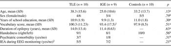
Ten participants with temporal lobe epilepsy (TLE), nine participants with idiopathic generalized epilepsy (IGE), and ten healthy control participants were tested for this study (Table 1). The healthy control participants did not report any neurological or psychiatric disorder. The study was approved by the local ethics committee and all participants gave written informed consent.
Characteristics of patients and control participants

Note
aANOVA; bChi-square test. TLE = temporal lobe epilepsy; IGE = idiopathic generalized epilepsy; EEG = electroencephalogram.
1 Data of one patient were not included into the group mean because he was not a native German speaker.
2 IEA = interictal epileptic abnormalities. Patients without IEAs during EEG monitoring were diagnosed as having TLE only if a sufficient number of auras or seizures could be classified as typical of a temporal focus.
All patients underwent a diagnostic 48- to 72-hour continuous video/electroencephalogram (EEG)-monitoring. Epilepsy was diagnosed when recurrent unprovoked seizures of an unquestionable epileptic nature could be verified from the patient’s history or during video/EEG monitoring. The diagnosis of TLE was based on reliable information about clinical seizure patterns typical of a temporal focus (aura type and other ictal clinical phenomena). Eight of the ten TLE patients showed structural abnormalities in the MRI, such as hippocampal sclerosis or atrophy, temporal lobe atrophy, or cavernoma. Four patients showed interictal epileptic abnormalities and/or MRI abnormalities in the left hemisphere, three in the right hemisphere, and one patient in both hemispheres. Because of the small sample size, data were not analyzed with respect to the lateralization of structural abnormalities and epileptic focus.
In addition, we included nine patients with idiopathic generalized epilepsy. Idiopathic generalized epilepsy was diagnosed in patients with a typical semiology of juvenile myoclonic epilepsy, absence seizures, or primary generalized tonic-clonic seizures, and generalized EEG patterns during video/EEG monitoring.
All patients took antiepileptic medications and the dosage of medication was not lowered during the EEG monitoring and the 24-hour retention phase (see experimental design). None of the patients had seizures during the retention phase.
The Benton Facial Recognition Test (Benton, Sivan, Hamsher, Varney, & Spreen, Reference Benton, Sivan, Hamsher, Varney and Spreen1994) was administered to test face perception without any memory demand. All TLE participants showed test scores within the normal range. One IGE patient had a score of 39, which is classified as “borderline” according to Benton et al. (Reference Benton, Sivan, Hamsher, Varney and Spreen1994), but cannot be regarded as a deficit in face perception.
Material and Procedure
Three hundred gray-scale photographs of unfamiliar human faces were used as stimulus material. Faces were assigned to one of three lists of pictures, which were parallel with regard to number of male/female faces and emotional expression. One list was used as the learning list, one as the distractor list for the immediate recognition test, and the third as the distractor list for the 24-hour recognition test. The assignment of lists to the experimental phases was counterbalanced across participants.
All participants took part in three study phases: a learning phase, an immediate recognition test, and a recognition test after 24 hours. During the study phase, participants were shown a set of 100 unfamiliar face stimuli. The order of stimuli was randomized across participants. Stimuli were presented in blocks of 20 faces each. A trial started with the presentation of a white fixation cross on a black background (1000 ms), followed by a face stimulus presented for 2000 ms. Participants were asked to judge the sex of the face and indicate their answer with a button press on a standard computer keyboard (left vs. right arrows). The sex judgment task ensured a similar processing strategy across participants. After each learning phase, participants counted backwards from 100 (naming every second number) to reduce retrieval from working memory.
During the immediate recognition phase, 40 faces were presented in a random sequence – 20 familiar faces seen before in the learning phase and 20 new, unfamiliar faces. The timing of stimuli was the same as in the learning phase except that a button press before 2000 ms terminated the presentation of the face stimulus. Participants were asked to judge by button press whether they had seen the face during the learning phase or not. The stress was on accuracy, but participants were told to respond as fast as possible. The cycle of learning and retrieval phases was repeated five times until participants processed 100 faces of the learning list and 100 new faces. After 24 hours, a second recognition test took place. The setup and instructions were the same as during immediate recognition, but 100 new faces were introduced as distractors.
Data Analysis
Memory performance was parameterized by combining hit rates (correct recognition of old faces) and false memory rates (erroneous recognition of new faces) to the sensitivity value d′. d′ was defined as z[p(hit)] – z[p(false memories)], where z scores were derived from the inverse cumulative normal distribution. This allows comparing recognition scores between groups not influenced by possible response biases (e.g., a tendency to say “yes”). For reaction time analyses, correct detections of “old” faces were analyzed. Trials with reaction times below 500 ms or more than two standard deviations above the individual mean reaction times were disregarded to exclude outliers.
For statistical analyses, analyses of variance (ANOVA) were run with the between participant factor Group and the within participant factors Time (immediate/24 hours). Significant interactions with the factor Group were further explored by planned contrasts.
RESULTS
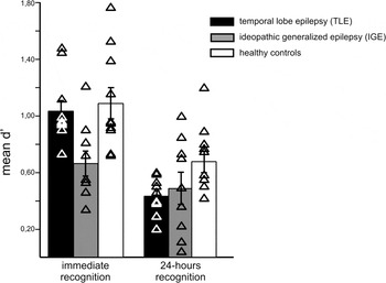
Patients with temporal lobe epilepsy did not show any deficit in the immediate recognition test, but their memory scores were reduced as compared to healthy controls after 24 hours (Figure 1). The patients with idiopathic generalized epilepsy showed a different pattern of results: Their memory scores were already reduced as compared to healthy controls and TLE patients in the immediate test, but they showed a smaller decrease after 24 hours.
Memory scores for unfamiliar faces (measured as d′) for the immediate and 24-hour recognition tests, shown separately for patients with temporal lobe epilepsy, idiopathic generalized epilepsy, and healthy participants. Error bars indicate the standard error of the mean. Results of single participants are depicted as triangles.

These differences between groups were supported by a significant Time x Group interaction, F(2, 26) = 8.05, p = .002, and a main effect of Group, F(2, 26) = 3.74, p = .038, in the overall ANOVA. In the immediate recognition test, the scores of patients with TLE did not differ significantly from those of the healthy control participants (contrast analysis p = .69). The performance of the IGE patients, however, was markedly reduced as compared to both the TLE group and the healthy control group (contrast analysis all p < .02). In the 24-hour test, TLE patients showed a worse performance as compared to the healthy control group (contrast analysis p = .014), but their performance did not differ from that of the IGE patients (p = .64). The difference between IGE patients and healthy controls was not significant (p = .19).
We also compared the decline of memory after 24 hours between groups by analyzing the difference between d′ after 24 hours and the immediate d′ score. There was a significant effect of Group on this difference score, F(2, 26) = 8.05, p = .002. Planned contrasts showed that TLE patients lost more items than IGE patients and healthy controls [contrast analysis –1 vs. 0.5, 0.5, i.e., TLE vs. (IGE and controls), p = .002]. In contrast, IGE patients showed less decline than TLE patients and healthy controls combined [contrast analysis –1 vs. 0.5, 0.5, i.e., IGE vs. (TLE and controls), p = .001].
Reaction time analysis for hits showed faster responses during the delayed 24-hour memory test as compared to the immediate memory test [main effect: Time F(1, 26) = 109.30, p < .001]. There was neither a significant main effect of Group nor a significant Time x Group interaction for reaction times (all p > .5).
DISCUSSION
The results of this study showed a double dissociation of short-term and long-term memory deficits in TLE and IGE patients, with a highly demanding test of memory for unknown faces. The immediate recognition score for TLE patients did not differ from age-matched healthy controls, but they showed a stronger decrease than the controls and IGE patients after a retention interval of 24 hours. In contrast, IGE patients were already impaired when asked to recognize faces a few minutes after encoding, but they showed less of a decline after 24 hours than TLE patients and controls.
The present results confirm results from former studies of our group showing selective long-term memory deficits in TLE patients, but short-term memory deficits in IGE patients (Bengner et al., Reference Bengner, Malina, Lindenau, Voges, Goebell and Stodieck2006). This is in line with the idea that short-term memory is a transient memory for an experience that requires consolidation to change into a more stable status of long-term memory (Nader, Reference Nader2003). The consolidation of these traces seems to depend on the medial temporal lobe.
Short-term memory, as tested in this study, has to be distinguished from the concept of working memory. Working-memory processes are constricted to more or less seven chunks of information during perception and initial processing, are vulnerable to distraction, and are active in the time range of seconds up to a minute (Baddeley, Reference Baddeley1992). In the present study, participants were engaged in a distracter task between the study phase and the immediate recognition test. Furthermore, immediate recognition itself took several minutes for completion. Thus, it is very unlikely that working-memory processes influenced immediate recognition. This point is important when comparing these results to former studies (e.g., Ranganath, Johnson, & D’Esposito, Reference Ranganath, Johnson and D’Esposito2003).
There are studies showing a memory impairment in patients with medial temporal lobe lesions when novel and complex material, like faces, were used, even when the retention interval lasted only for seconds (Olson, Moore, Stark, & Chatterjee, Reference Olson, Moore, Stark and Chatterjee2006). We used unknown faces and presented a larger number of faces as compared to former studies, and still, TLE patients showed no difference in relation to matched healthy controls in their short-term memory, but they showed a clear decrease after 24 hours. The latter argues against the interpretation that the present study did not have the power to detect memory deficits.
One reason for these conflicting results may be that former studies tested patients with damages from a variety of etiologies and more severe structural lesions in the temporal lobe than the patients in the present sample. However, most of the TLE patients in our study showed structural lesions in their magnetic resonance imaging (MRI) scans. Moreover, even subtle functional disturbances in TLE patients without structural lesions in MRI scans have been shown to induce deficits in long-term memory consolidation (Bengner et al., Reference Bengner, Malina, Lindenau, Voges, Goebell and Stodieck2006). The TLE group consisted of a mixed group of left and right temporal lobe seizure patients. Because of the rather small sample size, it was not possible to assess whether reported effects were mainly driven by TLE patients with a dominantly right temporal focus. Future studies should include quantitative measurements of lesions to further disentangle the relationship between severity and laterality of lesions and memory deficits.
In contrast to the TLE patients, a clear immediate memory deficit was seen in the IGE patients. These deficits have been linked to the frontal and prefrontal pathology underlying generalized seizures (Swartz et al., Reference Swartz, Halgren, Simpkins, Fuster, Mandelkern and Krisdakumtorn1996). It is well established that IGE is also related to thalamic dysfunction (Bernasconi et al., Reference Bernasconi, Bernasconi, Natsume, Antel, Andermann and Arnold2003). Thus, one may conclude that memory differences between IGE and TLE cannot be attributed to dissociated dysfunction in frontal and temporal regions, only. However, thalamic atrophy has also been reported for TLE patients (Natsume, Bernasconi, Andermann, & Bernasconi, Reference Natsume, Bernasconi, Andermann and Bernasconi2003). Future studies addressing short-term and long-term memory in TLE and IGE should include volumetric measures of prefrontal, thalamic, and temporal regions, and analyze whether volume reduction in these regions may explain the dissociation.
The results also have clinical significance. Most face recognition tests on the market test memory after a few minutes, only. These tests may underestimate memory deficits in TLE patients, although patients often express concerns about memory problems. The present study showed that a 24-hour delay is sensitive to memory deficits in this patient group.
In conclusion, the present study supports the idea of a functional dissociation of short-term memory and consolidation into long-term memory. Future studies should use different delays of minutes, hours, days, and weeks. Such a parametric design could provide a more fine-grained insight into the different time courses of pathological forgetting in IGE and TLE.
ACKNOWLEDGMENTS
The authors are grateful to Dr. W. Endl for face items. We would further like to thank the team of the Epilepsy Center Hamburg for their help with recruiting participants and data acquisition. None of the authors has any conflict of interest or a financial interest related to this article. There was no financial support.