Introduction
Older adults with amnestic mild cognitive impairment (aMCI) or late-life depression (LLD) are at greater risk of dementia than community-dwelling seniors (Steffens et al., Reference Steffens2006). A National Institute of Mental Health panel of experts specifically recommended that studies focus on clarifying the relationship between these two syndromes, particularly focusing on “whether mood symptoms might be included in diagnostic criteria sets (e.g. MCI and mood disorder), act as a modifier (e.g. MCI with depression), be excluded from the MCI diagnosis, or be seen as 1 of 2 distinct but not mutually exclusive diagnoses (e.g. MCI and major or minor depression)” (Steffens et al., Reference Steffens2006, p. 134).
To answer these questions, studies have focused on systematically comparing the neuropsychological profiles of both groups, particularly in terms of memory impairments, as this is typically the overlapping feature in both disorders. Dierckx et al. (Reference Dierckx, Engelborghs, De Raedt, De Deyn and Ponjaert-Kristoffersen2007) were among the first to specifically investigate how cognitive measures can be used to differentiate aMCI from LLD. Using neuropsychological tests, they found that cued recall was useful in distinguishing aMCI from LLD with 72% diagnostic accuracy, likely because cues help in minimizing the retrieval deficits often present in LLD (Crocco et al., Reference Crocco, Castro and Loewenstein2010). More recently, Mah et al. (Reference Mah, Wen, Grady, Anderson and Pollock2012) compared emotion perception in aMCI and LLD and reported that these syndromes may be differentiated on the basis of their behavioral and neural responses to emotional stimuli, possibly explained by neuroanatomical differences between groups.
These previous studies are a major step forward in understanding how aMCI and LLD relate to each other and have increased our understanding of their semiological characteristics. As described, most studies that have taken a neuropsychological perspective on this issue have focused on comparing either aMCI and LLD, or dementia and LLD, in an attempt to disentangle their cognitive features. But what about when aMCI patients exhibit comorbid depressive symptoms, as is the case in up to half of patients (Lopez et al., Reference Lopez, Becker and Sweet2005)? Only very recently have studies begun to examine how the cognitive profile of comorbid aMCI and depressive symptoms (aMCI/D+) relates to that of aMCI and LLD separately. This question bears considerable importance, because different cognitive profiles would suggest the existence of distinct neuropathological manifestations; preliminary results from a neuroimaging study indeed suggest different patterns of brain atrophy in aMCI, aMCI/D+, and LLD samples (Morin et al., Reference Morin, Mouiha, Pietrantonio, Duchesne and Hudon2012, but see Lee et al., Reference Lee2012). Furthermore, there is evidence indicating that the progression rate to dementia is significantly higher in aMCI/D+ than in aMCI (e.g. Richard et al., Reference Richard2013).
Interestingly, studies of aMCI/D+ have uncovered a cognitive profile in these individuals that is not simply the sum of that of aMCI and LLD. Rather, aMCI/D+ individuals present combined cognitive features of aMCI and LLD on some measures, whereas on others they may present features that are distinct. Greater executive dysfunction has been observed in aMCI/D+ than aMCI (Hudon et al., Reference Hudon, Belleville and Gauthier2008; Lee et al., Reference Lee2012), and aspects of semantic memory are impaired in aMCI/D+ but not in aMCI or LLD (Brunet et al., Reference Brunet2011; Callahan et al., Reference Callahan2014). On tests of episodic memory, participants with aMCI/D+ obtain higher cued recall scores than those with aMCI (Hudon et al., Reference Hudon, Belleville and Gauthier2008) but lower scores than are typically observed in LLD (Herrmann et al., Reference Herrmann, Goodwin and Ebmeier2007). However, not all studies have found memory differences between aMCI/D+ and aMCI, particularly when using free recall (Hudon et al., Reference Hudon, Belleville and Gauthier2008; Callahan et al., Reference Callahan, Simard, Mouiha, Rousseau, Laforce and Hudon2016). It is necessary to clarify the circumstances under which memory is differentially affected in these groups.
On nosological grounds, it is crucial to continue clarifying the cognitive similarities and differences between aMCI, aMCI/D+, and LLD to identify clinically homogeneous groups. One way to do this is using emotionally valenced information, as individuals with aMCI and LLD may exhibit different cognitive biases when processing emotional information. On the one hand, aMCI patients tend to remember more positive than negative (Marreneca et al., Reference Marreneca, Martins, Mendonça and Guerreiro2006; Werheid et al., Reference Werheid, Gruno, Kathmann, Fischer, Almkvist and Winblad2010, experiment 3; Callahan et al., Reference Callahan, Simard, Mouiha, Rousseau, Laforce and Hudon2016) or neutral (Brueckner and Moritz, Reference Brueckner and Moritz2009; Nieuwenhuis-Mark et al., Reference Nieuwenhuis-Mark, Schalk and de Graaf2009; Waring et al., Reference Waring, Seiger, Solomon, Budson and Kensinger2014; Gorenc-Mahmutaj et al., Reference Gorenc-Mahmutaj, Degen, Wetzel, Urbanowitsch, Funke and Schröder2015; Callahan et al., Reference Callahan, Simard, Mouiha, Rousseau, Laforce and Hudon2016) items on memory tests involving emotional stimuli (but not always, Mistridis et al., Reference Mistridis, Taylor, Kissler, Monsch, Kressig and Kivisaari2013; Parra et al., Reference Parra2013; Wang et al., Reference Wang2013). On the other hand, LLD patients tend to allocate more cognitive resources to processing negative stimuli compared to positive or neutral information (Broomfield et al., Reference Broomfield, Davies, MacMahon, Ali and Cross2007; but see Mah and Pollock, Reference Mah and Pollock2010). Importantly, however, none of these studies have tested memory for emotional items in LLD; all measured emotional biases in attention. Our group recently conducted the first study investigating emotional memory for word lists in LLD, and found that this group had enhanced recall of negative relative to neutral words, but only at immediate recall; after a 30-minutes delay, recall for both positive and negative words was enhanced relative to neutral words (Callahan et al., Reference Callahan, Simard, Mouiha, Rousseau, Laforce and Hudon2016). This was a surprising finding considering the wealth of evidence for a negative memory bias in depressed individuals (for a review, please see Gotlib and Joormann, Reference Gotlib and Joormann2010), but it is possible that the verbal nature of the stimuli may have dampened the negative bias and that more visually salient material (e.g. images) would evoke a stronger bias (Foland-Ross and Gotlib, Reference Foland-Ross and Gotlib2012).
This study compares episodic memory for emotional pictures in individuals with aMCI, aMCI/D+, LLD, and healthy controls (CONT). First, we hypothesize that all patient groups will show decreased overall memory performance (i.e. for all types of images) at immediate and delayed free recall compared to CONT participants. Second, relative to neutral images at immediate and delayed recall, we expect that images perceived as emotionally positive or negative will be more likely to be remembered by CONT participants. Participants with aMCI will be more likely to remember positive images, LLD patients will be more likely to remember negative images, and aMCI/D+ participants are expected to remember both positive and negative items better than neutral items.
Methods
Participants
Physicians in Quebec City (R. L. and M. D.) identified potential participants in their clinics who presented with memory and/or mood complaints. These patients were consecutively referred to the Neuropsychology of Aging Laboratory at Université Laval where they underwent full neuropsychological assessment, described below, to ascertain clinical diagnosis. Participants were diagnosed with aMCI (n = 58) using Petersen's (Reference Petersen2004) criteria, which include: (1) a subjective memory complaint, assessed using the Questionnaire de plainte cognitive (Thomas Antérion et al., Reference Thomas Antérion, Ribas, Honoré-Masson, Berne, Ruel and Laurent2003); (2) an objective memory impairment, based on performance >1.5 SD below age-adjusted norms on at least one standardized memory test in the neuropsychological battery (see section Materials); (3) essentially preserved general cognitive function; (4) preserved activities of daily living, assessed using the informant-rated version of the Alzheimer's Disease Cooperative Study-Activities of Daily Living (ADCS-ADL) scale (Galasko et al., Reference Galasko1997); and (5) no dementia. Cognitive deficits included episodic memory impairment in all cases.
Individuals with aMCI were divided into depressed and non-depressed subgroups on the basis of their score on the Geriatric Depression Scale (GDS; Yesavage et al., Reference Yesavage1983). Those with a score of ≥8 were classified as aMCI/D+, as this cut-off has been shown to be the most sensitive and specific for detecting depression in MCI (Debruyne et al., Reference Debruyne2009). Participants with GDS <8 were classified as non-depressed aMCI. Of note, individuals with aMCI/D+ could not meet DSM-IV-TR mood disorder criteria because their impaired social and/or occupational functioning was considered to be primarily a result of cognitive decline; in contrast, functional impairment was considered to be primarily a result of depressive symptoms in LLD participants, therefore meeting DSM-IV-TR mood disorder criteria. Although some studies have examined the relationship between depression and cognition using a continuous measure of GDS, the use of a splitting method here was based on the hypothesis that aMCI and aMCI/D+ are two distinct groups, phenotypically and perhaps also etiologically (Hudon et al., Reference Hudon, Belleville and Gauthier2008; Brunet et al., Reference Brunet2011; Morin et al., Reference Morin, Mouiha, Pietrantonio, Duchesne and Hudon2012; Callahan et al., Reference Callahan2014, Reference Callahan, Simard, Mouiha, Rousseau, Laforce and Hudon2016).
Participants with LLD (n = 24) met DSM-IV-TR (American Psychiatric Association, 2013) criteria for major depressive disorder, which include: (1) depressed mood and/or a loss of interest/pleasure in daily activities for at least two weeks; (2) at least five additional neurovegetative or suicidal symptoms; and (3) impaired social and/or occupational functioning as a result of depressive symptoms. Diagnostic criteria in all cases were confirmed by consensus of a four-member panel of researchers and experienced clinicians. All LLD participants scored above the standard GDS cut-off score of 11 (Yesavage et al., Reference Yesavage1983).
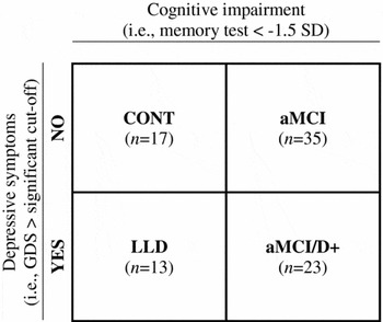
In order to isolate the independent contributions of cognitive impairment and depressive symptoms to emotional memory performance, we sought to avoid overlap in cognitive symptoms between LLD and aMCI participants. Thus, participants with LLD were only retained for the present study if they performed within normal limits (i.e. no scores below −1.5 SD) on the Dementia Rating Scale (DRS-2; Jurica et al., Reference Jurica, Leittan and Mattis2001), delayed free and cued verbal recall (Van der Linden, Reference Van der Linden2004), and Rey-Osterrieth Complex Figure Test 3-minute recall (ROCFT; Rey, Reference Rey1941) in the comprehensive neuropsychological battery described below. Eleven LLD participants were excluded on this basis. The result was a 2 × 2 factorial design (Figure 1) including individuals with cognitive impairment only (aMCI), depressive symptoms only (LLD), or both cognitive and depressive symptoms (aMCI/D+). Twenty-four healthy elderly adults (CONT) who reported no cognitive concerns or depressive symptoms were recruited through community advertisements, or were patients’ family members. Only those with no cognitive scores below −1.5 SD and no GDS score >11 were retained for the present study (n = 17).
Participant groups. CONT = Healthy controls; aMCI = amnestic mild cognitive impairment; aMCI/D+ = aMCI with depressive symptoms; LLD = late-life depression.

The final sample consisted of 35 aMCI, 23 aMCI/D+, 13 LLD, and 17 CONT participants. All participants were ≥55 years old and provided a comprehensive self-reported medical and psychiatric history, which was corroborated by knowledgeable informants when possible. None had a history of neurological disease, brain injury, stroke, psychiatric illness (other than depression), substance abuse, general anesthesia, intracranial surgery or oncologic treatment in the past six months, or untreated metabolic conditions or uncorrected visual/auditory impairments. Non-degenerative factors (e.g. cerebrovascular disease) were also ruled out based on this interview. Participants provided written consent after receiving a complete description of the study. The protocol was approved by the Research Ethics Board of the Institut universitaire en santé mentale de Québec.
Materials
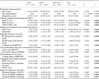
All participants underwent a comprehensive clinical and neuropsychological battery (Table 1). All tests within the battery, as well as the experimental task (described below), were administered by a single experimenter who was blind to participants’ diagnosis.
Mean (standard deviation) sociodemographic, neuropsychiatric and neuropsychological variables

Notes: CONT = Healthy controls; aMCI = amnestic mild cognitive impairment; aMCI/D+ = aMCI with depressive symptoms; LLD = late-life depression; MoCA=Montreal Cognitive Assessment; DRS=Dementia Rating Scale; ADCS-ADL=Alzheimer's Disease Cooperative Study-Activities of Daily Living; GDS=Geriatric Depression Scale; RL/RI=Rappel libre/rappel indicé à 16 items; ROCFT=Rey-Osterrieth Complex Figure Test; BORB=Birmingham Object Recognition Battery; WAIS-III=Weschler Adult Intelligence Scale III; BNT=Boston Naming Test; PPTT=Pyramids and Palm Trees Test; D-KEFS=Delis-Kaplan Executive Function System.
*p < 0.05 compared to the control group.
§p < 0.05 compared to the aMCI group.
†p < 0.05 compared to the aMCI/D+ group. Values given in bold indicate a statistically significant difference. MoCA data missing for 1 aMCI and 1 LLD; DRS data missing for 4 aMCI; Hachinski data missing for 6 aMCI, 1 aMCI/D+, 1 LLD; BORB data missing for 1 aMCI; Coding missing for 5 aMCI, 2 aMCI/D+ and 1 LLD; PPTT data missing for 1 aMCI; Stroop inhibition data missing for 1 aMCI and 2 aMCI/D+; Stroop switching data missing for 1 aMCI, 4 aMCI/D+, and 1 LLD. ADCS-ADL information version data missing for 3 aMCI; however, one of these participants completed the self-report version which was normal (67/68).
General cognitive functioning was assessed using the Montreal Cognitive Assessment (MoCA; Nasreddine et al., Reference Nasreddine2005) and the DRS-2 (Jurica et al., Reference Jurica, Leittan and Mattis2001). Depressive symptoms were evaluated using the GDS (Yesavage et al., Reference Yesavage1983), and vascular risk factors were estimated using Hachinski's scale (Hachinski et al., Reference Hachinski1975). The Rappel libre/Rappel indicé à 16 items (RL/RI-16; Van der Linden, Reference Van der Linden2004), a French-language test similar to the Free and Cued Selective Reminding Test (Grober et al., Reference Grober, Buschke, Crystal, Bang and Dresner1988), assessed episodic memory with the ROCFT (Rey, Reference Rey1941). Visuoperception and visuoconstruction abilities were assessed using the ROCFT copy score and the Birmingham Object Recognition Battery (BORB; Riddoch and Humphreys, Reference Riddoch and Humphreys1993) size-match subtest. The 15-item Boston Naming test (BNT; Mack et al., Reference Mack, Freed, Williams and Henderson1992), phonemic (T, N, P) and semantic (animals) fluency tasks (Consortium des Universités de Montréal et McGill, 1996) and the Pyramids and Palm Trees Test (PPTT; Howard and Patterson, Reference Howard and Patterson1992) assessed language and semantic memory skills. Processing speed and executive functioning were evaluated using the Delis-Kaplan Executive Function System (D-KEFS, Delis et al., Reference Delis, Kaplan and Kramer2001) Stroop subtest.
The computerized experimental task was presented using E-Prime 2.0 software (Psychology Software Tools, Pittsburgh, PA). Images were drawn from the International Affective Picture System (IAPS; Lang et al., Reference Lang, Bradley and Cuthbert2005). From the initial pool of 954 images, available valence and arousal ratings for 703 images were compiled from three published sources (Ito et al., Reference Ito, Cacioppo and Lang1998; Lang et al., Reference Lang, Bradley and Cuthbert2005; Libkuman et al., Reference Libkuman, Otani, Kern, Viger and Novak2007) and averaged. These images were tagged as “negative” (average valence < 4), “positive” (average valence > 5.99), or “neutral” (average valence 4–5.99). One hundred and fourteen images with valence ratings ±0.5 from a value of 5 were removed to ensure that “neutral” images were truly neutral. All images were then visually inspected, and 102 that depicted pornographic/erotic or violent scenes were removed. The remaining 487 images were sorted by arousal and valence, and 395 duplicates (i.e. images sharing a descriptor with at least one other image) were identified. Each was inspected and only those with the highest mean arousal and valence mean nearest the “absolute” valence value (positive=9, negative=1, neutral=5) were retained. A total of 10 positive, 10 negative, and 10 neutral images were selected from the remaining pool by a seven-member consensus panel of clinical researchers and graduate students who attempted to match images on visual complexity and content as closely as possible (please see the supplementary table for the full list of selected images at www.journals.cambridge.org/jid_IPG). This stimulus list length is similar to that used in other studies (e.g. 48 words used by Brueckner and Moritz, Reference Brueckner and Moritz2009; 32 words used by Nieuwenhuis-Mark et al., Reference Nieuwenhuis-Mark, Schalk and de Graaf2009; 15 words used by Mistridis et al., Reference Mistridis, Taylor, Kissler, Monsch, Kressig and Kivisaari2013; 24 faces used by Wang et al., Reference Wang2013; 24 images used by Gorenc-Mahmutaj et al., Reference Gorenc-Mahmutaj, Degen, Wetzel, Urbanowitsch, Funke and Schröder2015; 36 words used by Callahan et al., Reference Callahan, Simard, Mouiha, Rousseau, Laforce and Hudon2016). This list length was also chosen to avoid overwhelming or overtaxing the participants, particularly those with memory impairments. Valence ratings of each category were statistically confirmed to be significantly different from each other, F(2, 29) = 172.97, p < 0.001. Positive (M = 4.72, SD = 0.97) and negative (M = 5.05, SD = 0.87) images did not differ in their normative arousal rating on the 1 (low arousal) to 9 (high arousal) rating scale described in the manual (Lang et al., Reference Lang, Bradley and Cuthbert2005), p > 0.05. Both had higher arousal ratings than neutral images (M = 3.02, SD = 0.61), both p < 0.001.
Because participants may have found certain images disturbing, four negative images were pre-visualized by all participants who were told they could withdraw from the study at that point if they did not wish to continue. These images were selected in addition to the study sample, and were representative of the negative target stimuli. These practice trials also served to familiarize participants with the study procedure. One aMCI individual opted to discontinue after viewing the practice images and was not included in the final sample.
Procedure
During the study trial of the experimental task, participants were shown the 30 target images one by one in random order and asked to describe their content. Certain images were misperceived by some participants; for example, when shown an image of a lightbulb illuminating a blank wall (IAPS #7170), many participants described seeing “a pair of buttocks.” When this occurred, the experimenter did not provide clarification; all responses, including misperceptions, were recorded verbatim by the experimenter. Participants were then asked to decide, on a scale from 1 to 9, the extent to which they thought each image was negative/threatening (1) or positive/pleasant (9). There was no time limit, but response time was nonetheless recorded in order to control for its effect on subsequent recall. Participants were not told their memory would be tested, because implicit encoding may lead to better memory performance in participants with cognitive impairment (Son et al., Reference Son, Therrien and Whall2002). Immediately following the study phase, participants were asked to count upwards by 2 from 20 for 1 minute. They were then asked to orally recall the studied images for 2 minutes; these were recorded by the experimenter. Following a 20-minute delay, during which non-memory portions of the neuropsychological battery were administered, participants were again asked to recall the images for 2 minutes. For images to be counted as “recalled,” participants had to provide descriptive labels that were sufficiently similar to their original descriptions provided during encoding that the experimenter was able to identify them unambiguously. In the rare cases where it was unclear, the examiner prompted for a more detailed answer (“Please be more specific”). Images recalled from the practice trial were not counted.
Statistical analyses
Sociodemographic and clinical variables were compared between groups using one-way ANOVAs with Bonferroni correction. A Pearson χ 2test was performed to compare participants’ sex distribution. The Kolmogorov–Smirnov test indicated that the experimental data were non-normally distributed; rather than attempting to normalize the data through transformations, which alters the nature of the variables and makes results more difficult to interpret (Osborne, Reference Osborne2002), non-parametric analyses were performed.
To verify the hypothesis that patient groups would show decreased memory performance compared to CONT participants, total images recalled at immediate and delayed free recall were compared between groups using Kruskal–Wallis analyses. When between-group differences from this test were statistically significant, Mann–Whitney U tests were performed to compare performance pairwise between groups (total of six comparisons), and each time the alpha level was corrected to 0.008 (Bonferroni procedure=0.05/6).
Next, we sought to test the prediction that each group would show specific emotional biases in terms of the type of images they recalled. Of the 30 images presented at encoding, the mean percent of positive, negative and neutral images recalled was calculated for each group and compared using Friedman tests. Because previous research (e.g. Moritz et al., Reference Moritz, Voigt, Arzola and Otte2008) has shown that subjective interpretation of emotional stimuli is most strongly related to later recall, and because certain images had been misperceived during encoding, valence was based on participants’ subjective ratings. For instance, if a picture was given a valence score of 7 during the study phase and later recalled, it was counted as a “positive recall.” Ratings 1–4 were considered negative, ratings 6–9 were considered positive, and ratings of 5 were considered neutral. When Friedman tests were statistically significant, Wilcoxon tests were performed to compare performance pairwise between the valence groups, and each time the alpha level was corrected to 0.017 (0.05/3). This procedure was conducted for immediate and delayed recall scores separately. Post-hoc power analyses were conducted using G*Power v.3.1.9.2 (Faul et al., Reference Faul, Erdfelder, Lang and Buchner2007) and effect sizes (Cohen's d) are reported alongside the results, with d = 0.2, 0.5, and 0.8 representing small, medium, and large effect sizes, respectively (Cohen, Reference Cohen1988).
To further clarify the relationship between picture valence and memory performance, subsequent exploratory analyses were performed in which each image was considered as an independent observation associated with a valence rating (1–9) and a recall score (1 = recalled, 0 = not recalled). Two new variables were created: a “Positive gradient” and a “Negative gradient,” which represented the distance of subjective ratings from a perfect neutral score (5). For instance, a “Positive gradient” score of 1 was given to images that had been attributed a valence rating of 6. A “Negative gradient” score of 1 was given to images that had been attributed a valence rating of 4. Images with valence ratings of 9 or 1 were assigned a score of 4 on the “Positive” and “Negative” gradients, respectively. This approach allowed for the impact of positive and negative valence ratings on memory performance to be considered together in binary logistic regression models for each group. It also allowed for the consideration of the intensity of ratings, as increasing gradient scores were assumed to reflect higher valence intensity. Most importantly, it allowed for a more direct examination of the association between the rating given to a picture and the likelihood of that specific picture being subsequently recalled. Regression models included age, sex, education, and image exposure time as covariates, and Recall (yes/no) as the binary outcome measure.
All analyses were performed using SPSS v.23.0 Windows software. Significance thresholds were p < 0.05 except where corrected for multiple comparisons, as described above.
Results
Participant characteristics
Groups were matched on age, sex, and education (Table 1). Patients with aMCI and aMCI/D+ had lower general cognitive functioning than CONT and LLD participants, and aMCI/D+ and LLD reported more depressive symptoms than CONT and aMCI. LLD participants also endorsed more depressive symptoms than aMCI/D+ participants, and presented more vascular risk factors than CONT and aMCI. Consistent with diagnostic criteria, aMCI and aMCI/D+ participants had lower memory scores than CONT participants. aMCI and aMCI/D+ participants’ language and semantic memory scores were generally lower than those of CONT and LLD participants. In addition, aMCI participants presented longer inhibition latencies than CONT participants and longer switching latencies than LLD. All groups performed similarly on measures of visuoperception and visuoconstruction, processing speed, and phonemic fluency.
From the entire 30-item stimulus set, there were no significant group differences in the number of positive images identified by CONT (M = 11.35, SD = 3.97), aMCI (M = 12.89, SD = 4.27), aMCI/D+ (M = 10.83, SD = 4.64), and LLD (M = 11.54, SD = 1.94), F(3,87) = 1.352, p = 0.263; the number of negative images identified by CONT (M = 9.65, SD = 2.87), aMCI (M = 10.23, SD = 3.65), aMCI/D+ (M = 9.22, SD = 3.95), and LLD (M = 10.31, SD = 3.30), F(3,87)=0.466, p = 0.707; or neutral images identified by CONT (M = 9.00, SD = 4.12), aMCI (M = 6.89, SD = 4.09), aMCI/D+ (M = 9.96, SD = 4.88), LLD (M = 8.15, SD = 3.29), F(3,87) = 2.659, p = 0.053.
Overall memory performance
The groups differed significantly on the total number of images recalled at immediate (p = 0.006) and delayed (p < 0.001) recall. At immediate recall, Mann–Whitney U tests showed that CONT participants (M = 11.71, SD = 2.82) remembered more images than aMCI patients (M = 8.69, SD = 3.68, p = 0.002, d = 0.92), aMCI/D+ patients (M = 8.70, SD = 2.99, p = 0.003, d = 1.04), but not LLD patients (M = 11.15, SD = 4.38, p = 0.341, d = 0.15). At delayed recall, CONT participants (M = 11.35, SD = 2.34) remembered more images than aMCI patients (M = 7.34, SD = 4.25, p = 0.001, d = 1.17), aMCI/D+ patients (M = 6.65, SD = 2.81, p < 0.001, d = 1.82), but not LLD patients (M = 10.85, SD = 4.96, p = 0.483, d = 0.13). The aMCI/D+ group also recalled fewer images than LLD at delayed recall (p = 0.006, d = 1.04). There were no other differences between the number of pictures recalled by aMCI, aMCI/D+, and LLD patients at immediate or delayed recall (all p > 0.008).
Emotional memory biases
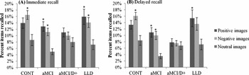
The different types of images recalled by each group are presented in Figure 2. At immediate recall, results from the Friedman test revealed significant differences in the type of images recalled by CONT (p = 0.007), aMCI (p = 0.003), and LLD (p = 0.001) participants. Wilcoxon signed-rank tests showed that negative (p = 0.009, d = 1.21), but not positive images (p = 0.077, d = 0.76) were more often recalled than neutral images by CONT, whereas aMCI participants recalled more positive (p = 0.009, d = 1.07) and negative images (p = 0.009, d = 0.99) relative to neutral ones. LLD participants similarly recalled more positive (p = 0.009, d = 1.21) and negative images (p = 0.009, d = 1.23) relative to neutral ones. The aMCI/D+ group showed no difference between the type of images recalled (p = 0.538).
Percent positive, negative and neutral items recalled immediately following stimulus presentation (A) or 30 minutes later (B). CONT = Healthy controls; aMCI = amnestic mild cognitive impairment; aMCI/D+ = aMCI with depressive symptoms; LLD = late-life depression. *p < 0.05 compared to neutral.

At delayed recall, the CONT (p = 0.008), aMCI (p < 0.001) and LLD (p = 0.003) groups showed differences in the type of images they recalled; again, the aMCI/D+ group did not (p = 0.665). Wilcoxon signed-rank tests showed that negative images (p = 0.010, d = 1.36) but not positive images (p = 0.054, d = 0.82) were more often recalled than neutral images by CONT, and that aMCI participants recalled significantly more positive (p < 0.001, d = 1.04) and negative images (p < 0.001, d = 1.02) than neutral ones. Positive (p = 0.003, d = 1.03) but not negative (p = 0.030, d = 0.97) were more often recalled than neutral images by the LLD group.
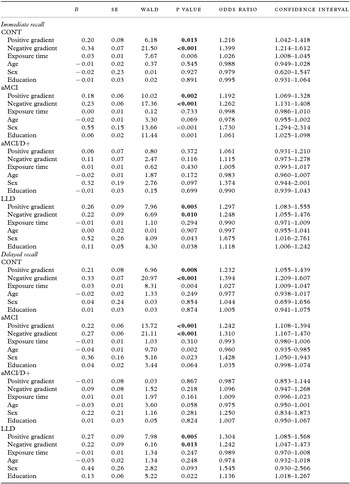
Results from exploratory binary logistic regression models arex presented in Table 2. In CONT, the model revealed significant associations between immediate recall and positive (B = 0.20, SE = 0.08, p = 0.013) and negative ratings (B = 0.34, SE=0.07, p < 0.001). In aMCI, immediate recall was associated with positive (B = 0.18, SE = 0.06, p = 0.002) and negative ratings (B = 0.23, SE = 0.06, p < 0.001). In LLD, immediate recall was associated with positive (B = 0.26, SE = 0.09, p = 0.005) and negative ratings (B = 0.22, SE = 0.09, p = 0.010). In all cases, the associations were positive, indicating that recall increased as gradient scores increased (i.e. images rated as most strongly positive or negative). Neither variable was a significant predictor of immediate recall in the aMCI/D+ group.
Regression coefficients from binary logistic regression models at immediate and delayed recall

Notes: CONT = Healthy controls; aMCI = amnestic mild cognitive impairment; aMCI/D+ = aMCI with depressive symptoms; LLD = late-life depression. Values given in bold indicate a statistically significant difference.
Delayed recall performance in CONT participants was associated with positive (B = 0.21, SE = 0.08, p = 0.008) and negative ratings (B = 0.33, SE = 0.07, p < 0.001). In aMCI, immediate recall was associated with positive (B = 0.22, SE = 0.06, p < 0.001) and negative ratings (B = 0.27, SE = 0.06, p < 0.001). In LLD, immediate recall was associated with positive (B = 0.27, SE = 0.09, p = 0.005) and negative ratings (B = 0.22, SE = 0.09, p = 0.013). Once again, all associations were positive. Neither variable was a significant predictor of immediate recall in the aMCI/D+ group.
Discussion
This study compared episodic memory for emotional pictures between individuals with aMCI, aMCI/D+, LLD, and CONT participants. Results first revealed that aMCI and aMCI/D+ groups showed decreased overall memory performance relative to CONT participants at immediate and delayed recall. This was in accordance with a priori hypotheses as well as with documented memory deficits in aMCI (Belleville et al., Reference Belleville, Sylvain-Roy, de Boysson and Ménard2008) and aMCI/D+ (Dierckx et al., Reference Dierckx, Engelborghs, De Raedt, De Deyn and Ponjaert-Kristoffersen2007; Hudon et al., Reference Hudon, Belleville and Gauthier2008; Johnson et al., Reference Johnson2013). Free recall performance was comparable between aMCI and aMCI/D+, which has also been reported previously (Hudon et al., Reference Hudon, Belleville and Gauthier2008; Callahan et al., Reference Callahan, Simard, Mouiha, Rousseau, Laforce and Hudon2016). One study reported worse free recall performance in depressed than non-depressed patients with MCI (Johnson et al., Reference Johnson2013), but results from that study may be confounded by the inclusion of non-amnestic patients in the sample.
Second, the results of this study pointed to specific relationships between emotional valence and episodic memory performance for different participant groups. Namely, emotional stimuli were better remembered by CONT, aMCI, and LLD participants relative to neutral items, whereas no benefit for emotional stimuli was apparent in the aMCI/D+ group. Previous studies have already shown that aMCI/D+ patients distinguish themselves from those with aMCI and LLD on measures of verbal memory (Callahan et al., Reference Callahan, Simard, Mouiha, Rousseau, Laforce and Hudon2016), semantic memory (Brunet et al., Reference Brunet2011; Callahan et al., Reference Callahan2014) and from those with aMCI on tests of executive functioning and cued recall (Hudon et al., Reference Hudon, Belleville and Gauthier2008). Here, we provide further evidence that the cognitive profile in aMCI/D+ individuals is distinct from that of aMCI and LLD, by showing that participants within each group presented different patterns of associations between valence and recall.
These findings have interesting and meaningful implications for the conceptualization of memory deficits in preclinical dementia. First, in response to experts’ recommendations to clarify the relationship between aMCI and depression (Steffens et al., Reference Steffens2006), our results converge with previous evidence (Hudon et al., Reference Hudon, Belleville and Gauthier2008; Brunet et al., Reference Brunet2011; Callahan et al., Reference Callahan2014, Reference Callahan, Simard, Mouiha, Rousseau, Laforce and Hudon2016) to suggest that depressive symptoms may act as a modifier of the cognitive phenotype of aMCI. Indeed, although depressed and non-depressed aMCI participants in this study were comparable in terms of their memory performance on clinical tasks, and indeed recalled comparable quantities of items on the experimental task, the quality of the information remembered differed significantly. In addition, considering that behavior is an indicator of brain vulnerability, results from this study add to those of previous work (Brunet et al., Reference Brunet2011; Morin et al., Reference Morin, Mouiha, Pietrantonio, Duchesne and Hudon2012; Callahan et al., Reference Callahan2014, Reference Callahan, Simard, Mouiha, Rousseau, Laforce and Hudon2016) to imply the likely existence of distinct neurobiological or neurofunctional manifestations in aMCI, aMCI/D+, and LLD. This necessitates further investigation using imaging measures, but we might speculate about plausible mechanisms by which depressive symptoms affect emotional memory. It is possible that performance in participants with aMCI/D+ is the result of neurobiological changes, potentially involving the hippocampus and/or the amygdala. Hippocampal atrophy has been associated with both aMCI and depression, independently (Morin et al., Reference Morin, Mouiha, Pietrantonio, Duchesne and Hudon2012) and correlates with recall of emotional pictures (Schultz et al., Reference Schultz, De Castro and Bertolucci2009). Similarly, amygdalar lesions are known to be present in early AD (Klein-Koerkamp et al., Reference Klein-Koerkamp, Baciu and Hot2012) and in depressed seniors (Burke et al., Reference Burke2011), and are thought to contribute to the disruption of emotional processes involved in emotional memory enhancement (Kensinger and Leclerc, Reference Kensinger and Leclerc2009). It is possible that hippocampal and/or amygdalar deterioration in MCI and depression may be additive in individuals with both these conditions (i.e. aMCI/D+), resulting in greater disruption of emotional memory enhancement effects.
Alternatively, it is possible that the presence of depressive symptoms in individuals who are already cognitively fragile (i.e. aMCI/D+) may interfere with the frontally-mediated up- or down-regulation strategies that appear to explain emotional memory enhancement in healthy older adults (Kensinger and Leclerc, Reference Kensinger and Leclerc2009). Indeed, both the integrity and function of the frontal lobes have been reported to be compromised in MCI participants with depressive symptoms, compared to those without depression (Pellegrino et al., Reference Pellegrino, Peters, Lyketsos and Marano2013) and executive dysfunction has been proposed by other authors to account for the lack of emotional enhancement effect sometimes observed in cognitively impaired participants (Klein-Koerkamp et al., Reference Klein-Koerkamp, Baciu and Hot2012). Based on data from the present study, this possibility seems less likely, as aMCI/D+ participants showed no significant executive impairment as measured by the Stroop task and a Phonemic fluency task; however, formal investigation of this hypothesis would require more thorough testing of various executive processes.
In addition to these theoretical implications, the qualitative differences in memory performance between aMCI and aMCI/D+ may suggest avenues for practical applications. For instance, a number of cognitive training programs have been developed for individuals with aMCI aimed at optimizing daily functioning through memory training and compensation techniques (e.g. Troyer et al., Reference Troyer, Murphy, Anderson, Moscovitch and Craik2008). A common strategy included in many of these programs is “elaboration,” that is encouraging deep encoding of information to be remembered. It is possible that emotion-focused elaborative encoding may be particularly helpful in improving memory performance in non-depressed individuals, whereas other types of semantic elaboration strategies might be explored for individuals presenting depressive symptoms.
The positive and negative images used in the experimental task had higher normative arousal ratings than did the neutral images, and results from exploratory follow-up analyses suggested that the perceived intensity of stimuli indeed seemed to have driven the emotional memory effect in aMCI, LLD, and CONT participants. Many prior studies support the role of intensity, also sometimes referred to as salience, in memory for emotional items (e.g. Moritz et al., Reference Moritz, Voigt, Arzola and Otte2008). A recent study examined emotional memory effects in participants with memory impairments (Gorenc-Mahmutaj et al., Reference Gorenc-Mahmutaj, Degen, Wetzel, Urbanowitsch, Funke and Schröder2015) and found that positive and negative images from the IAPS stimuli set were perceived as more intense than neutral items, which impacted subsequent memory performance, consistent with our findings.
Several additional results from this study are largely consistent with previous work. Namely, we replicate findings from prior studies of an emotional memory enhancement in healthy older adults (e.g. Kensinger et al., Reference Kensinger, Brierley, Medford, Growdon and Corkin2002). Some authors have reported improved memory for certain types of positive relative to negative information in seniors, contrary to our results (Mather and Carstensen, Reference Mather and Carstensen2005). However, this positivity bias has specifically been linked to goal-directed attentional processes, and the emotional enhancement effect is thought to extend to negative stimuli when cognitive processing is more automatic, particularly if the negative information is arousing (Mather and Carstensen, Reference Mather and Carstensen2005), which was the case in our sample. In addition, the emotional memory enhancement observed in the aMCI group is consistent with a number of studies showing better memory for emotional (positive and negative) relative to neutral stimuli (Brueckner and Moritz, Reference Brueckner and Moritz2009; Nieuwenhuis-Mark et al., Reference Nieuwenhuis-Mark, Schalk and de Graaf2009; Wang et al., Reference Wang2013; Gorenc-Mahmutaj et al., Reference Gorenc-Mahmutaj, Degen, Wetzel, Urbanowitsch, Funke and Schröder2015 immediate recall), though it is inconsistent with some prior reports of a positivity bias only in these patients (Marreneca et al., Reference Marreneca, Martins, Mendonça and Guerreiro2006; Werheid et al., Reference Werheid, Gruno, Kathmann, Fischer, Almkvist and Winblad2010, experiment 3; Gorenc-Mahmutaj et al., Reference Gorenc-Mahmutaj, Degen, Wetzel, Urbanowitsch, Funke and Schröder2015, delayed recall). These discrepancies may be a function of the heterogeneity within study samples, the type of stimuli used, the timing of recall, or the type of memory paradigm used.
On the other hand, some findings presented here conflict with prior work. Studies examining the relationship between depressive symptoms and cognition in younger adults have consistently shown a mood-congruent negative memory bias (Matt et al., Reference Matt, Vazquez and Campbell1992). In the present study, however, positive items were just as well-remembered as negative items by LLD participants (in some cases, more well-remembered). The aMCI/D+ group showed no negative bias whatsoever. It is possible that the mood-congruent negative cognitive bias reported in depressed young adults becomes less pronounced as these individuals get older or develop cognitive impairment (Mah and Pollock, Reference Mah and Pollock2010), perhaps due to age-related brain changes within the limbic system, which is known to modulate memory performance (Alexopoulos et al., Reference Alexopoulos2013). This may be particularly true on tasks involving visual material, although this needs to be verified in future studies directly comparing memory for emotional words and pictures in aMCI/D+ and LLD.
Limitations and future directions
In conclusion, the present study helps to clarify the relationship between aMCI, aMCI/D+, and LLD, and findings have important theoretical and practical implications. Growing evidence from this study and others suggests that the semiology of aMCI/D+ is sufficiently different from that of aMCI and LLD, at least from a cognitive point of view, and this may warrant further consideration for the diagnostic and prognostic significance of these semiological differences. One limitation of our findings was the relatively small size of the participant groups, which is a common issue in clinical research. Our power analyses indeed suggested that some of the marginally significant results would likely have reached statistical significance had our samples sizes been larger. Thus, our findings will need to be replicated by independent research groups, ideally with larger samples. A second important limitation is that data regarding the age of onset of the depressive symptoms or the number of prior depressive episodes in the LLD group were unfortunately not collected. Thus, an investigation of potential differences between early- and late-onset cases of LLD, or between duration and number of previous episodes, was not possible.
In addition, the present study did not aim to investigate neuroanatomical differences between aMCI, aMCI/D+, and LLD groups, and although the behavioral findings imply the possibility of such differences existing, imaging studies will be needed to directly explore this relationship. At this point, neuroimaging research is the next essential step in discovering whether these groups differ from a neuropathological standpoint, as behavioral evidence suggests. Establishing a clear nosological framework in which to consider aMCI, aMCI/D+, and LLD will foster research on appropriate treatment avenues for each condition. Tailored interventions are likely to be most effective in reducing the risk of conversion to AD as well as the disease's overall prevalence.
Conflict of interest
None.
Description of authors’ roles
B. Callahan designed the study and experimental task, collected the data, and wrote the manuscript. R. Laforce and M. Dugas contributed to participant recruitment and as well as to critical revision of the manuscript for important intellectual content. C. Hudon contributed to the study design, data analysis and interpretation, and critical revision of the manuscript for important intellectual content.
Acknowledgments
We thank Isabelle Tremblay, Sarah Tardif, Mélissa Dion, and Marie-Ève Gagnon for their help testing participants.
Supplementary material
To view supplementary material for this article, please visit https://doi.org/10.1017/S1041610216002179