Introduction
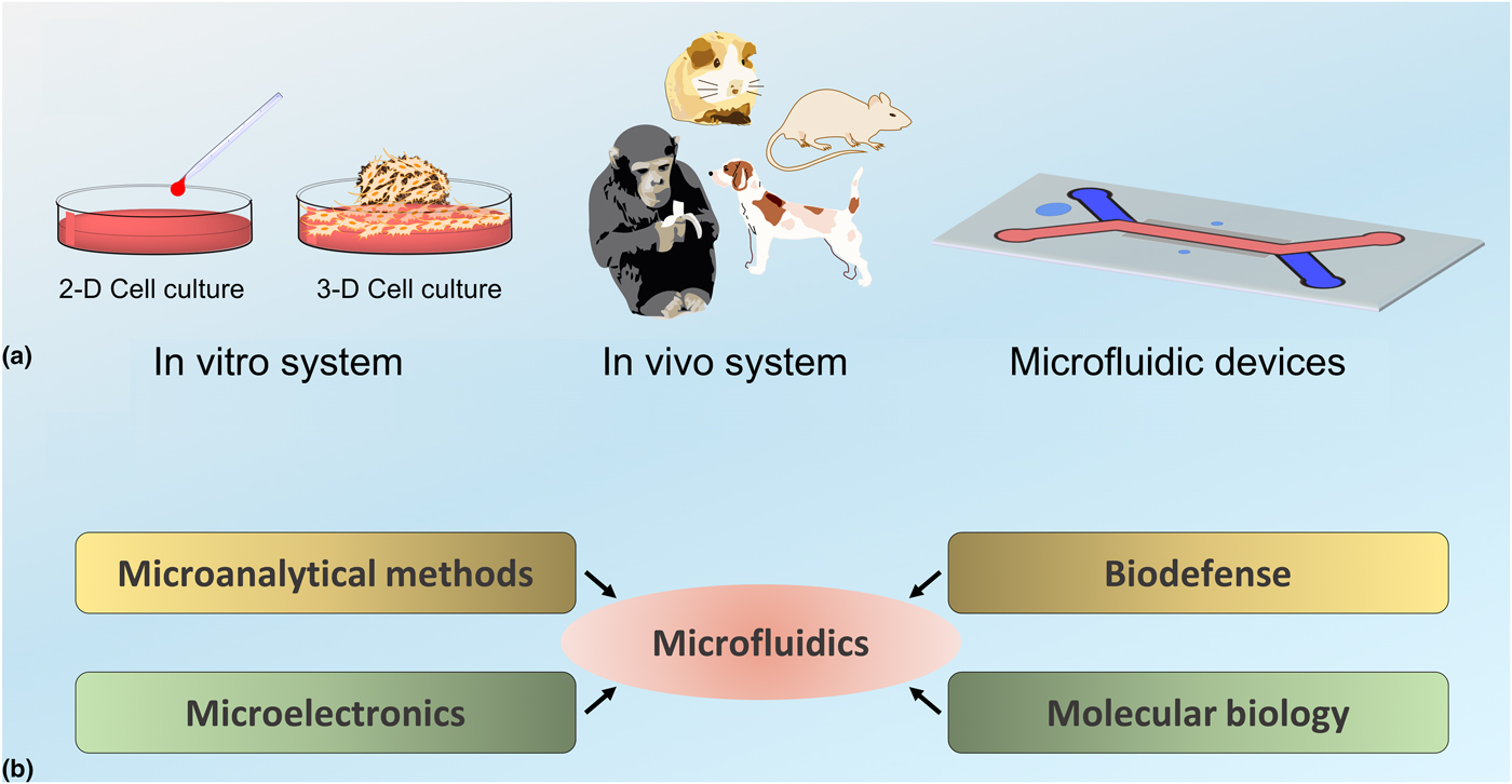
Miniaturization has already worked wonders in engineering new concepts and applications in microelectronics. This trend has inspired the cell architects to conceive novel ideas that were a mere science fiction a few decades ago.[Reference Kang, McNaughton and Espinosa1, Reference Zhang and Radisic2] One such incredible architecture is an organ-on-chip (OOC) device, which has metamorphosed from a cool concept to a practical solution using the state-of-art technology at the micron scale. Essentially, OOC attempts to fulfill the long-sought desire of researchers who have imagined to recreate tissues and organs by etching grooves on silicon or plastic wafers to engineer a microenvironment that mimics the complex and physiological conditions in real organs.[Reference Baker3] It is indeed a massive leap towards understanding the cell biology that is effusively reliant on the conventional in vitro and in vivo models, where the cell lines in 2D culture plates (in vitro) do not fully reflect the 3D architecture and in vivo behavior of cells.[Reference Greenman4, Reference Shamir and Ewald5] Further, 3D extracellular matrix and multicellular spheroids are also used to study the diverse functionality in the human body with biomedical, chemical, pharmaceutical, and environmental perspectives.[Reference Huh, Hamilton and Ingber6–Reference Duval, Grover, Han, Mou, Pegoraro, Fredberg and Chen8] All these methods serve as indispensable tools to investigate biologic processes at cellular and tissue levels through controllable environments, but these models do not reiterate the complex physiological functions of an organ in a human body.[Reference Huh, Kim, Fraser, Shea, Khan, Bahinski, Hamilton and Ingber9] Fig. 1(a) shows a simple pictorial representation of the available testing tools at our disposal for various material or product screening. Subsequently, in vivo methods involving animal models are the exclusive choice to emulate and predict human responses to antigens, drugs or chemicals. The animal testing yields crucial information about various interactions occurring inside the body while encountering foreign materials. However, they still have a few pitfalls like high cost, long study duration, and its inability to foresee human response quite often that lowers the credibility of the animal testing.[Reference Moraes, Mehta, Lesher-Perez and Takayama10] This state directly influences the outcome of new materials or products in biomedicine, biosensors, cosmetics etc., due to serious concerns over ethics and budgets for research development.
(a) Current testing tools for material or product screening, (b) contributions of other fields in the innovation of microfluidic devices.

Now, to fill all these voids in the current situation it is extremely important to create a simple and unique test platform that would mimic human in vivo conditions. In that context, OOC has a great potential to overcome all these limitations. In this prospective, we discuss the innovation, advances, applications, and market of OOC devices in biomedicine, biosensors, cosmetics, and drug discovery.
Innovation and architecture
The inception of OOC device was due to a prerequisite need for a surrogate testing tool, which is well equipped to negate the glaring discrepancies that exist among the current testing tools,[Reference Wagner, Materne, Brincker, Sussbier, Fradrich, Busek, Sonntag, Sakharov, Trushkin, Tonevitsky, Lauster and Marx11] and as an important step towards personalized precision medicine. The progress in OOC field dwells on the research and developments surrounding microfluidics and cell culture. More specifically the “miniaturization” revolution has led to a new coalition of bioengineers and cell biologists to innovate the OOC device.[Reference Huh, Kim, Fraser, Shea, Khan, Bahinski, Hamilton and Ingber9] But, why microfluidics? Firstly, microfluidic technology deals with the maneuvering of small quantities of fluids (10−9 to 10−18 L) within an artificially fabricated microsystem, where the channels have dimensions of ten to hundreds of micrometers. Secondly, it allows the integration, automation, and parallelization of biochemical processes apart from the miniaturization aspect. Overall, the main idea behind the microfluidic cell culture is to develop devices that can sustain cells in micro-scale volumes for various analysis and experiments.[Reference Halldorsson, Lucumi, Gomez-Sjoberg and Fleming12, Reference Meyvantsson and Beebe13] Fig. 1(b) provides an insight into the motivation and contributions of other fields in the innovation of microfluidics. The early inspiration to develop microfluidics came alongside the successful endeavor in molecular analysis systems that used capillaries for microanalysis of samples. It was closely followed by the developments in molecular biology and micro-devices to detect chemical or biologic threats for the defense sector. Though these fields provided ample motivation for the innovation of microfluidics, the major driving force for the expansion of microfluidics was owed to the contributions from microelectronic technologies.[Reference Whitesides14] One of the key encouraging factors that were indispensable for the innovation of microfluidics was the fascination for smaller devices that were extremely popular in microelectronics, made via photolithography and associated technologies. These microfabrication techniques form the primary backbone of microfluidics, which allows the fabrication of devices in desired shapes and positions at micron scales to provide an appropriate microenvironment for cell growth.[Reference Gauvin and Khademhosseini15, Reference Khademhosseini, Langer, Borenstein and Vacanti16] Early phases of microfluidics development were mostly reliant on silicon microfabrication and micromachining techniques that were expensive, complex, and limited to specialized microfabrication facilities, which subsequently paved way for polymer-based systems, like poly (dimethylsiloxane) (PDMS) microsystems.[Reference Huh, Hamilton and Ingber6, Reference Whitesides14] PDMS is well-suited for microfluidic device fabrication as it is optically transparent that permits real-time imaging of cellular responses and allows high gas permeability, which in addition can be helpful to provide adequate oxygen supply to cells. Since the late 90s, there have been several attempts to culture cells on such microfabricated chips, however, a significant breakthrough was achieved by Ingber's group in 2010 that revolutionized the OOC field.[Reference Liedl, Hogberg, Tytell, Ingber and Shih17, Reference Mammoto and Ingber18] Their study describes a biomimetic system that reconstitutes human lung functions on a chip, which was first of its kind.[Reference Huh, Matthews, Mammoto, Montoya-Zavala, Hsin and Ingber19] Following which, a sizable number of researchers have shown great interest to recreate different tissues or OOCs, which is attributed to the core traits of OOC that includes co-culturing of cells in spatially controlled manner, control over gradients and integration of flow.[Reference van Duinen, Trietsch, Joore, Vulto and Hankemeier20] Fig. 2(a) shows the microfluidic device fabrication methods using PDMS polymer and photolithography technique, which is used to transfer microscale patterns onto photosensitive materials by selective exposure to ultraviolet (UV) rays. A simple single channel microfluidic device was fabricated by replica molding with a blank PDMS block, where the inlets permit the flow of two different fluids, while the side channel was deployed to offer cyclic suction that provides a dynamic microenvironment for the cells as shown in Fig. 2(b).
An overview of simple microfluidic device fabrication methods. (a) Replica molding technique, and (b) single channel fabrication technique (Images reprinted with permission from S.N. Bhatia and D.E. Ingber.[Reference Bhatia and Ingber21]) Copyright 2014 Nature Biotechnology.

Recent advances and substantial understanding in microfluidics have immensely fueled the researchers to fabricate OOCs that provide an amicable niche for different cell types to emulate respective tissues or organs under in vivo conditions. So, OOC is essentially a microengineered device that accommodates live cells and provides a continuous and highly controlled supply of essential fluids to its chambers. The whole idea is to create basic functional entities that reiterate tissue or organ level functions.[Reference Bhatia and Ingber21] Initially, they were known as microfluidic cell culture analogs but later they were identified as OOC or multi-OOC. Now, the pioneers in this area have made significant progress in fabricating OOC devices, since the advent of a human lung-on-a-chip. Modern technologies and equipment have assisted scientists to create several distinct OOC like liver,[Reference Carraro, Hsu, Kulig, Cheung, Miller, Weinberg, Swart, Kaazempur-Mofrad, Borenstein, Vacanti and Neville22–Reference Knowlton and Tasoglu25] lung,[Reference Huh, Matthews, Mammoto, Montoya-Zavala, Hsin and Ingber19, Reference Huh26, Reference Benam, Villenave, Lucchesi, Varone, Hubeau, Lee, Alves, Salmon, Ferrante, Weaver, Bahinski, Hamilton and Ingber27] kidney,[Reference Jang and Suh28–Reference Jang, Mehr, Hamilton, McPartlin, Chung, Suh and Ingber30] gut,[Reference Kim, Huh, Hamilton and Ingber31–Reference Shim, Lee, Han, Nguyen, Park and Sung33] eye,[Reference Puleo, McIntosh Ambrose, Takezawa, Elisseeff and Wang34–Reference Estlack, Bennet, Reid and Kim36] brain,[Reference Wang, Wang, Zhu and Qin37] skin,[Reference Wufuer, Lee, Hur, Jeon, Kim, Choi and Lee38, Reference Mori, Morimoto and Takeuchi39] bone,[Reference Jusoh, Oh, Kim, Kim and Jeon40] bone marrow,[Reference Torisawa, Spina, Mammoto, Mammoto, Weaver, Tat, Collins and Ingber41] heart,[Reference Qian, Huang, Lin, Ivanovskaya, O'Hara, Booth, Creek, Enright, Soscia, Belle, Liao, Lightstone, Kulp and Wheeler42] etc., as illustrated in Fig. 3 and Table 1. Recently, the world is now witnessing a crucial transition from individual OOC to multi-OOC to realize the most ambitious vision of fabricating human body on a chip, which is speculated to ultimately render the contemporary testing tools (in vitro and in vivo) obsolete.
Organ-on-chip for different human organs. Modified from Ahadian et al.[Reference Ahadian, Civitarese, Bannerman, Mohammadi, Lu, Wang, Davenport-Huyer, Lai, Zhang, Zhao, Mandla, Korolj and Radisic43] with permission from Wiley VCH, 2017, Retina-on-a-chip image reprinted with permission from Springer Nature[Reference Dodson, Echevarria, Li, Sappington and Edd35] and Bone marrow-on-chip image reprinted with permission from Springer Nature.[Reference Torisawa, Spina, Mammoto, Mammoto, Weaver, Tat, Collins and Ingber41]

Summary of the organ-on-chip models.

Towards “Homo-chippiens”
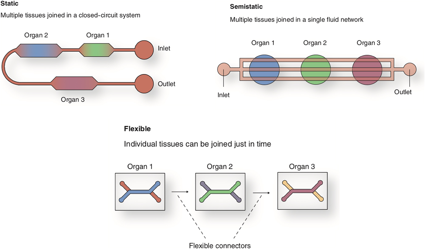
“Homo-chippiens” is an arbitrary term given to an entire human body on a chip.[Reference Reardon44] It would certainly take a remarkable effort to get there, especially with the existing challenges in multi-OOC that are to be secured. So, what are the existing challenges in multi-OOC research? Where do we stand as of today in multi-OOC research? These are the vital questions that subsequent sections of this review will attempt to answer. But firstly, what is a multi-OOC device? A multi-OOC platform integrates multiple distinct tissues-organs into one microfluidic device, which offers appropriate microenvironment to simulate identical conditions in the human body.[Reference Lee, Ha, Choi, Choi, Park and Sung45] So, it is the concept of integration that involves at least two or more tissues. Now, to create such a complex microsystem, it is desirable to have a concrete design that can be reproduced successfully, while it is able to precisely simulate the in vivo conditions of the human body. This quest lead researchers to develop three prominent design concepts for multi-OOC devices, which include (1) static, (2) semi-static, and (3) flexible systems as illustrated in Fig. 4. Among these systems, the flexible multi-OOC platform seems to have benefits since it can be used to recreate application specific multi-OOC systems.[Reference Rogal, Probst and Loskill46] Although, the multi-OOC concept is still naïve, yet there have been some major breakthroughs like fabricating two-organs,[Reference Wagner, Materne, Brincker, Sussbier, Fradrich, Busek, Sonntag, Sakharov, Trushkin, Tonevitsky, Lauster and Marx11, Reference Tsamandouras, Chen, Edington, Stokes, Griffith and Cirit47] three-organs,[Reference Skardal, Murphy, Devarasetty, Mead, Kang, Seol, Shrike Zhang, Shin, Zhao, Aleman, Hall, Shupe, Kleensang, Dokmeci, Jin Lee, Jackson, Yoo, Hartung, Khademhosseini, Soker, Bishop and Atala48] four-organs,[Reference Maschmeyer, Lorenz, Schimek, Hasenberg, Ramme, Hubner, Lindner, Drewell, Bauer, Thomas, Sambo, Sonntag, Lauster and Marx49, Reference Oleaga, Bernabini, Smith, Srinivasan, Jackson, McLamb, Platt, Bridges, Cai, Santhanam, Berry, Najjar, Akanda, Guo, Martin, Ekman, Esch, Langer, Ouedraogo, Cotovio, Breton, Shuler and Hickman50] and even up to seven and ten organs on a chip.[Reference Edington, Chen, Geishecker, Kassis, Soenksen, Bhushan, Freake, Kirschner, Maass, Tsamandouras, Valdez, Cook, Parent, Snyder, Yu, Suter, Shockley, Velazquez, Velazquez, Stockdale, Papps, Lee, Vann, Gamboa, LaBarge, Zhong, Wang, Boyer, Lauffenburger, Carrier, Communal, Tannenbaum, Stokes, Hughes, Rohatgi, Trumper, Cirit and Griffith51]
Multi-organ-on-chip design concepts with at least two distinct connected tissues (reproduced with permission from Future Science OA, 2017).[Reference Rogal, Probst and Loskill46]

Now, how does an OOC function and what are the inadequacies of the contemporary models? Inside a classical cell culture dish (in vitro), a number of factors critically differ from the actual human body conditions. For instance, the vital parameters like oxygen supply, nutrient supply, waste removal, and mechanical forces experienced by cells under in vivo conditions cannot be regulated under a regular in vitro setup. Moreover, the organs have inherent possession of physiologically required gradient concentrations of substances along with dissolved oxygen that is difficult to simulate in a normal in vitro setup, which explains the discrepancies among the two testing systems.[Reference Materne, Maschmeyer, Lorenz, Horland, Schimek, Busek, Sonntag, Lauster and Marx52] These inconsistencies led to the innovation of a surrogate testing tool, which has fulfilled the shortcomings of the conventional setup. Today, OOC research is gaining massive momentum in terms of publications and research funds. However, the individual OOC models still seem to be an incomplete testing tool largely due to the fact that tissues or organs in the human body are interconnected. So, it is critical to combine distinct tissues or organs on a single microfluidic device for various testing. Nevertheless, there are only limited multi-OOC platforms available today that mimic human tissues or model diseases.[Reference Skardal, Murphy, Devarasetty, Mead, Kang, Seol, Shrike Zhang, Shin, Zhao, Aleman, Hall, Shupe, Kleensang, Dokmeci, Jin Lee, Jackson, Yoo, Hartung, Khademhosseini, Soker, Bishop and Atala48]
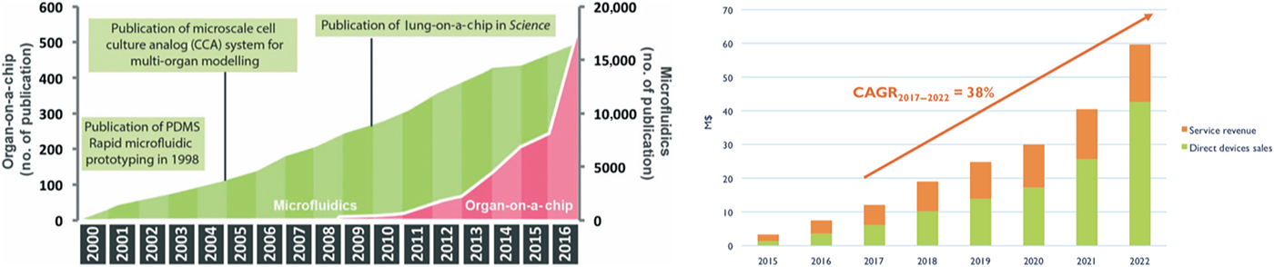
The rapid expansion of OOC promises potential applications in various fields at a lower cost and significantly lesser time compared with the in vivo models. Exhaustive reviews on individual OOC platforms are at our disposal today,[Reference Huh, Hamilton and Ingber6, Reference Halldorsson, Lucumi, Gomez-Sjoberg and Fleming12, Reference Bhatia and Ingber21, Reference Ewart, Dehne, Fabre, Gibbs, Hickman, Hornberg, Ingelman-Sundberg, Jang, Jones, Lauschke, Marx, Mettetal, Pointon, Williams, Zimmermann and Newham53, Reference Kaushik, Leijten and Khademhosseini54] compared with the multi-OOC platforms. The recent outstanding developments in -OOC and multi-OOC platforms offer incredible opportunities in modern healthcare toward the concept of “homo-chippiens”. Now, to develop and sustain a new concept it is extremely essential to explore the possible applications of the new model, which will provide an account on the longevity, reliability, ethical, and economic factors associated with it. Such investigations contemplating various scenarios have comprehensively backed the fabrication of numerous OOC platforms for distinct organ/tissue. Subsequently, OOC has the potential to become a multi-billion dollar industry in the years to follow. Already steps have been taken to promote OOCs research to the next level. But, why is it particularly exciting and soaring upwards rapidly? The answer lies in the unpredictability factor associated with the cell culture in dishes and animal testing, which serves as the main driving force for OOCs huge success. It is reported that a large proportion of drugs fail in the clinical trials either due to toxicity or lack of efficacy that is attributed to the inconsistency among the current testing models.[Reference Low and Tagle55] Further, the unavailability of many human disease models in vitro, limits their understanding and progress. So, in order to realize these issues, a first-ever prototype OOC was developed, which was known as micro cell culture analog (MCCA).[Reference Shuler, Ghanem, Quick, Wong and Miller56] This MCCA was a replica of physiologically-based pharmacokinetic (PBPK) model, where PBPK specified an organ (Liver). Here, MCCA cell compartments confined the appropriate cell populations and circulating media (blood alternate) was used to connect the cell compartments. Consequently, this prototype MCCA was scaled down to microscale using microfluidics technology and the cell compartments were connected by microchannels in a small device, which marked the beginning of OOC models.[Reference Sin, Baxter and Shuler57] Fig. 5 illustrates the recent trend in OOCs research publications and overall market scenario. From the figure, it is quite evident that OOC research and prospects have an upward trend, which is extremely essential for drug discovery, toxicology, and understanding human pathophysiology. In 2016, the market for OOCs was close to $7.5 million, which is expected to increase by 38–57% until 2022.[Reference Clerc and Villien58] These figures clearly indicate the urgency in replacing the contemporary testing tools owed to their consistent failure during clinical trials, which not only incurs the massive time and money loss but also raises ethical concerns regarding animal experiments.
Recent trends in organ-on-a-chip research. (a) Number of publications on Microfluidics and organ-on-a-chip research (image reprinted with permission from Royal Society of Chemistry).[Reference Zhang and Radisic2] (b) Overall market scenario for organ-on-a-chip devices (images adapted from Yole Development, Market technology report, 2017).

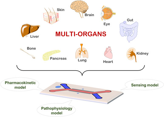
Currently, different OOC platforms have been fabricated based on the need for physiologically relevant and functional tissue/organ models. The fundamental expectation out of any testing system is targeted towards the point of care testing (POCT), which primarily speeds up the diagnosis at the vicinity of patient care. Further, POCT systems can be counted as versatile tools to realize the much required personalized therapy that centers around the concept of precision medicine.[Reference Borba, Castelletti, Filho and Martins59] The central theme of a POCT device architecture is a compact, self-containing, and portable miniaturized design that allows the analyses of complex samples at an affordable cost while providing sensitive, specific, user-friendly, robust/rapid, equipment-free prospects that can be delivered to those in need of it.[Reference Syedmoradi, Daneshpour, Alvandipour, Gomez, Hajghassem and Omidfar60, Reference Mabey, Peeling, Ustianowski and Perkins61] The OOC platforms have been tailored accordingly to effortlessly fit into your palm with enormous potential to be a supreme POCT device. An OOC device can thus precisely simulate the interactions of human organ/tissue with external factors. However, OOC platforms are still naïve and must be fine-tuned to achieve an ideal device that mirrors the in vivo human interactions with foreign materials, drugs or chemicals. The following sections will discuss the recent and unique OOC platforms for distinct organs/tissues that are available for pharmacokinetics (PK), pathophysiology and sensing applications.
Liver-on-chip platform as a PK model
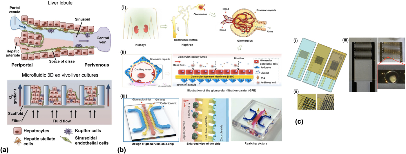
Liver-on-a-chip is one of the most familiar OOC model available today, perhaps due to its indispensable role in PK, where it is actively involved in various metabolisms and elimination of toxic wastes from the body. PK is a mathematical model used in pharmacology that deals with the fate of a drug in one's body like its absorption, distribution, metabolism, elimination, and toxicology.[Reference Bhatia and Ingber21] The classical anatomy of liver reveals that it is a triangular multi-lobed organ that extends across the abdominal cavity just below the diaphragm and above the right kidney. The lobules are the functional units of the liver that consist of multiple hepatic sinusoids as shown in the Fig. 6 (‘a’ top). Liver also consists of parenchymal and non-parenchymal cells that are separated from endothelial lining by the perisinusoidal space. The functions of the liver includes digestion of food, detoxification, biotransformation of drugs, glycogen storage, and plasma protein synthesis (predominantly albumin).[Reference Groger, Dinger, Kiehntopf, Peters, Rauen and Mosig62] Now, the current testing tools (in vitro and in vivo systems) fail to emulate the human metabolism or clearance, as there are species-specific differences among animals and human bodies that result in 26% of failure in clinical trials owed to hepatotoxicity.[Reference Beckwitt, Clark, Wheeler, Taylor, Stolz, Griffith and Wells63] Even the most successful liver models like liver slices and primary hepatocytes either face cytoarchitecture issues or show a decline in hepatic functions under conventional conditions.[Reference Khetani and Bhatia23] This situation appeals for the innovation of an ideal platform to emulate liver like functionalities precisely because many drugs fail in phase 3 clinical trials due to liver toxicity. A microfluidic model illustrated in Fig. 6 (‘a’ lower) gives a basic idea of a liver-on-chip model that shows 3D liver tissues subjected to fluid flow to facilitate shear stress and physiological oxygen gradient.[Reference Beckwitt, Clark, Wheeler, Taylor, Stolz, Griffith and Wells63] Perfusion of fluid through a microfluidic liver model is extremely beneficial as that would enable the expression of enzymes more efficiently than the 2D cultures, which is a very important trait to recapitulate the body conditions.[Reference Prot, Aninat, Griscom, Razan, Brochot, Guillouzo, Legallais, Corlu and Leclerc64] Recently, 3D models have been developed to overcome the drawbacks of 2D models that demonstrate sandwiched hepatocytes between two layers of gelled collagen, which preserves the polarity of hepatocytes.[Reference Lauschke, Hendriks, Bell, Andersson and Ingelman-Sundberg65] Subsequently, researchers came up with primary hepatocytes spheroid cultures, which maintained transcriptomic and proteomic signatures while maintaining viability and function for several weeks.[Reference Ewart, Dehne, Fabre, Gibbs, Hickman, Hornberg, Ingelman-Sundberg, Jang, Jones, Lauschke, Marx, Mettetal, Pointon, Williams, Zimmermann and Newham53, Reference Bell, Lauschke, Vorrink, Palmgren, Duffin, Andersson and Ingelman-Sundberg66] In another study, Bhise et al., developed a 3D human HepG2/C3A spheroids as a proof-of-concept model for the drug toxicity assessment, which was on par with the already published results obtained from in vivo and in vitro studies. In this study, a perfusable bioreactor and a direct write printer were interfaced to create the spheroid-laden hydrogel constructs, which was left under continuous perfusion and assessed for toxicity towards acetaminophen.[Reference Bhise, Manoharan, Massa, Tamayol, Ghaderi, Miscuglio, Lang, Shrike Zhang, Shin, Calzone, Annabi, Shupe, Bishop, Atala, Dokmeci and Khademhosseini67]
Distinct OOC models for pharmacokinetics, pathophysiology and sensing applications. (a) Liver-on-a-chip (images reprinted with permission from Elsevier),[Reference Beckwitt, Clark, Wheeler, Taylor, Stolz, Griffith and Wells63] (b) Glomerulus-on-a-chip (images reprinted with permission from the Royal Society of Chemistry),[Reference Wang, Tao, Su, Yu, Yu and Qin68] and (c) Pancreas-on-a-chip (images reprinted with permission from AIP publishing).[Reference Heileman, Daoud, Hasilo, Gasparrini, Paraskevas and Tabrizian69]
Glomerulus-on-chip as pathophysiology model
Now, besides the drug toxicity assessment, pathophysiology models are also extremely crucial in modern healthcare. Though science and technology have progressed many folds, still there are medical conditions that are not clear due to lack of appropriate models, e.g., the glomerulus.[Reference Ashammakhi, Elkhammas and Hasan70] The glomerulus is responsible for renal ultrafiltration—an ultra-selective filtering of blood plasma, which is size-, charge- and shape-selective to maintain body's electrolyte, pH, and blood hemostasis.[Reference Wang, Tao, Su, Yu, Yu and Qin68] Fig. 6(b), (i) and (ii) illustrate the glomerular structure and glomerular filtration barrier. The high selectivity occurs due to the complex structure of the glomerular barrier, the main components of which are the glomerulus epithelial cells, the podocytes, which have size-selective slit-shaped nanopores in their foot processes, and the glomerular endothelium, which has a charge-selective surface. In general, molecules larger than 4.2 nm cannot cross the glomerular filtration barrier (GFB) while those smaller than 2 nm can. Those falling in between these values are rejected or allowed to cross the barrier depending on their charge and shape. The glomerular basement membrane has a net negative charge. Proteins (negatively charged) tend to be retained in the blood flow. In contrast, the nephron reabsorbs water and filtered useful substances to return them to the circulatory system. This process is mediated both by osmotic pressure, as well as by active transport by the tubular epithelial cells. Most reabsorption is performed in the proximal tubule.[Reference Anil Kumar, Welsh, Saleem and Menon71] Recently, the advances in microfluidics and emerging novel OOC platforms have fortified the researchers to engineer glomerulus-on-chip devices that will allow them to probe and understand kidney biology, pathophysiology and drug interactions, which will ultimately benefit patients with glomerulopathies. It is of particular interest because recapitulating human functional podocytes is still a challenge, which explains the unavailability of glomerulus-on-chip devices. Recently, Musah et al., have directed the differentiation of human induced pluripotent stem cells into podocytes that express markers of a mature phenotype while also exhibiting primary and secondary foot processes. This OOC model of human glomerular function with mature human podocytes was used to demonstrate adriamycin-induced albuminuria and podocyte injury.[Reference Musah, Mammoto, Ferrante, Jeanty, Hirano-Kobayashi, Mammoto, Roberts, Chung, Novak, Ingram, Fatanat-Didar, Koshy, Weaver, Church and Ingber29] In another study, early-stage diabetic neuropathy was demonstrated with a glomerulus-on-chip device as shown in Fig. 6(b), (iii). Here, the device mimicked the GFB, glomerular endothelial cells, basement membrane, and podocytes. This device was used to probe the high glucose-induced diabetic neuropathy responses, which showed that hyperglycemia augmented the barrier permeability to albumin and glomerular dysfunction that led to proteinuria.[Reference Wang, Tao, Su, Yu, Yu and Qin68] Further, hypertensive neuropathy was simulated in another glomerulus-on-chip device. Glomerular hypertension promotes kidney disease progression by establishing high perfusion pressure, high filtration pressure and high transmembrane pressure in glomeruli. Here, a chip was fabricated that mimicked the physiological microenvironment with complex hemodynamic factors in glomeruli, which consisted of two channels lined by closely opposed layers of glomerular endothelial cells and podocytes. This device showed that cellular cytoskeletal rearrangement and damage to cells and their junctions that rises glomerular leakage triggered by the glomerular mechanical forces.[Reference Zhou, Zhang, Wen, Wu, Wang, Yang, Wang, Fang, Lin and Lin72]
Pancreas-on-a-chip as sensing model
The endocrine system is a collection of glands producing hormones which regulate metabolism, growth, and development. Among important insulin signaling pathway related diseases, Type II diabetes has emerged both as a genetically inherited and life-style disease, which is caused by a deficiency or diminished effectiveness of endogenous insulin. Nguyen et al., have reported an endocrine system on a chip for a diabetes treatment model using a microfluidic perfused 3D cell-culture chip, to potentially be able to screen drugs for the treatment of diabetes by measuring insulin release over time.[Reference Nguyen, van Noort, Jeong and Park73]
There are numerous POC glucose sensors available today, but there are negligible OOC models that mimic in vivo conditions with sensing capabilities. The endocrine system and pancreas are the typical examples, which lack adequate accurate and sensitive OOC platform. Pancreas serves as an accurate sensor of insulin production, processing, and secretion. These opponents interact with each other through the intra-islet insulin axis, besides other organs/tissues like the brain, liver, gut, insulin-dependent adipose, and muscle tissues. Altogether, the islet–organ/tissues axes described here form a highly sophisticated network that includes, but is not limited to, various signaling molecules, neuropeptides (brain-derived neurotrophic factor, NPY, melanin-concentrating hormone, gastrin-releasing peptide, VIP, and PACAP), hepatokines (betatrophin and HNFs), enteroendocrine hormones (the incretins GLP-1 and GIP, the decretins NmU and limostatin, gastrin, and CCK) as well as adipokines (leptin and adiponectin) and myokines (fibroblast growth factor-21 and IL-6) that mainly interact through GPCR signaling pathways, such as the cAMP cascade. In good health, the interplay between the pancreas and other organs or tissues ensure glucose homeostasis.[Reference Roder, Wu, Liu and Han74] However, impairments in the secretion of and/or sensitivity to insulin may result in metabolic diseases, such as Type II Diabetes. A breakthrough in pancreatic research was achieved with the fabrication of pancreatic islets on a microfluidic platform to assess its functionality with dielectric spectroscopy and response to glucose stimuli.[Reference Heileman, Daoud, Hasilo, Gasparrini, Paraskevas and Tabrizian69] Here, the pancreatic islets were immobilized on a microfluidic platform as shown in Fig. 6(c), under constant perfusion, to simulate physiological conditions. Dielectric spectroscopy was chosen for sensing islet membrane permittivity due to its real-time, non-invasive and rapid analysis attributes. This novel platform was able to track islet stimuli response during glucose challenge test and demonstrated islet membrane permittivity occurring due to the fusion of insulin vesicles during secretion. In another study, Nguyen et al., have recently reported an endocrine system-on-chip using a microfluidic perfused 3D cell-culture on a chip. Briefly, an endocrine system is a collection of glands producing hormones which regulate metabolism, growth, and development. Among important insulin signaling pathway related diseases, diabetes has emerged both as a genetically inherited and life-style disease, which is caused by a deficiency or diminished effectiveness of endogenous insulin. So, essentially it is a unique OOC platform that can potentially screen drugs for the treatment of diabetes by measuring insulin release over time. Insulin-secreting β-cells and L-cells are located in the pancreas and small intestines and trigger insulin secretion. Here, the fabricated endocrine system was shown as a useful tool in observing dynamical changes in endocrine hormones (GLP-1 and insulin) in a glucose-dependent environment.[Reference Nguyen, van Noort, Jeong and Park73]
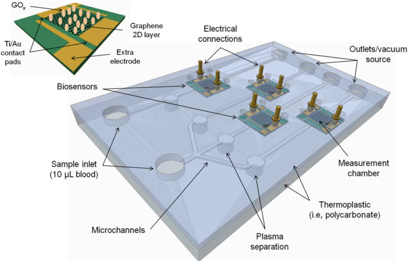
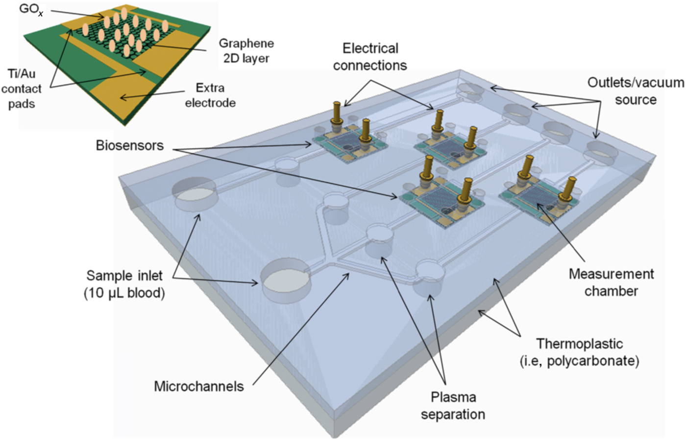
In a simplistic model, an artificial pancreas on a chip system combines a continuous glucose monitor and an insulin pump and uses a sophisticated algorithm to calculate the appropriate amount of insulin delivery based on the real-time glucose and insulin measurement by ultra-sensitive multiplex graphene-based microfluidic platform. Using an artificial pancreas system can significantly reduce the risk of hypoglycemia, a major concern for children and adults with Type 1 diabetes (T1D). In a limited study reported in T1D, results showed the artificial pancreas kept blood glucose levels in the normal range 60% of the time, compared with glucose levels by producing and releasing insulin and glucagon. Rapid, accurate, and quantitative measurement of biochemical analytes from body fluids are critical for accurate diagnosis of various metabolic disorders to improve disease management and clinical progress.[Reference Carlson, Mullen and Bergenstal75] So ideally, the goal would be to develop multiplexed sensors that use a drop of blood or urine (20–50 µL) to quantify multiple clinically important analytes simultaneously and answer specific questions for diagnosis as shown in Fig. 7.
Schematic of proposed sensor platform with four biosensors for Glucose, Insulin, Cholesterol, triglycerides (reproduced from S. Viswanathan, et al., Materials Today, 2015, Elsevier).[Reference Viswanathan, Narayanan, Aran, Fink, Paredes, Ajayan, Filipek, Miszta, Tekin, Inci, Demirci, Li, Bolotin, Liepmann and Renugopalakrishanan76]
Heart-on-a-chip
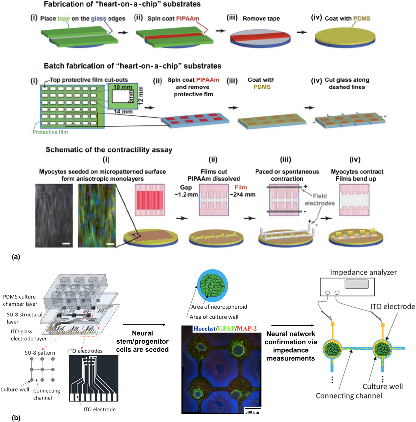
A pumping human heart assists the molecular transport of minerals and nutrients to provide energy for the entire human body. Recently, efforts have been made to develop ex vivo functional cardiac tissues to study the cardiac functions, tissue interactions, drug evaluation, toxicity, and regeneration due to the mounting burden of cardiovascular diseases and failure of the current animal testing models to accurately exemplify the human cardiotoxicity conditions. The advent and success of OOC prototypes, promises to recapitulate the morphological changes, tissue interactions, and functional complexities of human cardiac cells. Figure 8(a) illustrates a novel heart-on-a-chip construct based on the muscular thin film (MTF) technology that could be used to perform cardiac contractility and pharmacological studies in vitro.[Reference Grosberg, Alford, McCain and Parker77] This chip functioned alongside the existing 2D cell culture technique, with the myocytes arranged in an anisotropic organized layer. Further, fibronectin was printed as a microcontact onto a PDMS substrate where the ventricular myocyte of the neonatal rat could be seeded. This chip was able to monitor the contraction of eight MTFs simultaneously along with the evaluation of the effect of drugs on its contractile ability. Besides, studies related to the gene expression profiles of cellular structures and the contractile ability of cardiac tissue with a function of time were also performed. Likewise, a failing myocardial system on a chip was demonstrated with an engineered laminar ventricular tissue on a stretchable platform wherein, the stretchable chip was fabricated using MTF substrate coated with silicone membrane comprising of a temperature sensitive polymer and PDMS.[Reference McCain, Sheehy, Grosberg, Goss and Parker78] The substrates were micropatterned with fibronectin and seeded with ventricular myocyte of neonatal rat. The results revealed that such chips could be used to evaluate the genetic, structural, and functional response of a failing myocardium, leading to an early-stage drug discovery and the development of a heart-on-a-chip-based disease model. In a distinctive study, Xiao et al., developed a perfusable cardiac biowire system that could monitor the perfusion of pharmacological agents.[Reference Xiao, Zhang, Liu, Miklas, Gagliardi, Pahnke, Thavandiran, Sun, Simmons, Keller and Radisic79] In this study, they microfabricated a bioreactor that could generate 3D perfusable micro-tissue called the “biowires” using neonatal rat cardiomyocytes and human embryonic stem cells. These biowires were generated via polytetrafluoroethylene tubing template integrated with electrical field stimulation, using carbon rod electrodes. This platform was tested by introducing nitric oxide released from perfused sodium nitroprusside solution that was diffusing through the tubes. Moreover, this biowire was also used to improve the phenotype of cardiomyocytes via electrical stimulation under parallel and perpendicular stimulations. The pros of this construct include, (i) improved mimics of cardiac bundle structure and its alignment, (ii) easy customization of the device for high throughput screening and, (iii) the possibility of automatic topographical stimulations. More recently, a cardiac micro-physiological system (MPS) on a scale of 1 mm2 was demonstrated for drug screening applications that contained a standard multi-well plate, which could incorporate hundreds of cell culture channels.[Reference Mathur, Loskill, Shao, Huebsch, Hong, Marcus, Marks, Mandegar, Conklin, Lee and Healy80] This device was integrated with an isogenic GCaMP6f reporter cell line that provided mechanical and electrophysiological properties of micro cardiac tissue via high throughput analysis. This cardiac MPS was reported to be an ideal in vitro model that predicted the cardiac toxicity along with enhanced storing capability of viable and functional human induced pluripotent stem cell-derived cardiac tissue for several weeks. In an important study by Marsano et al., a low-cost microfluidic device was used to generate a 3D cardiac micro tissue that could directly visualize and quantitatively analyze in a controlled environment.[Reference Marsano, Conficconi, Lemme, Occhetta, Gaudiello, Votta, Cerino, Redaelli and Rasponi81] The mature 3D cardiac cells were generated from human induced pluripotent stem cell-derived cardiomyocytes and neonatal rat cardiomyocytes that efficiently predicted the signs of hypertrophic changes in cardiac phenotype via mechanical and stimulation studies.
(a) “Heart on a chip” assembly based on the muscular thin film that could perform in vitro cardiac contractility and pharmacological studies (image reprinted with permission from Grosberg et al., Copyright 2011 Royal Society of Chemistry),[Reference Grosberg, Alford, McCain and Parker77] (b) “Brain on a chip” to study neural communications and regeneration. (Image reprinted with permission from Liu et al., Copyright 2018 American Chemical Society.)[Reference Liu, Lee and Lei82]
Brain-on-a-chip
The brain is soft nervous tissue contained within the skull of humans, which exercises control over the other organs of the body via a well-orchestrated nervous system. It is composed of many complex units that include neurons, oligodendrites, microglia, and astrocytes that are crucial for brain functions. Recently, different in vitro brain models have been developed to understand its functions, neuronal interaction, and regeneration.[Reference van der Helm, van der Meer, Eijkel, van den Berg and Segerink83] Following which, brain-on-a-chip models have gained significant popularity to investigate various aspects of brain functions, neuronal interactions, axonal growth and transport, dendrite to neuronal signaling, neuronal electrical activity on and around the microelectrodes and neurological effect of drugs. One of the earliest forms of a brain-on-a-chip model comprised a microfluidic platform consisting of dual micropillar for embryonic stem cell culturing to control the shear stress and evaluate the cell docking.[Reference Lee, Kim, Borana, Chung and Chung84] The dual micropillar made of 16 circular-shaped outer micropillars that minimized the shear stress, and eight saddle-shaped inner micropillars that led to the efficient docking of embryonic cells was used. It was found that in a span of 6 days the embryonic stem cells differentiated into neural cells paving the way for a powerful method to screen single embryonic cells. Bang et al., reported a microfluidic platform filled with Matrigel, where a continuous flow of fluid was provided to mimic a 3D neural circuit.[Reference Bang, Na, Jang, Kim and Jeon85] The fluidic flow aligned the extracellular matrix along the flow direction, following which the primary rat cortical neurons were incorporated into the Matrigel at an average speed of 250 μm/day. These neurons developed axon bundles of 1500 µm length in 6 days, which were further confirmed by immunostaining for the expression of PSD-95 and synaptophysin. To understand the complex function of brain, during the occurrence of neurological disorder like Alzheimer's disease, a 3D brain-on-a-chip was developed with the 3D neurospheroids and an interstitial level of flow.[Reference Park, Lee, Jeong, Hyun, Lee and Lee86] This chip consisted of two chambers containing array of 50 cylindrical wells, and PDMS was used to form the concave microwell array via surface tension. These concave microwell arrays had uniform neurospheroids with cell–cell interactions and contacts in all directions, while a slow interstitial level of flow was maintained using an osmotic micropump system. This system was used to investigate the neurotoxic effect of amyloid-β exhibiting synaptic dysfunction and increased neural destruction. The results of this 3D in vitro brain model were comparable with the ones obtained from in vivo models, thus providing a better tool for better diagnosis and treatment. Recently, Lui et al. developed an artificial brain-on-a-chip to investigate the electrical stimulations, neural communications, and regeneration.[Reference Liu, Lee and Lei82] This construct paved way for an efficient disease model for various neurological diseases including Parkinson's disease. Here, the biochip platform consisted of nine 3 × 3 arrays of cultured wells embedded onto an ITO plate inter-connected via channels constructed using PDMS as shown in Fig. 8(b). Further, the polyelectrolyte films seeded with neurospheroids were placed onto the culture wells to form a multilayered neural network within a duration of 5 days. The micropatterns on the polyelectrolyte film promoted and guided the neurite outgrowth and neural differentiation. Immunohistochemistry techniques were used to reveal the differentiation and synaptic function of the fabricated artificial brain, while impedance measurements confirmed the electrical connections of the neural networks.
Multi-OOCs
One of the most requisite models to study cytotoxicity is the enterohepatic model, which would essentially simulate the circulation between liver and intestine. Any chemical or nutrient is first absorbed by the small intestine and is channelized to the liver through the portal vein. An OOC model can precisely dub these aspects and become an ideal PK/PD model that can boost the growth of drug development. To simulate such a complex mechanism, multi-OOC platforms serve as the main contenders, where in vivo functionalities can be simulated accurately compared with the existing setup to elucidate the fate of a drug in the body. Kimura et al., demonstrated that their liver-small intestine model could emulate anti-cancer drug activity on the target cells.[Reference Kimura, Ikeda, Nakayama, Sakai and Fujii87] Here, they co-cultured Caco-2 cells, HepG2 cells, and A549 cells for more than 3 days to mimic the physiological circulation and organ volume ratio for drug analysis. In a slightly more sophisticated version, four organs namely, the liver, small intestine, skin, and kidney were co-cultured for 28 days on a chip. Interestingly, this system simulated blood circulation and waste (collected from the kidney) removal through two separate pumps. This system exhibited three distinct fluid chambers and each maintained unique glucose concentration. It was noteworthy that the blood circuit had the highest glucose concentration and the kidney circuit has the lowest.[Reference Zhang and Radisic2, Reference Maschmeyer, Lorenz, Schimek, Hasenberg, Ramme, Hubner, Lindner, Drewell, Bauer, Thomas, Sambo, Sonntag, Lauster and Marx49] In another study by Xiao et al., a microfluidic culture model named EVATAR, that contained murine ovarian follicles was demonstrated to produce 28 days menstrual cycle hormone profile that controlled the human female reproductive tract and peripheral tissue dynamics in a single, dual and multi-unit microfluidic platforms.[Reference Xiao, Coppeta, Rogers, Isenberg, Zhu, Olalekan, McKinnon, Dokic, Rashedi, Haisenleder, Malpani, Arnold-Murray, Chen, Jiang, Bai, Nguyen, Zhang, Laronda, Hope, Maniar, Pavone, Avram, Sefton, Getsios, Burdette, Kim, Borenstein and Woodruff88] This is a very significant report since they simulate in vivo female reproductive tract along with the endocrine loops while maintaining the circulatory hemostasis between tissues. Moreover, there are only a handful of in vitro or in vivo models that reiterate the human female menstrual cycle. More recently, a functional OOC that emulated the feedback loop between pancreatic islets (insulin-secreting) and liver spheroids was demonstrated.[Reference Bauer, Wennberg Huldt, Kanebratt, Durieux, Gunne, Andersson, Ewart, Haynes, Maschmeyer, Winter, Ammala, Marx and Andersson89] The key highlights were the successful simulation of cross-talk between the pancreatic islet microtissues and liver spheroids, which was determined based on insulin and glucose regulation. They were also able to maintain the functional responses up to 15 days in an insulin-free media. Figure 9 illustrates the above mentioned multi-OOC platforms.
Multi-organ-on-chip platforms. (a) Four organ-on-a-chip model with liver, intestine, kidney, and skin (images reprinted with permission from Royal Society of Chemistry),[Reference Maschmeyer, Lorenz, Schimek, Hasenberg, Ramme, Hubner, Lindner, Drewell, Bauer, Thomas, Sambo, Sonntag, Lauster and Marx49] (b) human female reproductive tract model on a chip, (i) single microfluidic unit platform, (ii) two microfluidic units platform, (iii) five microfluidic units platform, and (iv) illustration of pump mechanism of the electromagnetic five microfluidic units platform (images reprinted with permission from Nature Communications),[Reference Xiao, Coppeta, Rogers, Isenberg, Zhu, Olalekan, McKinnon, Dokic, Rashedi, Haisenleder, Malpani, Arnold-Murray, Chen, Jiang, Bai, Nguyen, Zhang, Laronda, Hope, Maniar, Pavone, Avram, Sefton, Getsios, Burdette, Kim, Borenstein and Woodruff88] (c) two organ-on-chip platform with pancreatic islets and liver spheroids (images reprinted with permission from Nature Scientific Reports).[Reference Bauer, Wennberg Huldt, Kanebratt, Durieux, Gunne, Andersson, Ewart, Haynes, Maschmeyer, Winter, Ammala, Marx and Andersson89]

Challenges and future prospects
Undoubtedly, OOCs have delivered to its potential and also hold intriguing prospects for varied applications. However, the translation of OOCs from laboratory to market has been astoundingly low compared with the evolution it has actually attained in the laboratories. This may be due to the excessive attention given to the architecture and miniaturization of the OOC device rather than the commercialization perspectives. Perhaps it is extremely essential for the researchers to equally focus on the standardization, platform integration, and scalable economy to appeal a large market.[Reference Mohammed, Haswell and Gibson90] Now, this includes both biologic and technical aspects of OOC such as organ scaling, necessity for a universal media, stem cell sourcing, vascularization, inclusion of immune components, fate of drug bound to PDMS, sterile connection platforms, bubble-free media circulation, flow-rate regulation between distinct platforms, inclusion of biosensors, dissolved oxygen and nutrients in various platforms.[Reference Low and Tagle91] The use of clinical biomarkers and analytical systems, such as sequencing, microarray and/or mass spectroscopy can validate the organ models and the clinical reference points. But, the availability of the minute quantity of cells and media warrant the highest possible sensitivity of the analytical systems employed for the cause.[Reference van Duinen, Trietsch, Joore, Vulto and Hankemeier20] Apart from these the ability to successfully link distinct organs together to precisely mimic the in vivo physiological conditions to evaluate its response to a drug has to be validated. Currently, several researchers are working on multi-OOC platforms, while some have shown great prospects. However, a “homo-chippien” concept is still far from being a reality.
So far we have seen a transition from the 2D cell cultures to 3D and OOC models. This transition has routed the attention of several researchers towards the concept of “homo-chippiens”. We envision that in near future a much-sophisticated microfluidic technology (high throughput platforms) with 3D cell cultures will be used to validate the biologic development and physiologically relevant models. Also, the focus will shift towards the making of user-friendly devices with enhanced assay ability, which will ultimately pave way for the personalized precision medicine.
Conclusion
Multi-OOC is a solid platform that has promised to deliver the ambitious dream of the body on-chip, rather “homo-chippiens”. However, Multi-OOC may lack the morphology of native organs and subcutaneous implant are likely to face rejection.[Reference Tasciotti, Cabrera, Evangelopoulos, Martinez, Thekkedath, Kloc, Ghobrial, Li, Grattoni and Ferrari92] While Multi-OCC as an external patch connected to mobile devices is likely to be approved by FDA. Researchers have immensely contributed to the development of OOC platforms in the past decade. Several funding agencies like National Institutes of Health (NIH), Food and Drug Administration (FDA), Defense Advanced Research Projects Agency (DARPA), and The European Union have actively funded for OOC research, which clearly demonstrates the worldwide recognition given to OOC platforms. Besides, there are a lot of pharmaceutical and cosmetic companies that are really interested to commercialize this technology.[Reference Balijepalli and Sivaramakrishan93] However, the biologic and technical challenges need to be addressed to ensure scaling this technology to the next level. Moreover, the researchers should find a realistic way and focus to transfer this technology from laboratory to market. Given the immense popularity and potential of OOC platforms to precisely predict the fate of drugs in the human body, it can indeed be a commercial drug testing tool and may be a well-suited candidate to replace the current testing tools, though it has to be approved by the regulatory authorities. Especially, when one of the prime factors such as gender is often overlooked during drug screening process, which has a significant impact on the drug efficacies, pathophysiology, and toxicity to different individuals. Under most circumstances, only male tissues/cells/animals/subjects are chosen over their female counterparts.[Reference Nawroth, Rogal, Weiss, Brucker and Loskill94] This can be negated by the use of multi-OOC platforms that will substantially encourage personalized medicine. Eventually, it would be safe to opine that “OOC” can certainly blaze new trails that the world has never seen before.
Acknowledgment
Authors would like to thank MEXT, Japan, for the financial support under the Monbukagakusho fellowship during the research. Part of this study was supported by a grant for the program of the strategic research foundation at private universities S1101017, organized by the MEXT, Japan, since April 2012. VR acknowledges support from NIH, Foundation Edmund Rothschild, Paris, France.