
I. INTRODUCTION
In 2007, the ground-breaking work of Takahashi et al.Reference Takahashi, Tanabe, Ohnuki, Narita, Ichisaka, Tomoda and Yamanaka1 reported on the generation of induced pluripotent stem cells (iPSCs) from adult human fibroblasts and other human somatic cells. iPSCs are generated from somatic cells by transfecting four transcription factors (Oct4, Sox2, Klf4, and c-Myc) in fibroblasts.Reference Takahashi and Yamanaka2 This paved the way for the possibility to use mature differentiated cells and revert them back to pluripotency, that is, an embryo-like state. As a result, the present decade is seeing vast perspectives for human disease modeling, regeneration of tissues and organs, and drug discovery.Reference Ohnuki and Takahashi3 iPSCs have thus shown in a short time-lapse strong promise to accelerate disease treatment and limitless possibilities as they allow the generation of representative samples of any tissue in a patient’s body.
Prior to iPSCs, embryonic stem cells (ESCs) have allowed the generation of genetically engineered mice which are used as human disease models. Although, animal models have contributed enormously to the better understanding of disease mechanisms, they have limitations in correctly mimicking human diseases. These limitations arise from existing variability in the genetic makeup of the animal models compared with humans and different species have distinct genetic makeup and hence sometimes unique proteins. iPSCs can act as a more suitable alternative as they provide identical human cell environment and are functional in vitro as well as in vivo after transplantation.Reference Singh, Kalsan, Kumar, Saini and Chandra4 In addition, a patient specific iPSC allows the development of personalized therapeutics regimen/drug.
Nanotechnology can have an augmenting effect on iPSC-driven tissue engineering for reconstruction through the use of 2D or 3D scaffolds.Reference Ohnuki and Takahashi3 This novel approach involves the combination of stem cells and biologically inspired scaffolds. Modifying the nanotopography of scaffolds would enhance or guide cell differentiation.Reference Ramalingam and Rana5
Bone tissue engineering (BTE) has to address numerous challenges such as bone fracture at various sites and osteodegenerative diseases in aging adults. The use of engineered bone tissue is becoming a preferred option compared to conventional bone grafts as they prevent disease transmission. Combining biomaterials, cells, and growth factor is the trend in BTE to accelerate bone repair and regeneration. In this review, we critically analyze in vivo bone repair models and the osteogenic differentiation of iPSCs. We next look at the contribution of nanotopographical scaffolds coupled with iPSCs to accelerate and enhance regeneration of bone tissue. The ability of iPSCs to address age-related bone diseases compared to mesenchymal stem cell (MSC) is also discussed.
II. BONE BIOLOGY
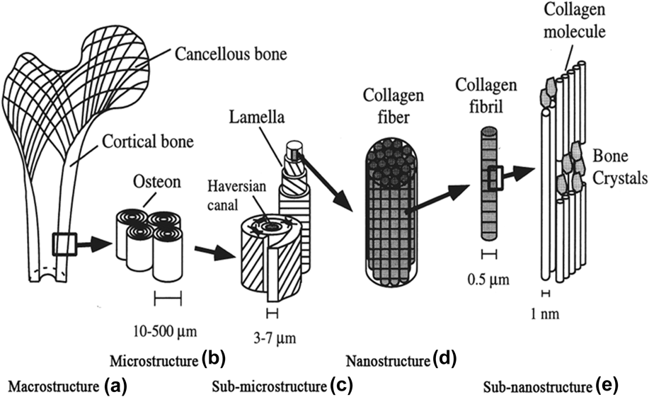
Bone is a dynamic, highly vascularized tissue with the unique capacity to heal and remodel without the formation of scars. Bone is present in two architectural forms in the adult body, namely trabecular (also called cancellous or spongy bone) and cortical (also known as compact bone) bone. Trabecular and cortical bones form around 20% and 80% of the total skeleton, respectively, but their proportions may vary depending on their location in the skeleton. In humans, trabecular bone has a porosity of 50–90% with an average density of approximately 0.2 g/cm3.Reference Kingham and Oreffo6,Reference Keaveny, Morgan, Niebur and Yeh7 Cortical bone has a much denser structure with a lower porosity of 3–12% and an average density of 1.80 g/cm3.Reference Barrère, Mahmood, de Groota and van Blitterswijka8,Reference Cooper, Matyas, Katzenberg and Hallgrimsson9 As a result of the differences in porosity, the modulus and ultimate compressive strength of trabecular bone is around 20 times inferior than that of cortical bone.Reference Sikavitsas, Temenoff and Mikos10,Reference Temenoff, Lu, Mikos, Davies and Vacanti11 The bone matrix consists of two major components: an inorganic mineral part constituting hydroxyapatite (HA) which contributes 65–70% to the matrix and an organic part, composed of glycoproteins, proteoglycans, and sialoproteins, comprises the remaining 25–30% of the total matrix.Reference Sommerfeldt and Rubin12 Bone shows a distinct hierarchical structural organization of its constituents on numerous levels including macrostructure (cancellous and cortical bone), microstructure (haversian systems, osteons, and single trabeculae), submicrostructure (lamellae), nanostructure (fibrillar collagen and embedded minerals), and subnanostructure (molecular structure of constituent elements, such as mineral, collagen, and noncollagenous organic proteins)Reference Henkel, Woodruff, Epari, Steck, Glatt, Dickinson, Choong, Schutz and Hutmacher13 (Fig. 1).
Hierarchical structural organization of bone: (a) cortical and cancellous bone; (b) osteons with haversian systems; (c) lamellae; (d) collagen fiber assemblies of collagen fibrils; (e) bone mineral crystals, collagen molecules, and noncollagenous proteins. Reproduced with permission from Ref. Reference Rho, Kuhn-Spearing and Zioupos20 Copyright 1998 Elsevier.

The maintenance and resorption of healthy bone results from the interaction of three cell types, namely osteoblasts, osteocytes, and osteoclasts.Reference Ducy, Schinke and Karsenty14–Reference Nijweide, Burger, Nulend, Van der Plas, Bilezikian, Raisz and Rodan16 Osteoblasts are cells which are differentiated from MSCs but they are not terminally differentiated cells. Osteoblasts have two fates: they become embedded in their own bone matrix and become osteocytes, or they undergo apoptosis.Reference Jilka, Weinstein, Bellido, Parfitt and Manolagas17 Osteoblasts are responsible for skeletal architecture via deposition of the bone matrix and regulation of osteoclast activity.Reference Safadi, Xu, Smock, Kannan, Selim, Odgren, Marks, Owen and Popoff18 Osteocytes play a key role in mechanotransduction and are involved in the calcification of the osteoid matrix and blood-calcium homeostasis. Osteoclasts are multinucleated cells derived from fusion of mononuclear hemopoietic precursors. Their primary function is to secrete acids and proteolytic enzymes, which erode bone ECM under the influence of chemical cues.Reference Mackie15,Reference Hadjidakis and Androulakis19
III. CURRENT IN VIVO MODELS FOR BONE TISSUE ENGINEERING
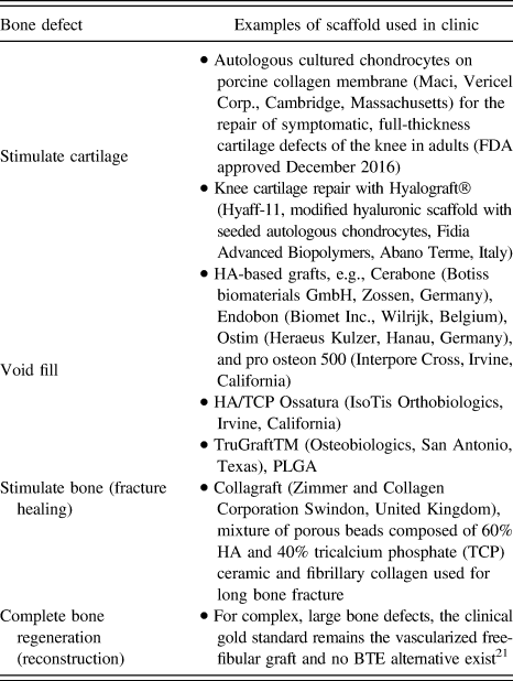
BTE is required to address therapeutic challenges ranging from trauma to bone degeneration (Table I). Most of them make use of bone tissue-mimicking materials to accelerate healing.
Bone defects and some clinical applications of scaffolds.

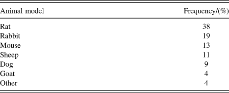
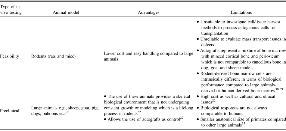
Before reaching clinical application, the scaffold has to undergo in vitro as well as in vivo assessment. In vitro models have been considered until now as inadequate in recreating the complex in vivo environment and thus in vivo models are used to give a reproducible approximation of the clinical setting. These in vivo models are used in an attempt to answer two main objectives: the performance of the scaffold and material in the clinical setting and ways by which it will accelerate healing. Usually, the animals used have rapid breeding and detection of biological events under study. Animal models play a critical role in terms of scaffold testing for feasibility and bioactivity studies and for preclinical translational testing. The relative frequency of use of different animal models in the treatment of critical and noncritical sized gap defects is summarized in Table II.
Relative frequency of use of animal models in the treatment of critical and noncritical sized gap defects.Reference O’Loughlin, Morr, Bogunovic, Kim, Park and Lane22

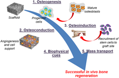
To the best of our knowledge, no ideal single animal model is currently available whereby bone tissue regeneration may be assessed reliably and reproducibly. Indeed, the ideal in vivo model for BTE needs to fulfill several criteria. In particular, it should mimic the physiological environment and orthopedic situation as closely as possible. Moreover, it should provide quantifiable parameters to assess the success and functional performance of the newly formed bone. In fact, the selection of animal models for BTE is closely related to the therapeutic modality that is under investigation. The therapeutic modalities related to BTE may be summarized into five categories as depictedReference Muschler, Nakamoto and Griffith23 in Fig. 2, in particular osteogenesis,Reference Stedman24 osteoconduction,Reference Burchardt25,Reference Muschler, Lane, Habal, Reddi and Saunders26 osteoinduction,Reference Burchardt25,Reference Muschler, Lane, Habal, Reddi and Saunders26 mass transport, and biophysical cues.
Five therapeutic modalities investigated in BTE.

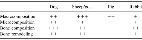
As highlighted before, one of the major criteria in the selection of animal models is close mimicking of the underlying bone biology of humans. Differences in bone composition, density, and quality vary between different species (human, dog, sheep, pig, and rabbit) (Table III). It was found that compared to other animal models, the bone composition (ash weight, hydroxyproline, extractable proteins, and IGF-1 content) of dogs matches closely with that of human bone.Reference Aerssens, Boonen, Lowet and Dequeker27 In terms of the bone anatomy, morphology, healing, and remodeling, pig is considered to be a close representative of the human bone.Reference Thorwarth, Schultze-Mosgau, Kessler, Wiltfang and Schlegel28 In addition, compared to the human bone, the rodent skeleton undergoes constant remodeling throughout their lives, the trabecular bone content is limited and the haversian remodeling (concentrically oriented lamellar bone containing a centrally located blood vessel, also known as osteons) by tunneling osteoclasts do not occur in rodents. On the other hand, larger and relatively more trabecular bone mass with haversian remodeling can be found in rabbits. The bone marrow of rabbits is fatty and thus different in terms of physical properties compared to the human bone marrow.Reference Yamamoto, Irisa, Sugioka and Sueishi29,Reference Miyanishi, Yamamoto, Irisa, Yamashita, Jingushi, Noguchi and Iwamoto30 The bone composition and biology of dog, sheep, goat, and pig have been shown to be very similar to those of humans.Reference Pearce, Richards, Milz, Schneider and Pearce31,Reference Reinwald and Burr32 However, the differences in physical size, anatomy, species specific physiology, etc. make some animals better suited for the study of individual areas.
Summary of four key attributes in terms of similarity between animal and human bone.Reference Pearce, Richards, Milz, Schneider and Pearce31

+ least similar, ++ moderately similar, +++ most similar.
Feasibility testing of scaffolds is often carried out in small animals such as mice and rats, whereby outcomes are determined after relatively short periods of time ranging from days to weeks. However, in some cases for instance (i) when the surgical procedure involved cannot be performed reproducibly in small animals, e.g., spinal fusion procedures or (ii) when the size of the scaffold exceeds the volume capacity of the small animal or (iii) when modeling effects of large diffusion distances in the survival of transplanted cells, feasibility studies need to be conducted in larger animal models such as rabbits, dogs, sheep, or goat.Reference Muschler, Raut, Patterson, Wenke and Hollinger33 Table IV summarizes the advantages and limitations associated with the use of small and large animals for BTE applications.
Summary of advantages and limitations associated with the use of small and large animals for BTE.

The effect of aging on bone regeneration has been studied using rabbits, rats, and mice models. As age increases, chondrogenic potential of the periosteum decreased in rabbits,Reference O’Driscoll, Saris, Ito and Fitzimmons34 the rate of regain of mechanical competence after fracture decreased in rats,Reference Meyer, Tsahakis, Martin, Banks, Harrow and Kiebzak35 and the rate of cellular differentiation and repair decreased in mice.Reference Naik, Xie, Zuscik, Kingsley, Schwarz, Awad, Guldberg, Drissi, Puzas, Boyce, Zhang and O’Keefe36 The rate of revascularization also decreases with increasing age as seen in tibial fractures created in juvenile (4-week-old), middle-aged (6-month-old), and elderly (18-month-old) mice.Reference Lu, Hansen, Sapozhnikova, Hu, Miclau and Marcucio37
IV. iPSCs AND BONE REGENERATION
iPSCs have several similarities with human ESCs, namely in terms of their morphology, unlimited self-renewal potential, and gene expression profiles.Reference Ramalingam and Rana5 Patient-specific cells with high pluripotency may be obtained by reprogramming the patient’s own-cells thus reducing the risks of immunological rejection. Furthermore, due to the vast availability of reprogrammable cells, iPSCs may be easily obtained without any invasive procedures and their use is associated with fewer ethical issues in contrast to ESCs. Reprogramming cells to pluripotency requires a shift from oxidative to glycolytic metabolism. It has been shown for acquisition of pluripotency that this shift occurs early during reprogramming of human cells and requires hypoxia-inducible factors, HIF1α and HIF2α.Reference Mathieu, Zu, Xing, Sperber, Ferricio, Agoston, Kuppusamy, Moon and Ruohala-Baker40,Reference Mordhorst, Murphy, Ross, Samuel, Salazar, Ji, Behura, Wells, Green and Prather41 Experiments using fibroblast indicated that the majority of the increased metabolic genes during early reprogramming are hypoxia-responsive genes. This reprogramming has been shown to be more efficient when performed under hypoxic conditions.
Due to major limitations associated with autografts and allografts, the clinical use of engineered bone is becoming more and more common.Reference Frohlich, Grayson, Wan, Marolt, Dorbnik and Vunjak-Novakovic42 Since bone is highly vascular, engineering new bone from other cell sources in addition to bone cells is a promising strategy to create vascular and nerve compartments.Reference Meijer, de Bruijn, Koole and van Blitterswijk43 MSCs isolated from adult tissues are the most commonly used stem cells in BTE applications. However, the functionality of MSCs in regeneration as well as their self-renewal ability is reduced with aging.Reference Kretlow, Jin, Liu, Zhang, Hong, Zhou, Baggett, Mikos and Cao44,Reference Stenderup, Justesen, Clausen and Kassem45 Moreover, their proliferation and differentiation capabilities decrease during in vitro culture expansion.Reference Jesudason and Clifton46
To limit the tumorigenicity associated with pluripotency of iPSCs, in vitro differentiation of iPSCs toward the MSCs and osteoprogenitor cells prior to transplantation is necessary. Embryoid bodies (EBs) are generally formed from iPSCs as an intermediate step to generate osteoprogenitors during osteogenic differentiation.Reference Bilousova, Jun, King, Langhe, Chick, Torchia, Chow, Klemm, Roop and Majka47–Reference Wang, Liu and Zhao49 Other studies have applied different strategies to skip this step in the osteogenic induction of iPSCs.Reference de Peppoa, Marcos-Campos, Kahler, Alsalman, Shang, Vunjak-Navakovic and Marolt50–Reference Zou, Luo, Chen, Wang, Ding, Petersen, Kang, Dagnaes-Hansen, Zeng, Lv, Ma, Le, Bessenbacher, Bolund, Jensen, Kjems, Pu and Bünger52 Furthermore, the direct differentiation of iPSCs into osteoblast phenotypes has also been reported.Reference Jin, Kim, Kim, Won, Yoo, Choi, Hyun and Kim53,Reference Pan, Yu, Zhai, Lee, Zhao, Liu, Wang, Tee, Shi, Cui and Bao54
Recent studies have shown that iPSCs could be used as an alternative source of stem cells in bone regeneration because of their potential to overcome limitations associated with MSCs. For example, the differentiation of iPSCs into two types of iPSC-derived MSCs (iMSCs), namely, early (aiMSCs) and late (tiMSCs), outgrowing cells could be achieved following short-term exposure to transforming growth factor-β.Reference Sheyn, Ben-David, Shapiro, De Mel, Bez, Ornelas, Sahabian, Sareen, Da, Pelled, Tawakocli, Liu, Gazit and Gazit55 Compared with bone marrow-derived MSCs (BM-MSCs), aiMSCs displayed more of a stem cell phenotype, whereas tiMSCs acquired more of a differentiated osteoblast phenotype. tiMSCs helped in the in vivo bone regeneration following orthotropic injection into radial defects but does not allow the cells to induce ectopic bone formation.Reference Sheyn, Ben-David, Shapiro, De Mel, Bez, Ornelas, Sahabian, Sareen, Da, Pelled, Tawakocli, Liu, Gazit and Gazit55
A. Impact of iPSC origin on osteogenic differentiation potential
As mentioned earlier, iPSCs may be derived from various differentiated adult somatic cells and therefore the effect of cell origin on the osteogenic differentiation potential of iPSCs is a highly questionable aspect. There are contradictory findings in the literature with regard to whether iPSCs possess a memory for their tissue of origin, and whether this leads to an increased propensity to develop back into their original cell type.Reference Kim, Doi, Wen, Ng, Zhao, Cahan, Kim, Aryee, Ji, Ehrlich, Yabuuchi, Takeuchi, Cunniff, Hongguang, McKinney-Freeman, Naveiras, Yoon, Irizarry, Jung, Seita, Hanna, Murakami, Jaenisch, Weissleder, Orkin, Weissman, Feinberg and Daley56 Various studies have described the presence of the epigenetic memory of iPSCs for their somatic tissue of origin that can influence the in vitro differentiation capacity of the iPSC.Reference Polo, Liu, Figueroa, Kulalert, Eminli, Tan, Apostolou, Stadtfeld, Li, Shioda, Natesan, Wagners, Melnick, Evans and Hochedlinger57 For example, Ishiy et al. showed that the osteogenic differentiation potential of human deciduous teeth (SHED) derived iPSCs (iPS-SHED) was higher than that of iPSCs derived from human dermal fibroblasts (iPS-FIB)Reference Ishiy, Fanganiello, Griesi-Oliveira, Suzuki, Kobayashi, Morales, Capelo and Passos-Bueno58 as indicated by improved alkaline phosphatase (ALP) activity and calcium mineralization. Moreover, Hynes et al. generated iPSCs from three different somatic tissues namely gingiva, periodontal ligament (PDL), and lung.Reference Hynes, Menicanin, Mrozik, Gronthos and Bartold59 In vivo subcutaneous implantation of iPSC-seeded osteoconductive scaffolds in mice resulted in enhanced formation of mineralized structures for iPSCs generated from PDL in contrast to the other two cell lines. In addition, de Peppo et al.Reference de Peppoa, Marcos-Campos, Kahler, Alsalman, Shang, Vunjak-Navakovic and Marolt50 investigated the differences in osteogenic potential of iPSCs derived from dermal fibroblasts (11c and 1013A) and BMSCs. Significantly higher expression of ALP gene and calcium deposition were noted for both BC-1 and 1031A-derived iPSCs. On the other hand, 11c-derived iPSCS showed poor osteogenic potential. In vivo implantation of BC1-derived iPSCs displayed areas of dystrophic calcification, in comparison to 1013A-derived iPSCs which resulted in the formation of uniform bone-like tissue with matrix-embedded cells.Reference de Peppoa, Marcos-Campos, Kahler, Alsalman, Shang, Vunjak-Navakovic and Marolt50
By contrast, the study by Nasu et al. reported that cell origin did not influence the osteogenic differentiation potential.Reference Nasu, Ikeya, Yamamoto, Watanabe, Jin, Matsumoto, Hayakawa, Amano, Sato, Osafune, Aoyama, Nakamura, Kato and Togushida60 Indeed, no difference in the osteogenic gene expression profiles of BMSC-derived iPSCs and dermal fibroblast-derived iPSCs was found.
Overall, findings from most studies suggest that even after iPSCs are induced into MSC-like cells, they may still maintain epigenetic memory of their tissue of origin.
Sparks et al. compared the osteogenic potential of two human iPSC lines (RIV9 and RIV4) to human H9 ESCs.Reference Sparks, Martinez, Soto and zur Nieden61 The latter gave rise to neural-crest–derived osteoblasts at a much faster rate. The H9 cells resulted in neural-crest–derived osteoblasts, whereas the RIV9 cells used both the mesodermal and a neural crest route at a lower rate. Further studies are required to determine the bone regeneration potential of mesodermal-derived osteoprogenitors compared to hiPSC-derived neural crest cells.
B. Effect of the use of supplements in osteogenic medium and/or transduction agents on osteogenic differentiation potential
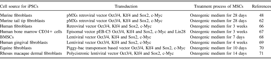
Several research groups have investigated the possibility of differentiating iPSCs into osteoblasts via supplementing the culture medium with osteogenic agents or via the use of transduction strategies. Table V summarizes few studies whereby osteoprogenitor cells were successfully obtained from iMSCs cultured under osteogenic differentiation conditions without scaffolds. Addition of TGF-β, IGF-1, and FGF-β to the osteogenic medium improves the osteogenic capacity of iPSCs. As reported by Li et al., TGF-β1 and 3 derived-cells possessed higher potential to differentiate into osteoblasts compared to bFGF or BMP-2 derived-cells. TGF-β1 in combination with retinoic derived-cells seeded onto HA/TCP ceramics deposited typical bone when implanted in vivo.Reference Li and Niyibizi62
Summary of studies whereby iMSCs successfully differentiated into osteoprogenitor cells.

Another alternative which may be used to enhance the osteogenic differentiation of iPSCs involves gene transduction of the cells via potent osteogenic transcription factors. For instance, Liu et al. reported on the transduction of BMP-2 in hiPSC-derived MSCs for the first time via the use of lentiviral vectors.Reference Liu, Chen, Zhao and Xu63 The ALP activity of BMP2-transduced hiPSC-MSCs was shown to be 1.8 times higher than that of untransduced hiPSC-MSCs after 14 days in vitro culture. Furthermore, when seeded on RGD-functionalized calcium phosphate cement (CPC) scaffold, COL1A1 and osteocalcin (OC) expressions were 1.9 times and 2.3 times, respectively, that of untransduced cells at 14 days. In addition, transduction of Runx2 into mouse iPSCs (miPSCs) improved their osteogenic differentiation by increasing the functions of osteoblast markers Runx2, osterix, bone sialoprotein, collagen I (COL1), OC, ALP activity, and bone mineral deposition.Reference Tashiro, Inamura, Kawabata, Sakurai, Yamanishi, Hayakawa and Mizuguchi64 Similarly, when miPSCs were transduced with SATB2, the cells displayed elevated ALP activity and up-regulated key osteogenic genes as well as increased mineral nodules.Reference Ye, Xu, Gao, Yan, Zhao, Tu, Zhang, Duan, Sommer, Mostoslavsky, Kaplan, Wu, Zhang, Wang and Chen65
C. Role of mechanical strain on cell differentiation
Even though it has been shown that mechanical stimuli play important roles in the proliferation and differentiation of adult stem cells, only few studies on their effects on iPSCs have been reported.Reference Vining and Mooney72 The differentiation of human pluripotent stem cells (hPSCs) is not efficient enough to generate large populations of clinical-grade cells. The possibility to manipulate the biophysical environment to produce mechanical stimuli can influence the differentiation efficiency.Reference Ireland and Simmons73 The elastic modulus of hESCs has been reported in the range of 0.05–10 and 0.37–1.2 kPa, as measured by atomic force microscopy (AFM) and creep cytocompression. The differences elastic modulus values depend on the differentiation stage of these cells or the lineage. hiPSCs were found to have similar moduli to that of hESCs however with stiffness lower than differentiated cells, including fibroblasts, articular chondrocytes, MSCs, and adipose-derived stem cells.
Thus specific mechanical strain at different stages of cell differentiation as well as adjusted depending of cell lineage can influence differentiation of iPSCs.
The first study to report on enhancement of reprogramming efficiency into iPSC using mechanical stimuli used human dermal fibroblasts infected with retrovirus expressing the four reprogramming factors OCT4, SOX2, KLF, and c-MYC (OSKM). The cells were subjected to equiaxial stretching (3 or 8% for 2, 4, or 7 days) and seeded on feeder cells (STO).Reference Kim, Kang, Park, Han, Kim, Shin and Shin74 It was found that the equiaxial stretching enhanced reprogramming efficiency without affecting the viral transduction rate. Since no enhancement of the infection rate was found, it indicated that mechanical stretching had positive effects on reprogramming rather than on infection.
V. CONCERNS WITH iPSCs FOR TE
However, despite the promises of iPSCs, some recent studies have indicated that in vitro iPSC models do not fully mimic the human situation. For instance, hiPSC-derived cardiomyocytes (CMs) could not accurately replicate the native morphology and function of CMs in the adult human heart due to the complexity of structure and function of the heart.Reference Sarić, Halbach, Khalil and Er75,Reference Lee, Klos, Bollensdorff, Hou, Ewart, Kamp, Zhang, Bizy, Guerrero-Serna, Kohl, Jalife and Herron76 In an attempt to better replicate the in vivo myocardium microenvironment, various approaches such as electric stimulus, cyclic stretch, and chemical molecules have been applied.Reference Zhu, Blazeski, Poon, Costa, Tung and Boheler77–Reference Ruan, Tulloch, Razumova, Saiget, Muskheli, Pabon, Reinecke, Regnier and Murry79 Other challenges include specific differentiated protocols and immune-response of iPSCs.Reference Ruan, Tulloch, Razumova, Saiget, Muskheli, Pabon, Reinecke, Regnier and Murry79 Tumorigenicity is also possible due to the use of retrovirus or lentivirus as the vectors.Reference Li, Zhu and Young80 Hence, safety issues are another major setback of iPSCs in the clinical setting. However, following the new reprogrammed protocols without exogenous DNA integrationReference Kim, Kim, Moon, Chung, Chang, Han, Ko, Yang, Cha, Lanza and Kim81,Reference Zhou, Wu, Joo, Zhu, Han, Lin, Trauger, Bien, Yao, Zhu, Siuzdak, Schöler, Duan and Ding82 or with only small molecule compounds,Reference Hou, Li, Zhang, Liu, Guan, Li, Zhao, Ye, Yang, Liu, Ge, Xu, Zhang, Zhao and Deng83 the risks of tumor formation could be decreased.
For successful translation, major challenges have to be overcome for the regeneration of complex 3D tissues or organs such as the cardiac tissue and heart using iPSC alone.Reference Li, Zhu and Young80 For instance, allogenic iPSC-derived CMs (iPSC-CMs) had lower cTnT (Cardiac troponin T) than the adult heart showing immaturity of the iPSC-CMs.Reference Shiba, Gomibuchi, Seto, Wada, Ichimura, Tanaka, Ogasawara, Okada, Shiba, Sakamoto, Ido, Shiina, Ohkura, Nakai, Uno, Kazuki, Oshimura, Minami and Ikeda84 In addition to the combination of stem cells and scaffolds for tissue/organ regeneration, 3D and 4D bioprinting technologies allows the fabrication of complex structures close to native human tissues/organs.Reference Li, Zhang, Akpek, Shin and Khademhosseini85 Thus combining iPSCs, biomaterials, and bioprinting technologies is perhaps the next-generation therapeutic method.
VI. NEW POSSIBILITIES: SCAFFOLDS FOR GUIDING STEM CELLS IN BONE RECONSTRUCTION AND SKELETAL DISEASE MODELING
Combining iPSCs with biomaterial scaffolds provides a promising strategy for engineering tissues and organs primarily because they are nonimmunogenic, easy to harvest, and possess the ability to differentiate into a variety of cell types with specialized functions. In particular, for patients with extensive end-stage organ failure and with limited proliferative capacity in culture, iPSCs are considered to be an alternative cell source.Reference Ramalingam and Rana5 Cellular interaction of iPSCs with nanoscale features on three-dimensional (3D) scaffolds plays a crucial role in controlling cell behavior and other physiological functions of iPSCs. Indeed as reported by Hoveizi et al., iPSCs attached and proliferated better on nanofibrous scaffold compared to the corresponding solution cast film, indicating that iPSCs can sense the surface physical properties of scaffolds and regulate their behaviors accordingly.Reference Hoveizi, Ebrahimi-Barough, Tavakol and Nabiuni86
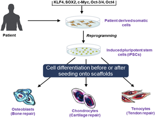
iPSC-seeded engineered scaffolds have been successfully applied in different areas of tissue engineering such as bone,Reference Ardeshirylajimi, Dinarvand, Seyedjafari, Langroudi, Adegani and Soleimani87 cartilage,Reference Liu, Nie, Xu, Niu, Guo, Yin, Guo, Li, Wang and Zhang88 tendon,Reference Liu, Hindieh, Leong and Sun89 etc. Differentiation of iPSCs derived from patients after or prior to scaffold seeding can be used to generate the appropriate cell lineages (Scheme 1). However, in this review, the focus will be on BTE. Biomaterials used for scaffolds and the scaffold itself provide physicochemical cues for the differentiation of iPSCs into specific cell lineages.Reference Higuchi, Kumar, Ling and Alarfaj90 Elasticity, topography, and electrical and mechanical forces are the main physical parameters influencing growth. For instance, cell adhesion and mechanosensing process of stem cells depends on elasticity of biomaterials, whereas topographic cues at the nano- and microscales induce stem-cell differentiation into specific cell lineages.
Potential of iPSCs for bone, cartilage, and tendon repair.

A. Bone TE: iPSC-seeded scaffolds
A wide range of studies have focused on the use of iPSCs in combination with scaffolds (inorganic as well as polymeric) for bone regeneration. We attempt here to classify these studies according to their outcome on bone tissue formation.
1. Enhanced differentiation of iPSCs into bone phenotype and enhanced bone formation
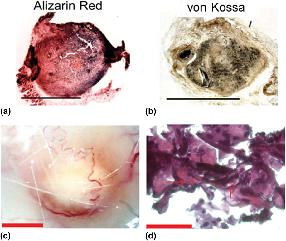
iPSCs were successfully differentiated into mesenchymal lineages of bone, cartilage, and fat after 12 weeks following the subcutaneous implantation of iPS-derived osteoblast-seeded gelfoam scaffold.Reference Bilousova, Jun, King, Langhe, Chick, Torchia, Chow, Klemm, Roop and Majka47 Histological analysis indicated mineralization [Figs. 3(a) and 3(b)], persistence of an osteoblast phenotype, and microvascularization in the implant [Figs. 3(c) and 3(d)]. Furthermore, Wang et al. implanted RGD-functionalized iPSC-seeded CPC in vivo. The results indicated that the hiPSC–CPC constructs generated new bone that was 2.8-fold that of CPC control without cell seeding.Reference Wang, Liu, Zhao, Weir, Sun, Chen, Man and Xu91 In another study, iPSC-derived mesenchymal stem cells (iPSMSCs) encapsulated within alginate microbeads were mixed with CPC paste and filled into cranial defects in rats.Reference Wang, Song, Weir, Sun, Zhao, Simon and Xu92 Cells maintained good viability inside microbeads after injection and the microbeads were able to release the cells which had more than 10-fold increase in live cell density after 14 days. New bone area fractions were (38.9 ± 18.4)% for the CPC-OS-iPSMSC group which was 2–3 times that of (15.6 ± 11.2)% the CPC control at 12 weeks. In addition, inorganic HA-based scaffolds have been used to support the differentiation of iPSCs into osteoblasts. In particular, it was recently reported that the morphology of nano-HA (sphere versus rod) influences the osteogenic differentiation of human gingival fibroblast-derived hiPSCs.Reference Ji, Tong, Huang, Wang, Lin, Cao, Zhang, Dong, Qin and Hu93 Scaffolds consisting of sphere-shaped HA significantly increased hiPSC proliferation and osteogenic differentiation in vitro. These scaffolds also led to large bone formation in vivo in contrast to scaffolds containing rod-shaped which generated tiny bone.
Histochemical alizarin red (a) and von Kossa (b) staining confirmed the presence of matrix and calcium within the nodules and hematoxylin the presence of osteoblasts; After 12 weeks, implants were identifiable (c) as surrounded by vasculature. Paraffin sections stained with H&E demonstrated the vascular supply within the implant, as evidenced by the presence of red blood cells in the lumens of microvessels [(d), arrows] using phase-contrast microscopy. Adapted with permission from Ref. Reference Bilousova, Jun, King, Langhe, Chick, Torchia, Chow, Klemm, Roop and Majka47 Copyright 2011 Wiley.

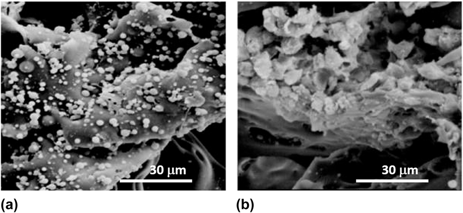
Natural polymers such as silk have also been shown to successfully form new bone following in vivo implantation in mice.Reference Ye, Xu, Gao, Yan, Zhao, Tu, Zhang, Duan, Sommer, Mostoslavsky, Kaplan, Wu, Zhang, Wang and Chen65 Nuclear matrix protein SATB2-overexpressing iPSCs were seeded on silk scaffolds (pore size 500–600 μm, disk-shaped, 4 mm diameter, and 2 mm thick) (Fig. 4). The iPSCs grew along the pores of the scaffolds, tightly to each other and reached confluence with abundant fibril networks of extracellular matrix deposited on the scaffolds. Thus the scaffold facilitated iPSCs’ initial attachment onto the surface, spreading, and subsequent growth. These iPSC-seeded scaffolds were transplanted into critical-size calvarial bone defects created in nude mice. 5 weeks post operation, enhanced new bone formation and mineralization was observed in the SATB2 group compared to scaffold alone or iPSCs cultured without osteogenic supplements where little osseous healing was detected and no appreciable new bone formation was found.
Scanning electron microscopic evaluation of the silk scaffold microstructure and cells’ state (a) 4 h after the iPS cells were seeded onto the scaffold; cells were seen attached to the inner surface of the scaffold. (b) 5 days after seeding, the iPSCs grew tight to each other and reached confluence with abundant fibril networks of extracellular matrix deposited on the scaffolds. Scale bar = 30 μm. Reproduced with permission from Ref. Reference Ye, Xu, Gao, Yan, Zhao, Tu, Zhang, Duan, Sommer, Mostoslavsky, Kaplan, Wu, Zhang, Wang and Chen65 Copyright 2011 Elsevier.

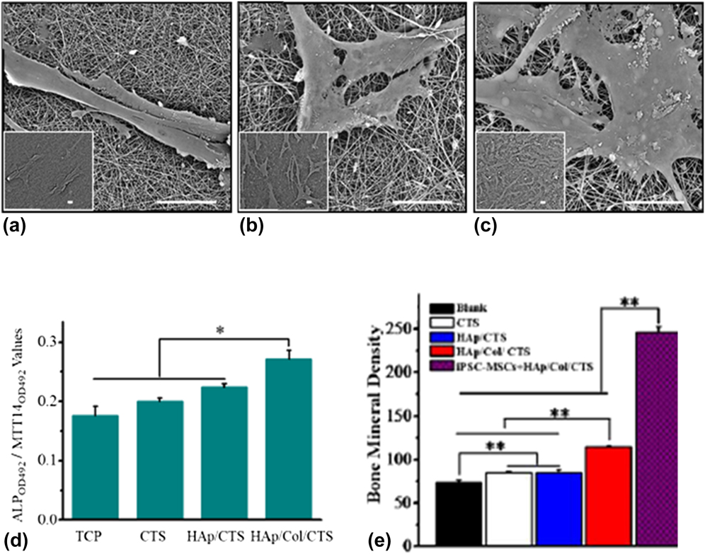
Recently, electrospun scaffolds consisting of hydroxyapatite/collagen/chitosan (HAp/Col/CTS) were used to investigate their effects on the osteogenic differentiation of iPSC-MSCs.Reference Xie, Peng, Zhao, Wang, Yuan, Yang, Li, Lou and Zhang94 Compared to different TCPS, CTS, and HAp/CTS surfaces, the HAp/Col/CTS scaffold was found to act as a better support for the attachment and proliferation [Figs. 5(a)–5(c)] as well as osteogenic differentiation of iPSC-MSCs. Indeed, considerably higher secreting activities of osteogenesis protein markers, ALP, and Col were noted on the HAp/Col/CTS scaffold compared to the others [Fig. 5(d)]. Furthermore, iPSC-MSCs in combination with the electrospun scaffolds were implanted into mouse cranial defects to investigate their efficacy for in vivo bone regeneration. Examinations by computed tomography (CT) imaging, bone mineral density, and hematoxylin eosin (HE) staining suggested that the cell-scaffold construct of iPSC-MSCs + HAp/Col/CTS could effectively promote bone regeneration and the bone mineral density of the iPSC-MSCs + HAp/Col/CTS group was found to be nearly 2-fold higher than that of others after 6 weeks [Fig. 5(e)].
SEM micrographs of iPSC-MSCs on CTS, HAp/CTS, and HAp/Col/CTS nanofibers after culturing for 4 days (a–c). Scale bar = 20 μm; ALP activity with normal medium at 14 days (d) and bone mineral density of the defects after 6 weeks implantation in vivo (e). Adapted with permission from Ref. Reference Xie, Peng, Zhao, Wang, Yuan, Yang, Li, Lou and Zhang94 Copyright 2016 Elsevier.

Nanofibrous scaffolds of polyethersulfone (PES) was successfully used for the osteogenic differentiation of hiPSC-derived EBs as observed by significantly higher expressions of osteogenic-related genes (runx2, COLI, OC, and osteonectin), ALR, and ALP activities in PES-seeded human iPSCs compared with TCPS.Reference Ardeshirylajimi, Hosseinkhani, Parivar, Yaghmaie and Soleimani95
Bone morphogenetic proteins (BMPs) can accelerate bone formation and periodontal regeneration. BMP-6–treated iPSCs (RUNX2, OPN, and OCN) compared to cells treated with osteogenesis medium alone showed that osteogenesis-related gene expressions were higher in BMP-6–treated iPSCs and expression of the cementum-related gene, CAP, was significantly upregulated.Reference Chien, Chang, Wang, Chuang, Yang, Tai, Wang, Liu, Li, Chen, Kao, Chen and Lo96 These promising results lead to the use of an injectable and thermosensitive chitosan/gelatin/glycerol phosphate hydrogel-iPSCs-BMP-6 complex in animal models of maxillary-molar defects (Fig. S1). The BMP-6-hydrogel released BMP-6 in a sustained manner and enabled the iPSCs to maintain their biology and regular growth. The hydrogel acted as a 3D bioscaffold for iPSCs to be engrafted. Within six weeks post implantation, the iPSC-BMP-6-hydrogel group demonstrated a rapid bone bridging with a large volume of regenerated bone tissue within periodontal defects. Significant mineralization with increased bone volume, trabecular number, and trabecular thickness were also observed.
2. Increased vascularization
Vascularization of TE bone constructs remains a major barrier to the clinical translation of BTE. Amongst the various strategies proposed to enhance neovascularization within constructs,Reference Goonoo97 Zhang et al. proposed the use of a novel triculture system of hiPSCs, human umbilical vein endothelial cells (HUVECs), and pericytes.Reference Zhang, Hu, Liu, Reynolds, Bao, Wang, Zhao and Xu98 In vivo studies demonstrated highest amount of new bone formation as well as blood vessel formation following implantation of the tri-culture group compared to the biculture system and the control. This indicates that direct contact between bone forming and vessel forming cells is necessary for angiogenesis and osteogenesis. Following in vivo implantation of iPSC-seeded CPC, new blood vessels were found residing in the newly formed bone. The new blood vessel density in hiPSC–CPC constructs was significantly higher than that in the CPC control.Reference Liu, Chen, Zhang, Thein-Han, Hu, Reynolds, Bao, Wang, Zhao and Xu99 HUVECs co-cultured with hiPSC-MSCs formed microcapillary-like structures in vitro and promoted mineralization of hiPSC-MSCs on CPC scaffolds. 12 weeks post-implantation in a cranial bone defect model in nude rats, the co-culture on the CPC scaffold group achieved the greatest new bone area percentage of 46.38 ± 3.8% compared to controls (CPC alone, HUVEC-seeded CPC, and hiPSC-MSC-seeded CPC), for example, 10.61 ± 1.43% of CPC control.
Using tissue-engineered constructs to repair large bone defects is impeded by the lack of vascular networks compromising the survival of seeded cells. Very recently, co-seeding of HUVECs with hiPSC-MSCs on the calcium phosphate scaffold (CPC) for prevascularization was achieved.Reference Wang, Liu, Zhao, Weir, Sun, Chen, Man and Xu91
3. Coupling matrix remodeling and immunomodulation
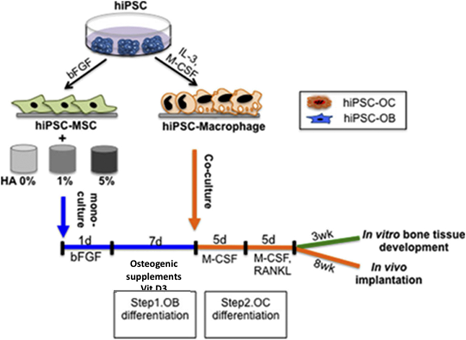
Coordination between cytokines and regulatory molecules secreted from immune cells is required to mimic functional and structural intricacies of native bone. Apart from bone formation, the generation of a functional tissue is essential in clinic and to achieve this, the bone substitutes should be able to foster the connection between bone and immune cells. Jeon et al. recently reported on a 3D model of human bone by co-culturing differentiated hiPSC-derived MSCs and macrophages into osteogenic (OB) and osteoclastogenic (OC) lineages within HA-based scaffolds. (Fig. 6).Reference Jeon, Panicker, Lu, Chae, Feldman and Eliseeff100 The scaffolds were engineered so as to provide a suitable environment for hiPSC-MSCs to differentiate into osteoblasts and deposit bone-like matrix and to activate osteoclasts. The HA-based 3D porous polyester composite scaffold allowed the co-culture hiPSC-MSCs and macrophages committed to OB and OC lineages, mimicking in vivo bone environment and crosstalk between bone cells. They showed that the incorporation of immune cells responsible for remodeling significantly improves bone formation in vitro and in vivo compared with monoculture of osteogenic cells. Eight weeks after implantation in athymic nude mice, explanted scaffolds from monocultured hiPSC-MSCs were filled with fibrous connective tissue, but without bone matrix deposition. Co-culture constructs formed more mature bone with lamellar structure, organized collagen fibers, more OC and type I collagen, and osteocytes embedded in the mineralized extracellular matrix.
hiPSC-MSCs were seeded and allowed to attach to the HA-based composite scaffolds in MSC growth medium for 1 day. Medium containing osteogenic supplements was used to commit these cells to the OB lineage for 7 days, after which it was seeded with hiPSC-macrophages and incubated without osteogenic supplements, containing M-CSF and RANKL, to differentiate into both OB and OC lineages for 10 days. hiPSC-derived multicellular bone constructs were either grown in vitro or implanted subcutaneously into athymic nude mice for 8 weeks. Reproduced with permission from Ref. Reference Jeon, Panicker, Lu, Chae, Feldman and Eliseeff100 Copyright 2016 Springer Nature under Creative Commons.

4. Promote cartilage repair via chondrogenesis
Repair of cartilage loss is difficult due to its poor blood supply and regenerative capability. In clinic, treatments for cartilage loss include autologous chondrocyte transplantation procedure, arthroscopic lavage and debridement, subchondral bone microfracture technique, and osteochondral allograft. They are however often associated with problems such as donor site morbidity, loss of chondrocyte phenotype during in vitro expansion, fibrocartilage formation, and cartilage degeneration. The use of iPSC is thus being explored to promote cartilage repair through chrondrogenesis. To this effect, scaffolds can be used to enhance the chondrogenesis of iPSCs.
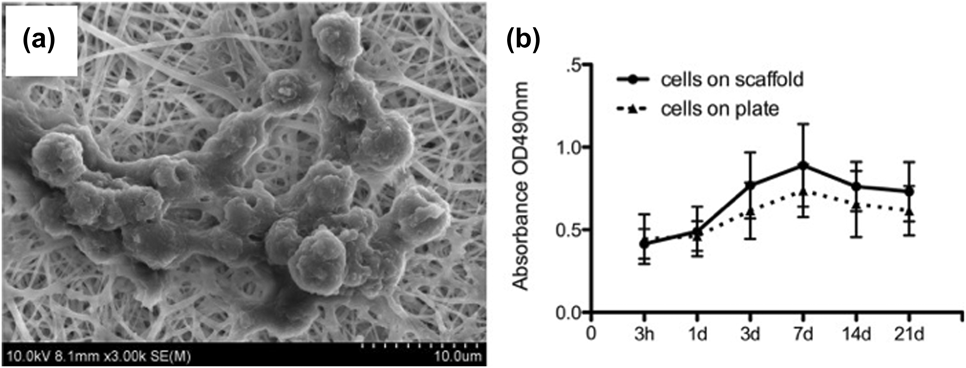
A 3D nanofibrous scaffold made up of super-hydrophilic and durable mechanic polycaprolactone (PCL)/gelatin showed good results on the chondrogenesis of the iPSCs (Fig. 7) and the restoration of the cartilage defect in vivo compared to the scaffold alone.Reference Liu, Nie, Xu, Niu, Guo, Yin, Guo, Li, Wang and Zhang88 The PCL and gelatin-composite scaffolds had randomly oriented nonwoven fibers with an open porosity and interconnected pores (mean fiber diameter = 305 ± 72 nm, scaffold thickness = 50 ± 5.56 µm, porosity = 96%). In the New Zealand rabbit cartilage defect model, the scaffold–cell complex exhibited higher cartilage-specific gene expression and protein levels, as well as subchondral bone regeneration. The scaffold with the cells showed better repair than scaffold without cells, as indicated by a greater number of chondrocytes and upregulated expression of collagen II and aggrecan. The nanofibrous scaffolds covered the defect area, provided the implanted cells with mechanical and spatial support, cartilage-mimicking microenvironment, and facilitated exchange of nutrients and waste, which promoted iPSC differentiation and bioactivity.
(a) Cluster of iPSCs on the scaffold. (b) Proliferation of the iPSCs cultured on the scaffolds and on the plate was determined using CCK-8 tests; the results revealed that the scaffolds promoted cell growth. Reproduced with permission from Ref. Reference Liu, Nie, Xu, Niu, Guo, Yin, Guo, Li, Wang and Zhang88 Copyright 2014 PLoS under Creative Commons.

5. Promoting directed differentiation of hiPSCs to osteoblastic phenotype without EB formation
For the successful application of polymeric scaffold-containing stem cells, the role of the scaffold as a platform for survival and osteodifferentiation is crucial. The cell–nanofiber communication is a determinant parameter for translational possibilities.
Unlike other stem cells, hPSCs can self-renew and differentiate into major lineages of somatic cells. However, the culturing and differentiation of these cells are performed on Matrigel. The latter is obtained from Engerbreth–Holm–Swarm mouse sarcomas with basement membrane components, inhibitors, growth factors, and a range of unknown proteins. These together are considered as limiting as they can give rise to microbial/viral contamination, immunogenicity, and variability of experimental results. Thus nanofibrous scaffolds provide the possibility of having a well-defined, replicable, xeno-free, and safe platform for controlled and uniform differentiation of these cells.
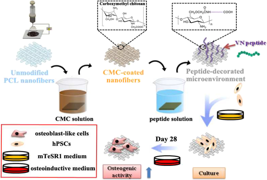
Some studies using nanofibrous polymeric scaffolds such as poly(L-lactic acid)/poly-benzyl-L-glutamate/collagen (PLLA/PBLG/Col) ternary nanofibrousReference Ravichandran, Venugopal, Sundarrajan, Mukherjee and Ramakrishna101 and PESReference Ardeshirylajimi, Hosseinkhani, Parivar, Yaghmaie and Soleimani95 have been reported as promoting greater osteogenic differentiation and maturation of stem cells. However, Matrigel was used to pretreat the nanofiber surfaces to enhance hPSC attachment and proliferation. Another drawback was that osteogenic differentiation of hPSCs transited through the embryonic body (EB) formation such that a well-defined scaffold matrix–cell interaction could not be determined. One recent study reported on the use of chemically defined peptide-decorated PCL nanofibrous scaffold without Matrigel for the osteogenic differentiation of hPSCs without the induction of differentiation by EB formation (Fig. 8). The latter is known to contain different types of cells.Reference Deng, Yang and Wie102 Undifferentiated hPSCs formed tightly packed colonies with defined colony borders and expressed pluripotency markers on the scaffolds. The size of the colonies increased with time, whereas no cell adhered on control PCL nanofiber scaffolds. The differentiated cells displayed a strong expression of bone-specific ECM proteins such as COL1, OPN, and OCN.
Schematic illustration of the fabrication of peptide-decorated nanofibrous microenvironment and in vitro culture and directed osteogenic induction of hiPSCs on the fibrous niche. Reproduced with permission from Ref. Reference Deng, Yang and Wie102 Copyright 2017 American Chemical Society.

B. Modeling of skeletal diseases
Osteodegenerative diseases such as osteoporosis (OP) and fragility fracture affect osteoporotics and a broad spectrum of the elderly population. Several factors may promote bone disorders related to loss of bone mass and decreased bone density. The primary drugs in clinical practice are anti-OP agents that inhibit bone resorption, such as bisphosphonates, which however cause numerous adverse effects. Long-term use of bisphosphonates has demonstrated antifracture efficacy, but it cannot heal bone fracture and promote repair.Reference Watts and Diab103 Parathyroid hormone (PTH), the first bone anabolic drug approved for the treatment of OP, has shown the ability to induce fracture healing in animal studies.Reference Jorgensen and Schwarz104 In humans, PTH has demonstrated accelerated healing of various experimental fractures and of distal radius and pelvic fractures. The exact mechanism of action has not been elucidated, but it is believed to occur via an increased recruitment and differentiation of chondrocytes and enhancement of endochondral ossification.Reference Larsson and Fazzalari105 Large randomized clinical trials are needed to fully understand their effects on fracture healing.Reference Hegde, Jo, Andreopoulou and Lane106,Reference Vannucci and Luisa107
iPSCs may also be used to create models of diseases affecting bones as many genetic bone disorders have limited treatment possibilities due to the unavailability of appropriate in vivo models and inaccessibility of native bones.Reference Csobonyeiova, Polak, Zamborsky and Danisovic108 iPSC-derived disease models from patients with genetic mutations will allow understanding of the origins and pathologies of diseases. In this context, iPSCs have been used to model infrequent genetically influenced disorders, namely fibrodysplasia ossificans progressiva (FOP) and metatropic dysplasia.
FOP is a genetic disorder in which muscle tissue and connective tissue such as tendons and ligaments are gradually replaced by bone, forming bone outside the skeleton that constrains movement.109 The latter is caused by gene mutations in the type I receptor activin receptor-like kinase 2 (ACVR1/ALK2), which is a part of the bone morphogenic protein (BMP) signaling. A “disease in a dish” model of FOP was successfully developed to investigate differences in individual steps of endochondral bone ossification between FOP dermal fibroblast-derived iPSCs and control iPSCs.Reference Matssmoto, Hayashi, Schlieve, Ikeya, Kim, Nguyen, Sami, Baba, Barruet, Nasu, Asaka, Otsuka, Yamanaka, Conklin, Toguchida and Hsiao110,Reference Matsumoto, Ikeya, Hino, Horigome, Fukuta, Watanabe, Nagata, Yamamoto, Otsuka and Toguchida111 As expected, increased mineralization and chondrogenesis of FOP iPSCs was observed in vitro. Interestingly, the addition of a small molecule inhibitor of BMP signaling (DMH1) could suppress mineralization phenotypes109 in FOP iPSCs. In another study, FOP hiPSCs were generated by episomal vectors from kidney cells isolated from urine of FOP patients.Reference Cai, Orlova, Cai, Eekhoff, Zhang, Pei, Pan, Mummery and Dijke112 The mutation in ALK2 reduced the differentiation efficiency and maintenance of FOP hiPSCs into ECs probably due to elevated BMP signaling in FOP hiPSC-ECs, which resulted in downregulation of the VEGFR2 expression that mediated VEGF-induced proliferation and survival of ECs. In addition, heterogeneous BMP/SMAD signaling and mineralization were noted in FOP hiPSCs and derivative cells, suggesting that intrinsic genetic and/or epigenetic features of donor cells may influence properties of hiPSCs and the progeny.Reference Cai, Orlova, Cai, Eekhoff, Zhang, Pei, Pan, Mummery and Dijke112
Craniometaphyseal dysplasia is characterized by progressive thickening of bones in the craniofacial region and a widening of metaphysis in long bones.Reference Chen, Wang, Strecker, Koczon-Jaremko, Boskey and Reichenberger113 Mutation for the autosomal dominant CMD occurs in the ankylosis gene and in Connexin 43 for the recessive form.Reference Hu, Chen, de Almeida, Tiziani, Raposo Do Amaral, Gowrishankar, Passos-Bueno and Reichenberger114 Osteoblast and osteoclast defects have been noted in mice with CMD mutations.Reference Chen, Wang, Jiang, Aguila and Reichenberger115 Recently, human iPSCs were successfully obtained from the peripheral blood of donors suffering from CMD using a simple and effective method.Reference Chen, Fukuda, Fusaki, Iida, Hasegawa, Lichtler and Reichenberger116
Marfan syndrome (MFS) is a heritable connective tissue disorder caused by mutations in the gene coding for FIBRILLIN-1 (FBN1), an extracellular matrix protein.Reference Chen, Fukuda, Fusaki, Iida, Hasegawa, Lichtler and Reichenberger116 MFS displays major manifestations in the ocular, skeletal, and cardiovascular systems. To date, most knowledge of MFS has been obtained by extrapolation of studies in the mouse Fbn1 null/transgenic models.Reference Granata, Serrano, Bernard, McNamara, Low, Sastry and Sinha117–Reference Neptune, Frischmeyer, Arking, Myers, Bunton, Gayraud, Ramirez, Sakai and Dietz122 However, the derivation of human iPSCs from MFS patients now offers a unique opportunity to examine key features of this syndrome based on a human genome background. Quarto et al. successfully created a human MFS “model” phenocopying human embryonic MFS stem cells, strongly supporting the hypothesis that these are bona fide disease phenotypes resulting from the FBN1 mutations and not simply sporadic cell-culture artifacts.Reference Quarto, Mastrogiacomo, Cancedda, Kutepov, Mukhachev, Lavroukov, Kon and Marcacci123
Overall, as shown by several research groups, hiPSCs may be used to investigate the molecular mechanisms underlying the pathology of several osteodegenerative diseases, as well as to identify potential therapeutic drugs.
As described above, iPSC-derived models have successfully recapitulated abnormalities of some skeletal diseases in vitro. Biomaterial-scaffolds can enhance the differentiation processes to create more homogeneous and physiologically relevant in vitro disease models and to induce disease phenotypes.Reference Salazar-Noratto, Barry and Guldberg124
VII. FUTURE PERSPECTIVES: iPSC AND OSTEODEGENERATIVE DISEASES IN THE AGING ADULT
Most advances in the medical field have contributed to increasing the human life expectancy. As a result, age-related diseases have now become a reality in developed and developing countries and will constitute a huge economic burden. There is and will be a strong need for therapies both to alleviate age-related diseases and provide a good quality of life. These diseases include metabolic syndrome and diabetes, atherosclerosis, neurodegenerative diseases, OP, and cancer.Reference Boyette and Tuan125 Skeletal problems due to bone degeneration [OP and osteoarthritis (OA)] in aging population require the use of improved therapeutic products as the healing rate in aging adults is different from young adults. For instance, more than 55 million people in northern America are affected by OP and low bone mineral density.Reference Kiernan, Davies and William126 This resulted in over 2 million fragility fractures and costs USD 20 billion per year. Amongst these, in the United States, osteoporotic hip fractures was over 300,000 yearly leading to 31,000 associated deaths and disability in survivors.
Bone degeneration such as OP and OA is often seen with increasing age. In OA, degradation and loss of the articular cartilage is a central feature that is sometimes attributed to “wear and tear”. The knee is commonly affected by OA and is thought to account for the majority of disability from OA. The prevalence of radiographic OA increases with each decade of life from 33% among those aged 60–70 to 43.7% among those over 80 years of age. The tendon is also subjected to early degenerative changes since both the collagen and noncollagenous matrix components of tendons show qualitative and quantitative changes. Despite progress in orthopedic surgery, there is still a lack of efficient treatment options for age-related degenerative diseases. Tissue engineering approaches, combining tenoblasts cells (tendon-derived cells) or osteoblasts (bone-derived cells), scaffolds, and environmental factors, have recently demonstrated tremendous clinical potential for regeneration of tendons or large bone defects.
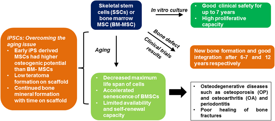
It is believed that skeletal stem cells or BM-MSCs give rise to cartilage, bone, marrow fat, and hematopoiesis-supportive stroma, in a well-ordered sequence during embryonic and postnatal development.Reference Jones, Giannoudis and Kouroupis127 The overall decrease in the number and function of BM-MSCs is believed to be the root-cause of age-related bone lossReference Kiernan, Davies and Stanford128 Clinical studies have used autologous-cultured BM-MSCs for large bone defect repair. Good clinical safety of cultured BM-MSCs for a follow-up period of up to 7 years and uncultured BM-MSCs with a follow-up of up to 20 years have been reported. Clinical trials using BM-MSCs implanted on polymeric scaffolds for large long bone defectsReference Quarto, Mastrogiacomo, Cancedda, Kutepov, Mukhachev, Lavroukov, Kon and Marcacci123,Reference Marcacci, Kon, Moukhachev, Lavroukov, Kutepov, Quarto, Mastrogiacomo and Cancedda129 and bone deficienciesReference Jaeger, Herten, Fochtmann, Fischer, Hernigou, Zilkens, Hendrich and Krauspe130 showed new bone formation and good integration after 6–7 and 12 years, respectively. Thus, the use of adult stem cells to repair degenerated tissues of the musculoskeletal structures is promising due to their high proliferative capacity, ability to differentiate into other cell types and their innate immunomodulatory properties. However, there is growing evidence that many diseases of aging may reflect adult stem cell exhaustion. For instance, a decreased in vitro life span of cells from the elderly was found when compared with cells obtained from young donors (24 ± 11 population doublings [PD] versus 41 ± 10 PD, P < 0.05) and the mean PD rate was lower in old donor cells (0.05 ± 0.02 PD/day) compared with young donor cells (0.09 ± 0.02 PD/day) (P < 0.05).Reference Aldahmash131 Aging is associated with decreased maximal life span and accelerated senescence of bone marrow stromal cells.Reference Sheyn, Ben-David, Shapiro, De Mel, Bez, Ornelas, Sahabian, Sareen, Da, Pelled, Tawakocli, Liu, Gazit and Gazit55 MSC from old donors exhibited a decreased maximal life span compared with cells from young donors and their differentiation potential and proliferation capacity have also been reported to be age-dependent.Reference Roobrouck, Ulloa-Montoya and Verfaillie132,Reference Steinert, Ghivizzani, Rethwilm, Tuan, Evans and Noth133
A meta-analysis of MSC-based OA clinical trials, showed a short follow-up from six months to two years, except for one trial with a five-year follow-up of four cases.Reference McCarraher and Fournier-Bell134 One major problem identified with MSCs during these studies is their tendency to differentiate into fibro-like tissue instead of hyaline cartilage, the major component of articular cartilage.
The use of iPSCs to regenerate aged/damaged tissue is promising as they exhibit advantages of MSCs and ESCs while donor age does not seem to impact on reprogramming efficiency. In mice models, it has been shown that the reprogramming potency in aged mice seems to be impaired to a greater extent in BMSC than in dermal fibroblasts.Reference Li, Collado, Villasante, Strati, Ortega, Canamero, Blasco and Serrano135,Reference Cheng, Ito, Nishio, Xiao, Zhang, Suzuki, Okawa, Murohara and Isobe136 In humans, studies have shown that aging does not drastically impair the ability of human cells to reprogram into iPSCs.Reference Mahmoudi and Brunet137 As discussed previously, bone vasculature plays a vital role in bone development and fracture repair.Reference Levi, Hyun, Montoro, Lo, Chan and Hu138 Comparison of osteogenic progenitor cells derived from both hESCs and hiPSCs seeded on gelatin sponge showed similar osteogenic differentiation efficiency in vivo (a rat femur nonunion fracture) but hiPSC showed markedly higher expression of angiogenic factors, such as vascular endothelial growth factor and basic fibroblast growth factor.Reference Zou, Chen, Quanbeck, Bechtold and Kaufman139 An in vitro study demonstrated that the osteogenic capacity of the iPSCs compared with MSCs isolated from human adipose tissue showed that iPSCs had increased proliferation compared to AT-MSCs with higher ALP activity and mineralization.Reference Ardeshirylajimi, Soleimani, Hosseinkhani, Parivar and Yaghmaei140
A recent study showed that iPSCs differentiated into MSCs could be used for bone regeneration.Reference Sheyn, Ben-David, Shapiro, De Mel, Bez, Ornelas, Sahabian, Sareen, Da, Pelled, Tawakocli, Liu, Gazit and Gazit55 Exposure to transforming growth factor-β was used to direct iPSCs toward MSC differentiation and two types of iMSCs were obtained, early (aiMSCs) and late (tiMSCs) outgrowing cells. The aiMSCs demonstrated higher osteogenic potential than tiMSCs and BM-MSCs. Upon orthotopic injection into radial defects, all cell types regenerated bone and contributed to defect repair.
Significant progress on the osteogenesis of iPSCs using scaffolds has been achieved. For example, the use of an osteogenic scaffold micro-niche containing HA, poly-L-lactic acid, and BMP-2 showed a high survival rate and differentiated osteogenic cells with very low teratoma formation.Reference Roobrouck, Ulloa-Montoya and Verfaillie132
The iPSC has also been successfully seeded on CPC used to fill complex-shaped bone defects.Reference Tang, Chen, Liu, Weir, Cheng and Xu67 The mesenchymal differentiation of the iPSC-MSCs showed potential to differentiate into adipocytes, chondrocytes, and osteoblasts. They differentiated into the osteogenic lineage and synthesized bone minerals and the latter increased with time.
Furthermore, human MSC-like cells generated from iPSCs had the capacity to facilitate periodontal regeneration in a rat periodontal defect model, including newly formed fibrous tissue, newly formed mineralized tissue, and newly formed PDL-like tissues.Reference Hynes, Menicanin, Han, Marino, Mrozik, Gronthos and Barthold141
Overall, these studies demonstrate the high potential of iPSCs in overcoming BM-MSC deficiencies associated with aging (Scheme 2). Biomaterial-scaffolds enhance and guide the differentiation of iPSCs which is crucial in age-related diseases. The possibility to apply the lock and key concept on scaffolds and cells would open up new perspectives in this field.
Role of BM-MSCs in bone regeneration in clinic and iPSC overcoming BM-MSC deficiencies with aging.

Supplementary Material
To view supplementary material for this article, please visit https://doi.org/10.1557/jmr.2018.132.
ABBREVIATIONS

ACKNOWLEDGMENTS
The authors are grateful to the Mauritius Research Council for a Postdoctoral Fellowship to NG under the Nanotechnology program at the research unit and for supporting Biomaterials and Drug Delivery Research at CBBR. ABL is also indebted to the University of Mauritius for providing indirect funding to the research unit.