Case report
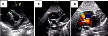
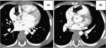
A 5-year-old boy was referred to our department for evaluation of a heart murmur. He had no relevant past medical history, was asymptomatic from a cardiac point of view, and was under no medication. On auscultation, a continuous murmur loudest at the left fourth intercostal space, with no radiation, was evident. On examination, no other relevant findings were found. His chest X-ray revealed an increased cardiothoracic ratio (65%), with right-sided prominence heart and normal pulmonary vascular markings. The electrocardiogram at rest showed right-axis deviation (QRS complex axis of +100°), associated with right atrium and right ventricle hypertrophy. Findings on two-dimensional transthoracic echocardiogram included a moderately dilated right atrium and right ventricle, a giant aneurysm measuring 10 mm (Z-score 19.6, Fig 1a) at the proximal portion of right coronary artery, normal origin and dimensions of the left coronary artery and a fistulous tract originating in the aneurysm and apparently draining into the right atrium next to the atrial septum (Fig 1b). On colour Doppler, a continuous turbulent flow into the right atrium was observed (Fig 1c). Biventricular function was preserved with normal regional motility, and the pulmonary artery pressures were normal. In order to clarify the coronary anatomy, a cardiac computed tomography angiogram (Fig 2) and cardiac catheterisation were performed, both confirming the above findings, as well as another aneurysm at the distal end of the fistula, measuring 15 mm. The Qp/Qs was 2.
Transthoracic echocardiogram showing an aneurysm of the proximal portion of the right coronary artery measuring about 10 mm (a). A 5-mm fistulous tract draining into the right atrium next to the atrial septum was identified (b), and related to a continuous turbulent flow (c). LCA = left coronary artery; RA = right atrium; RCA = right coronary artery.

Cardiac computed tomography angiogram showing a significantly dilated proximal right coronary artery (arrow, a), related to an anomalous communication compatible with a coronary fistula measuring 19 mm at its maximum diameter (arrow, b), that drains into the right atrium. Ao = ao root; LPA = left pulmonary artery; PT = pulmonary trunk; RCA = right coronary artery; RPA = right pulmonary artery.

The patient underwent surgical fistula ligation and closure of the fenestrated atrial septal defect. His post-operative period was uncomplicated and he was discharged home 1 week after surgery on double antiaggregation therapy, with aspirin (5 mg/kg/day) and dipyridamole (1 mg/kg/day).
Currently, 6 months post surgery, he remains asymptomatic. His last transthoracic echocardiogram showed no residual atrial septal or fistulous shunt, with almost normal right-sided chamber dimensions. However, his right coronary artery remains very enlarged (7 mm, Z-score 12.2), for which the patient remains on double antiaggregation therapy.
Discussion
Congenital coronary artery fistulas are defined as a direct communication between an epicardial coronary artery and a cardiac chamber, great vessel or other vascular structures, bypassing the myocardial capillary bed.Reference Kim, Ko and Yoon 1 , Reference Esmaeilzadehl, Khaledifar and Usefi 2 These abnormal communications are rare in childhood, with an incidence of 1–2% in the general population.Reference Chowdhury, Rizhvi and Sheil 3 A coronary artery aneurysm is defined as a dilatation of coronary artery with a diameter greater than 1.5 times the diameter of the adjoining normal coronary artery. It is also rare, with a reported incidence of ∼1%.Reference Dogan, Ozaydin and Altinbas 4
The fistulae may be isolated (55–80%), or associated with other congenital cardiac anomalies (20–45%) or may be superimposed on coronary artery disease (35%). The coronary artery fistulas may be single (74–90%) or multiple (10–16%). The origin of the fistulous communication can be the right coronary artery (50–60%), left anterior descending coronary artery (25–42%), or circumflex coronary artery (18%). Its origin from the main left coronary artery is rare.Reference Chowdhury, Rizhvi and Sheil 3 As observed in our case, in more than 90% of the cases, the fistula terminates in the right side of the heart (40% in the ventricle, 30% in the atrium, and 20% in the pulmonary artery).Reference Chowdhury, Rizhvi and Sheil 3 It rarely terminates on the left side of the heart, but when it does the majority enters the left atrium.Reference Park 5
No definite pathogenetic explanation has been described, but coronary artery fistulas may develop upon enlargement of the capillary network during embryogenesis, or when the main coronary arteries remained attached to the pulmonary trunk at the time of their segmentation.Reference Esmaeilzadehl, Khaledifar and Usefi 2
Patients younger than 20 years are mostly asymptomatic. However, the incidence of symptoms and complications increases over time, depending on the location, the amount of left to right shunt, and the presence of coronary steal phenomenon – due to blood shunting away from the myocardium.Reference Kim, Ko and Yoon 1 , Reference Boening, Scheewe and Fischer 6 The symptoms and complications of untreated patients include dyspnoea, arrhythmias, congestive heart failure – when drainage occurs into the right system, resulting in pulmonary congestion and hypertension – subacute infective endocarditis, aneurysm formation, myocardial infarction, myocardial rupture, or sudden death.Reference Kim, Ko and Yoon 1 – Reference Chowdhury, Rizhvi and Sheil 3 The predominant physical finding is a continuous murmur at the second/third right or left parasternal border, representing the systolic–diastolic flow from the fistulae.Reference Kim, Ko and Yoon 1
A chest X-ray may show cardiomegaly and pulmonary plethora.Reference Chowdhury, Rizhvi and Sheil 3 The electrocardiogram is usually normal.Reference Park 5 Two-dimensional transthoracic echocardiogram can identify a dilated coronary artery, with Doppler evidence of turbulent flow at the termination site. However, it is operator dependent, and evaluation may be incomplete because of suboptimal acoustic window.Reference Gulati, Ramamurthy and Sharma 7 , Reference Zahn, Smallhorn and Egger 8 The diagnostic “gold standard” remains coronary angiography, which allows haemodynamic assessment, shunt quantification, fistula characterisation, and description of the coronary artery morphology. However, it is an invasive examination; selective catheterisation of the fistulas may be difficult and the distal coronary artery may not be well evaluated because of the steal phenomenon, occurring at the proximal fistula.Reference Chowdhury, Rizhvi and Sheil 3 , Reference Gulati, Ramamurthy and Sharma 7 Cardiac computed tomography angiogram and magnetic resonance imaging are alternative diagnostic methods.
Owing to the potential adverse events and the low spontaneous closure rate (1–2%), the current management is elective closure of the fistulas during childhood, even in asymptomatic patients.Reference Chowdhury, Rizhvi and Sheil 3 , Reference Boening, Scheewe and Fischer 6 Surgical treatment is also advocated in symptomatic patients, those with large shunts (Qp/Qs > 1.5), in cases with tortuous or aneurysmatic fistulae and concomitant cardiac pathology.Reference Esmaeilzadehl, Khaledifar and Usefi 2 , Reference Mandegar, Saidi and Roshanali 9 The most frequent surgical technique includes fistula ligation at its most proximal and distal ends, without jeopardising flow in the normal arteries.Reference Park 5 The surgical mortality in patients younger than 20 years is 1–2%. Significant surgical complications – arrhythmias, myocardial ischaemia, and complications related to cardiopulmonary bypass – occur in 15% of all the patients, but in only 7% of young patients.Reference Boening, Scheewe and Fischer 6
If the fistula is simple and single, transcatheter percutaneous closure can be an alternative option to surgery and has been increasingly used, especially among children.Reference Chowdhury, Rizhvi and Sheil 3 , Reference Boening, Scheewe and Fischer 6 , Reference Mandegar, Saidi and Roshanali 9 Complications include device migration, and currently there are no long term follow-up data regarding clot propagation and recurrence of the fistulas.Reference Chowdhury, Rizhvi and Sheil 3 , Reference Boening, Scheewe and Fischer 6 In our case, device closure was not attempted as the coronary anatomy was considered unsuitable and also because there was concomitant congenital heart disease, both of which corrected during the same surgical procedure.
Acknowledgements
None.
Financial Support
This research received no specific grant from any funding agency, commercial or not-for-profit sectors.
Conflicts of Interest
None.