Introduction
The reserve hypotheses provide explanations for the incoherence between brain pathology and the clinical manifestation of the pathology. Whereas the brain reserve hypothesis predicts that larger brain size passively provides resilience to brain damage until a threshold is reached (Satz, Reference Satz1993; Stern, Reference Stern2009), the cognitive reserve (CR) construct hypothesizes that the brain actively copes with brain damage by using compensatory strategies and recruiting alternative neural networks (Stern, Reference Stern2002, Reference Stern2012). Although the two reserve concepts focus on different sources of resilience, the proxies used to operationalize reserve may be identical as cognitively stimulating activities affect the underlying brain anatomy (Lazarov et al. Reference Lazarov, Robinson, Tang, Hairston, Korade-Mirnics, Lee, Hersh, Sapolsky, Mirnics and Sisodia2005; Van Praag et al. Reference Van Praag, Shubert, Zhao and Gage2005). Evidence suggests that both concepts synergistically contribute to reserve (Mortimer et al. Reference Mortimer, Snowdon and Markesbery2003; Brickman et al. Reference Brickman, Siedlecki, Muraskin, Manly, Luchsinger, Yeung, Brown, Decarli and Stern2011). Education, leisure activities and occupation are the most commonly used reserve proxies (Harrison et al. Reference Harrison, Sajjad, Bramer, Ikram, Tiemeier and Stephan2015). Other measures such as intracranial volume (ICV) (Wolf et al. Reference Wolf, Julin, Gertz, Winblad and Wahlund2004), head circumference (Mortimer et al. Reference Mortimer, Borenstein, Gosche and Snowdon2005), socio-economic status (Karp et al. Reference Karp, Kareholt, Qiu, Bellander, Winblad and Fratiglioni2004), intelligence quotient (IQ) (Alexander et al. Reference Alexander, Furey, Grady, Pietrini, Brady, Mentis and Schapiro1997; Steffener et al. Reference Steffener, Reuben, Rakitin and Stern2011) and processing resources (Boyle et al. Reference Boyle, Wilson, Schneider, Bienias and Bennett2008) have also been found to alter the relationship between brain pathology and cognitive functioning.
Alzheimer's disease has been considered a model disease for the reserve concepts given its predictable disease course and limited variability in pathological expression (Stern, Reference Stern2002). However, the concepts may be generalizable to all situations where an individual endures brain disease/pathology, for example Parkinson's disease dementia (Quinn et al. Reference Quinn, Rossor and Marsden1986), HIV-related cognitive dysfunction (Stern et al. Reference Stern, Silva, Chaisson and Evans1996; Basso & Bornstein, Reference Basso and Bornstein2000; Pereda et al. Reference Pereda, Ayuso-Mateos, Gomez Del Barrio, Echevarria, Farinas, Garcia Palomo, Gonzalez Macias and Vazquez-Barquero2000) and multiple sclerosis (Sumowski et al. Reference Sumowski, Chiaravalloti, Wylie and Deluca2009). Barnett et al. (Reference Barnett, Salmond, Jones and Sahakian2006) reviewed the evidence of CR in neuropsychiatric diseases and concluded that reserve is also a resilience factor in at least some psychiatric disorders. Most evidence of a protective role of reserve stems from studies of schizophrenia and only one previous study (Forcada et al. Reference Forcada, Mur, Mora, Vieta, Bartres-Faz and Portella2015) has attempted to elucidate the role of reserve in bipolar disorder (BD). In that study, a composite proxy of CR was found to be predictive of psychosocial functioning (Forcada et al. Reference Forcada, Mur, Mora, Vieta, Bartres-Faz and Portella2015), but as no measure relating to brain pathology was included in that study, it is still undecided whether the reserve concept is applicable to BD.
Meta-analyses of structural magnetic resonance imaging (MRI) studies of BD report volume reductions in whole brain and prefrontal cortex (PFC), increased volume of the globus pallidus and lateral ventricles, and increased rates of white matter lesions (Kempton et al. Reference Kempton, Geddes, Ettinger, Williams and Grasby2008; Arnone et al. Reference Arnone, Cavanagh, Gerber, Lawrie, Ebmeier and Mcintosh2009). Although not replicated, there is also evidence that patients with BD type I have increased ventricular cerebrospinal fluid (CSF) volume (Maller et al. Reference Maller, Thaveenthiran, Thomson, McQueen and Fitzgerald2014). In line with this, meta-analyses indicate that memory, attention/speed and executive functions are the most commonly impaired domains in euthymic BD (Torres et al. Reference Torres, Boudreau and Yatham2007; Arts et al. Reference Arts, Jabben, Krabbendam and Van Os2008). Volumes of deep white matter hypointensities (WMH), ventricular CSF and volume of PFC might thus comprise suitable measures of global brain burden for investigating the CR hypothesis in BD.
The aim of this study was to test the applicability of the CR concept in BD. More specifically, we investigated the ability of CR to lessen the influence abnormalities in total volumes of deep WMH, ventricular CSF and PFC on memory, speed/attention and executive functions in patients with BD type I and II.
Method
The St. Göran Bipolar Project is an ongoing longitudinal study of BD (Ryden et al. Reference Ryden, Thase, Straht, Aberg-Wistedt, Bejerot and Landen2009). Briefly, all patients were enrolled at the bipolar out-patient unit at the Northern Stockholm Psychiatric Clinic (Sweden). The inclusion criteria for the current study were ⩾18 years of age, fulfilling the Diagnostic and Statistical Manual of Mental Disorders, 4th edition (DSM-IV) criteria for BD type I or II, not otherwise specified, and schizo-affective disorder BD type. Exclusion criteria were incapability of providing informed consent and inability to complete the standard clinical assessment. For the current study, only patients with BD subtype I or II (n = 75) who had completed a neuropsychological as well as an MRI examination were included.
Clinical assessment and diagnostic procedures
All examinations were performed when the patients were in a stable euthymic mood. Euthymia was based on clinical judgment and also warranted a score below 14 on both the Montgomery–Åsberg Depression Rating Scale and the Young Mania Rating Scale. The standardized interview protocol Affective Disorders Evaluation (ADE; Sachs et al. Reference Sachs, Thase, Otto, Bauer, Miklowitz, Wisniewski, Lavori, Lebowitz, Rudorfer, Frank, Nierenberg, Fava, Bowden, Ketter, Marangell, Calabrese, Kupfer and Rosenbaum2003) was conducted by a psychiatrist or a psychiatrist in training. The ADE guides the interviewer through a systematic assessment of the patient's current mental state, past history and diagnosis according to DSM-IV criteria as per the Structured Clinical Interview for DSM-IV (American Psychiatric Association, 2000). Psychiatric co-morbidity was assessed using the Mini International Neuropsychiatric Interview (M.I.N.I.) (Sheehan et al. Reference Sheehan, Lecrubier, Sheehan, Amorim, Janavs, Weiller, Hergueta, Baker and Dunbar1998). A best-estimate diagnostic decision was made by a panel of experienced board certified psychiatrists specialized in BD based on the following information sources: patient records, ADE, M.I.N.I. and interviews with next of kin when possible. Disease severity was assessed on a seven-point Likert scale by means of the Clinical Global Impression Severity (CGI-S) tool (Guy, Reference Guy and Guy1976).
Control subjects
Age- and sex-matched, population-based healthy controls (n = 82) were included in the current study for the purposes of brain volume (BV) normalization and standardization of cognitive tests. The controls were randomly selected by Statistics Sweden. Responders were subjected to a preliminary telephone screening to rule out substance abuse, severe mental health problems and neurological problems. Controls were included in the study if none of the following was observed during the assessment: any current psychiatric disorder, neurological disease (excluding mild migraine), or substance abuse problems. Other exclusion criteria were untreated endocrine disorders, pregnancy, and BD in first-degree relatives or a family history of schizophrenia. Past minor depressive episodes, mild eating disorders, isolated episodes of panic disorder or obsessive–compulsive disorder that had remitted spontaneously or with brief psychotherapy did not warrant exclusion. Patients and controls underwent identical examinations with the exception of the BD-specific assessments.
Ethics
The study was approved by the Regional Ethics Committee in Stockholm. The authors assert that all procedures contributing to this work comply with the ethical standards of the relevant national and institutional committees on human experimentation and with the Helsinki Declaration of 1975, as revised in 2008. After complete description of the study; all enrolled patients and controls consented orally and in writing to participate in the study.
MRI and processing
MRI scans were acquired at the MR Research Center, Karolinska University Hospital, Stockholm. Coronal three-dimensional T1-weighted images were acquired with a spoiled gradient echo recall sequence (3D-SPGR, repetition time = 21.0 ms, echo time = 6 ms, field of view = 18 cm, flip angle = 30°, acquisition matrix = 256 × 256 × 128, voxel size: 0.7 × 0.7 × 1.8 mm3) using a 1.5-Tesla MRI medical scanner (General Electric Signa Excite 1.5 T) equipped with an eight-channel head coil. Additional axial fluid attenuation inversion recovery T2-weighted scans were acquired for examination by a senior radiologist to exclude patients with significant anatomical abnormalities and neuropathology.
The same investigator manually reoriented all T1-weighted images to the anterior–posterior commissure line using Matlab version 8.1 (R2013a) and SPM8. Measures of ICV, total BV, volumes of deep WMH and PFC (see below for details), as well as ventricular CSF were obtained using automated segmentation and cortical surface reconstruction methods provided by FreeSurfer v5.1 (Dale et al. Reference Dale, Fischl and Sereno1999; ; Fischl et al. Reference Fischl, Sereno and Dale1999, Reference Fischl, Salat, Busa, Albert, Dieterich, Haselgrove, Van Der Kouwe, Killiany, Kennedy, Klaveness, Montillo, Makris, Rosen and Dale2002, Reference Fischl, Salat, Van Der Kouwe, Makris, Segonne, Quinn and Dale2004a , b; Fischl & Dale, Reference Fischl and Dale2000). In brief, the procedure includes intensity normalization, removal of non-brain tissue, segmentation of cortical grey, subcortical white, deep grey matter volumetric structures, as well as triangular tessellation of the grey/white matter interface and the pial surface (white matter/CSF boundary). All surface reconstructions were then visually inspected and, where necessary, corrected manually by trained operators using editing tools provided by FreeSurfer, including corrections of erroneous skull stripping, white matter and grey matter segmentations. FreeSurfer also allows an automated parcellation of the cortical surfaces into 34 anatomical regions of interest (ROIs; Desikan atlas) (Fischl et al. Reference Fischl, Van Der Kouwe, Destrieux, Halgren, Segonne, Salat, Busa, Seidman, Goldstein, Kennedy, Caviness, Makris, Rosen and Dale2004b ; Desikan et al. Reference Desikan, Segonne, Fischl, Quinn, Dickerson, Blacker, Buckner, Dale, Maguire, Hyman, Albert and Killiany2006). For each ROI, measures were obtained from subjects’ native space. The PFC volume was created by combining the superior frontal rostral, caudal middle frontal, pars opercularis, pars triangularis and pars orbitalis subregions from the left and right hemispheres.
Volumes of WMH, CSF and PFC were then normalized by using a regression-based approach (Jack et al. Reference Jack, Twomey, Zinsmeister, Sharbrough, Petersen and Cascino1989). In short, normalization was performed by adjusting the absolute volumes for the deviation in ICV according to the linear regression equation for absolute volumes v. ICV in the healthy control group. The underlying assumption of this approach is that these regressions represent the expected relationships between ICV and the ROIs.
Cognitive outcome measures
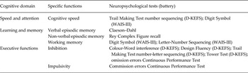
The cognitive domains – memory, executive, and attention/speed functions – that have been consistently identified as impaired in BD were evaluated in this study. Several cognitive functions were assessed within each domain to obtain a comprehensive approximation of the participants’ level of cognitive functioning. The individual test scores were z-transformed (mean = 0; s.d. = 1) on the basis of controls’ performance and then combined into cognitive domains based on their mutual measurement properties consistent with reference literature (Table 1) (Spreen & Strauss, Reference Spreen and Strauss1991; Lezak et al. Reference Lezak, Howieson, Bigler and Tranel2012).
Cognitive domains and functions measured

D-KEFS, Delis–Kaplan executive function system; WAIS-III, Wechsler's Adult Intelligence Scale version III.
CR measure
The Vocabulary subtest of the Wechsler's Adult Intelligence Scale version III (WAIS-III), which measures word knowledge and concept formation (Wechsler, Reference Wechsler1997), was used as a proxy for CR. The Vocabulary subtest is the subtest with the highest correlation with the WAIS-III full-scale IQ score (Wechsler, Reference Wechsler1997).
Statistical methods
The primary aim of this study was to determine the ability of CR to lessen the influence of deep WMH, ventricular CSF and PFC volume abnormalities on memory, speed/attention and executive functions in patients with BD.
Preliminary analyses were conducted to ensure no violation of linearity, multicollinearity, normality and homoscedasticity. The independent variables were centred (score – mean) prior to being entered in the model. All models were evaluated visually and analytically.
Linear regression was conducted in two steps (hierarchical/sequential regression). At step 1, we assessed the initial association between the brain ROI and the cognitive variables as well as the covariates age, sex and CGI-S. At step 2, we added the interaction term for the brain ROI and the CR measure (e.g. WMH × CR) whilst controlling for all associations at step 1. The interaction term directly tests the ability of CR to reduce the influence of brain pathology on the examined cognitive domain. The measure of clinical severity, CGI-S, was included to ensure that the reserve measure was applicable at all levels of clinical severity.
We report the adjusted r 2 of the model, β values, standard error and the standardized β values. False discovery rate correction was applied at p = 0.05 (Q value), as described by Benjamini & Hochberg (Reference Benjamini and Hochberg1995), to correct for multiple comparisons. Analysis of variance, or where applicable χ2 (Pearson χ2 for 2 × 3 tables and χ2 with Yates’ continuity correction for 2 × 2 tables), was used for group comparisons. All analyses were carried out using SPSS version 22 (IBM Corp., USA).
Results
Sample characteristics
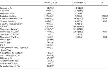
Most patients were diagnosed with BD type I (68%) (Table 2). The majority of the patient sample was female (58.6%) and the average age was 36.5 (s.d. = 12.2) years. The mean CGI-S score was 4.5 (s.d. = 0.8). Patients had an average of 14.2 (s.d. = 1.9) years of education. Refer to Table 2 for additional descriptive variables.
Sample demographics and characteristics

Data are given as mean (standard deviation) unless otherwise specified.
ICV, Intracranial volume; BV, brain volume; PFC, prefrontal cortex; CSF, whole-brain ventricular cerebrospinal fluid volume; WMH, total volume of deep white matter hypointensities; CGI-S, Clinical Global Impression Severity.
Patients and controls did not differ in terms of sex, age and years of education. A higher performance was observed for the controls in all the examined cognitive domains and the most notable difference was observed in the executive domain (p ⩽ 0.0005). However, the proxy for CR, the WAIS-III Vocabulary subtest, did not differ between the groups. Gross anatomical measures did not differ between patients and controls. But the PFC volume was lower in the patient group (F 1,155 = 9.3, p = 0.003) and the WMH volume was higher in patients compared with controls (F 1,154 = 10.5, p = 0.001).
The CR hypothesis
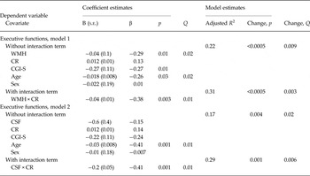
We first examined the associations of the brain ROIs with performance in each cognitive domain. These and all subsequent models were controlled for sex, age and CGI-S. At step 1, we found that an increase of 1 s.d. of total WMH volume (0.7 cm3) was associated with 0.29 s.d. (z = 0.26) lower executive performance (Table 3). No significant associations were found between WMH volume and any other cognitive domains. Volume of total-brain ventricular CSF and volume of PFC were not significantly associated with any cognitive domain at step 1. CR was significantly associated with memory/learning (all models β ⩾ 0.40, p ⩽0.0005) and speed/attention functions (all models β ⩾0.42, p ⩽0.0005) – but not with executive functions – in all of the initial models.
Sequential linear regression of executive performance as a function of brain volumes, cognitive reserve and their interactions a

s.e., Standard error; Q, false discovery rate-corrected p value; WMH, total volume of deep white matter hypointensities; CR, cognitive reserve; CGI-S, Clinical Global Impression Severity; CSF, whole-brain ventricular cerebrospinal fluid volume.
a Only significant models are displayed.
In step 2, the interaction term for each brain ROI and CR was added to each of the initial models. When the interaction term for total WMH volume and CR was added to the initial model, an increment of 1 s.d. in CR reduced the influence of WMH volume on executive performance by 0.38 s.d. There were no significant interactions between CR and WMH volume in the memory and attention/speed domains. There was also a significant interaction between whole-brain ventricular CSF volume and CR (p = 0.001). The burden of CSF on executive function was reduced by 0.41 s.d. for each 1 s.d. increment in CR. However, as the whole-brain ventricular CSF volume was not initially associated with executive function, this interaction must be interpreted with caution. No significant interactions were observed between ventricular CSF volume and CR in any other cognitive domain. Also, no interaction effects were observed between PFC volume and CR.
Additional analyses
Because there is concern that normalization of brain measures can influence the associations between subregions and overall brain size, and might also be dependent on the normalization procedure (Luders et al. Reference Luders, Narr, Thompson, Rex, Woods, Deluca, Jancke and Toga2006), all regression models were re-performed. To that end, we used unscaled MRI measures with and without ICV as a covariate in a secondary set of analyses. These analyses essentially resulted in identical main findings (data not shown).
Moreover, all interaction models were repeated with educational years as the measure of CR instead of IQ. There were no significant interactions for any of the models using educational years as a proxy for CR.
Discussion
The concept of CR has been suggested to be relevant to virtually all situations where brain function is disrupted. Here, we investigated if the concept is applicable to BD. As numerous studies report brain abnormalities and cognitive impairment in BD, we hypothesized that higher CR would lessen the influence of brain abnormalities on cognitive functions in patients with BD. In agreement with this hypothesis, we found that the interactions between CR and volumes of deep WMH/ventricular CSF were associated with executive functioning in BD regardless of clinical severity. These findings place the current study in line with others that have found higher intellectual capacity to alter the relationship between brain pathology and clinical presentation across a number of clinical disorders.
Consistent with what we and others have reported previously (Bourne et al. Reference Bourne, Aydemir, Balanza-Martinez, Bora, Brissos, Cavanagh, Clark, Cubukcuoglu, Dias, Dittmann, Ferrier, Fleck, Frangou, Gallagher, Jones, Kieseppa, Martinez-Aran, Melle, Moore, Mur, Pfennig, Raust, Senturk, Simonsen, Smith, Bio, Soeiro-De-Souza, Stoddart, Sundet, Szoke, Thompson, Torrent, Zalla, Craddock, Andreassen, Leboyer, Vieta, Bauer, Worhunsky, Tzagarakis, Rogers, Geddes and Goodwin2013; Palsson et al. Reference Palsson, Figueras, Johansson, Ekman, Hultman, Ostlind and Landen2013; Sparding et al. Reference Sparding, Silander, Palsson, Ostlind, Sellgren, Ekman, Joas, Hansen and Landen2015), the sex- and age-matched healthy controls had higher average scores than BD patients in all cognitive domains. By contrast, the score on the CR proxy measure did not differ between the groups. Also, the control and patient groups did not differ significantly on the gross anatomical measures ICV and BV. But in agreement with previous reports (Arnone et al. Reference Arnone, Cavanagh, Gerber, Lawrie, Ebmeier and Mcintosh2009), patients had significantly lower PFC volumes and more WMH compared with the healthy controls.
Our initial analysis showed that a higher volume of deep WMH was associated with lower executive performance, which accords with earlier studies (Oertel-Knochel et al. Reference Oertel-Knochel, Reinke, Alves, Jurcoane, Wenzler, Prvulovic, Linden and Knochel2014; Poletti et al. Reference Poletti, Bollettini, Mazza, Locatelli, Radaelli, Vai, Smeraldi, Colombo and Benedetti2015). In the next step, we found that the influence of WMHs on executive functions was diminished as a function of increased CR. Although this has not been previously observed in BD, studies of other disorders have observed the same phenomenon (Deary et al. Reference Deary, Leaper, Murray, Staff and Whalley2003; Dufouil et al. Reference Dufouil, Alperovitch and Tzourio2003; Brickman et al. Reference Brickman, Siedlecki, Muraskin, Manly, Luchsinger, Yeung, Brown, Decarli and Stern2011). For example, Brickman et al. (Reference Brickman, Siedlecki, Muraskin, Manly, Luchsinger, Yeung, Brown, Decarli and Stern2011) studied a community-based sample of healthy older adults and found that individuals with higher reserve had a higher WMH burden, indicating that those with higher reserve were better at compensating for brain pathology than those with lower reserve. WMH abnormalities has been proposed as a biomarker for BD as it has been observed in patients with BD as well as their relatives (Ahearn et al. Reference Ahearn, Steffens, Cassidy, Van Meter, Provenzale, Seldin, Weisler and Krishnan1998), but this finding has not been consistently replicated (Gunde et al. Reference Gunde, Blagdon and Hajek2011). WMHs most likely represent a reduction in white matter density caused by neuropil atrophy, ischaemia-related microangiopathy and/or demyelination (Ovbiagele & Saver, Reference Ovbiagele and Saver2006). The cause of WMH in BD, however, remains elusive, and future studies including diffusion tensor imaging procedures, or the simultaneous analyses of T2-weighted images (Yoshita et al. Reference Yoshita, Fletcher and Decarli2005) might advance our understanding of the nature of white matter abnormalities in BD.
The finding that CR lessens the influence of ventricular CSF volume on executive function is uncertain as there was no association between CSF and executive functions at step 1. As total volumes of ventricular CSF and WMH might reflect central atrophy, the influence of the former would be more indirect and could account for the lack of a significant association. Although speculative, the absence of an initial association could be explained by the possibility that the reserve capacity mediates even the slightest association between CSF and executive functions.
Despite the prominent role of the PFC in the cognitive domains examined (Miller & Cohen, Reference Miller and Cohen2001; Kesner & Churchwell, Reference Kesner and Churchwell2011), neither the initial models nor their interactions were significant. A possible explanation for the lack of associations may be that the PFC as an entity is too large for observing any relation to cognitive functions (Strakowski et al. Reference Strakowski, Delbello and Adler2005), which might have more focal correlates. For example, Lopez-Larson et al. (Reference Lopez-Larson, Delbello, Zimmerman, Schwiers and Strakowski2002) found that patients with BD only had grey matter reductions in subregion-specific areas of the PFC in comparison with healthy controls. Widespread PFC abnormalities have nonetheless been reported in BD (Rimol et al. Reference Rimol, Nesvag, Hagler, Bergmann, Fennema-Notestine, Hartberg, Haukvik, Lange, Pung, Server, Melle, Andreassen, Agartz and Dale2012; Abe et al. Reference Abe, Ekman, Sellgren, Petrovic, Ingvar and Landen2016). Others have also reported non-specific PFC changes in BD to be related to sustained attention (Sax et al. Reference Sax, Strakowski, Zimmerman, Delbello, Keck and Hawkins1999). Nonetheless, the only cognitive domain that was positively associated with CR was executive functions. As this was the domain in which we found the largest performance difference between patients and healthy controls – approximately 0.6 s.d. – it is possible that the cognitive impairment in the other domains was too small to benefit from increased CR, or that the effects were too small to be detected considering our sample size.
Educational attainment and occupational status are the most commonly used CR measures given their flexibility to account for lifetime accumulation of knowledge (Valenzuela & Sachdev, Reference Valenzuela and Sachdev2006; Stern, Reference Stern2009). However, onset of BD might disrupt scholastic achievement and reduce educational quality (Perlis et al. Reference Perlis, Dennehy, Miklowitz, Delbello, Ostacher, Calabrese, Ametrano, Wisniewski, Bowden, Thase, Nierenberg and Sachs2009; Post et al. Reference Post, Leverich, Kupka, Keck, McElroy, Altshuler, Frye, Luckenbaugh, Rowe, Grunze, Suppes and Nolen2010). There are also several reports that indicate a discrepancy between reported level of education and academic ability in psychiatric populations (Iltis et al. Reference Iltis, Misra, Dunn, Brown, Campbell, Earll, Glowinski, Hadley, Pies and Dubois2013). Furthermore, the relatively low age of our sample adds to the possibility that there is a discrepancy between native ability and educational attainment as well as occupational status. Therefore, we defined CR as the score on the WAIS-III Vocabulary subtest. Measures of IQ have previously been used to gauge CR in various populations (Stern et al. Reference Stern, Zarahn, Hilton, Flynn, Delapaz and Rakitin2003, Reference Stern, Zarahn, Habeck, Holtzer, Rakitin, Kumar, Flynn, Steffener and Brown2008; Alchanatis et al. Reference Alchanatis, Zias, Deligiorgis, Amfilochiou, Dionellis and Orphanidou2005; Alosco et al. Reference Alosco, Spitznagel, Raz, Cohen, Sweet, Van Dulmen, Colbert, Josephson, Waechter, Hughes, Rosneck and Gunstad2012; Galioto et al. Reference Galioto, Alosco, Spitznagel, Stanek and Gunstad2013; Kontis et al. Reference Kontis, Huddy, Reeder, Landau and Wykes2013) and might yield more accurate estimates of CR than education when it is unclear if the latter can be considered representative (Stern, Reference Stern2012). As educational years might also underestimate native ability, pre-morbid IQ has been considered to be a more reliable estimate (Schmand et al. Reference Schmand, Smit, Geerlings and Lindeboom1997; Rentz et al. Reference Rentz, Huh, Faust, Budson, Scinto, Sperling and Daffner2004). Given that patients with BD have unimpaired IQ (Torres et al. Reference Torres, Boudreau and Yatham2007), we believe that the estimation of CR for the current sample is probably a fair reflection of the patients’ ability to compensate for disruption in face of brain abnormalities. This is further corroborated by the fact that our post-hoc analysis using educational years as a proxy for CR yielded negative results.
This study has some limitations to consider. First, the sample size did not permit inclusion of some potential confounders. Although the determination of WMH using FreeSurfer in combination with T1-weighted images is acceptable, it has its limitations, such as possible region-specific underestimation of WMH (Olsson et al. Reference Olsson, Klasson, Berge, Eckerstrom, Edman, Malmgren and Wallin2013). Then again, anatomical mapping of WMH might lead to inaccurate estimates of the entire distribution of WMH (Yoshita et al. Reference Yoshita, Fletcher and Decarli2005). Finally, the lack of follow-up data prevents us from testing whether reserve also mediates cognitive trajectories as a result of brain abnormalities/changes.
In conclusion, this study suggests that the concept of CR is applicable in BD. The interactions between CR and whole-brain volumes of WMH/ventricular CSF account for executive functioning in BD. These findings might imply that higher reserve capacity in BD alters the relationship between brain pathology and clinical presentation.
Acknowledgements
We are grateful to the research participants and healthy volunteers for participation in this research study. We acknowledge the contribution of the staff at the Northern Stockholm Psychiatry Clinic and the St. Göran research team for recruitment and evaluation of the study participants. Björn Hultman is acknowledged for designing the psychological test battery; and Haydeh Olofsson and Mathias Kardell for data management support. Marie Tegnér and Yords Österman are recognized for performing the MR scanning.
This research study was supported by grants from the Swedish Medical Research Council (K2014-62X-14647-12-51 and K2010-61P-21568-01-4), the Swedish Foundation for Strategic Research, the Swedish Brain Foundation, the Swedish Federal Government under the LUA/ALF agreement (ALF 20130032, ALFGBG-142041), the Psychiatric Foundation, and the Fredrik and Ingrid Thuring Foundation.
Declaration of Interest
None.