Introduction
Computed tomography (CT) is the primary modality for imaging the paranasal sinuses. The causes of unilateral sinus opacification on CT can be divided into inflammatory and neoplastic diseases.Reference Eckhoff, Cox, Luk, Maidman, Wise and DelGaudio1 The odds of neoplasia associated with unilateral sinus opacification are significantly greater compared to bilateral opacification.Reference Eckhoff, Cox, Luk, Maidman, Wise and DelGaudio1 The proportion of cases with unilateral maxillary sinus opacification that represented neoplasia and malignancy according to a pooled analysis of 366 cases was 18.3 per cent and 7.1 per cent respectively.Reference Knisely, Holmes, Barham, Sacks and Harvey2 However, this was not a true pooled analysis, as the data were not meta-analysed to account for the weighting of studies. Nonetheless, the European Position Paper on Rhinosinusitis and Nasal Polyps 2020 stated that clinicians should be wary of conservative management for isolated maxillary sinus opacification, and to exercise a low threshold for early surgical management.Reference Fokkens, Lund, Hopkins, Hellings, Kern and Reitsma3
Some have suggested that histological confirmation is obligatory in all cases of unilateral maxillary sinus opacification,Reference Lee4 whilst others advocate biopsy in the setting of suspicious clinical findings.Reference Chen, Chen, Chang and Huang5 We hypothesised that the neoplasia rate associated with unilateral maxillary sinus opacification was low based on our local anecdotal experience, and therefore a protocol to biopsy all unilateral maxillary sinus opacification may lead to many unnecessary invasive investigations. We aimed to determine the rate of inflammatory and neoplastic causes for unilateral maxillary sinus opacification, and establish the proportion of patients who underwent functional endoscopic sinus surgery (FESS) at our organisation.
Materials and methods
We conducted a retrospective, longitudinal, observational study at a district general hospital in the West Midlands, UK. The Strengthening the Reporting of Observational Studies in Epidemiology (‘STROBE’) statement for reporting observational studies was followed.Reference von Elm, Altman, Egger, Pocock, Gøtzsche and Vandenbroucke6
A list of patients who had undergone CT of the sinuses over a 12-month period in 2018 was obtained from the radiology department. The CT images were viewed in axial and reformatted coronal views to determine unilateral maxillary sinus opacification. The images were given a Lund–Mackay score (by authors KD and RA), and any discrepancies were resolved by discussion (with ZM and RS). The maxillary sinus was given a score of 1 for partial opacification and 2 for complete opacification. Only unilateral maxillary sinus opacification cases (with or without ipsilateral paranasal sinus involvement) were included in our study.
The exclusion criteria were scans with clear maxillary sinuses or bilateral opacification. Patients who did not have a consultation with an otolaryngologist were also excluded to ensure all cases had a clinical diagnosis.
The following clinical parameters were extracted from the hospital electronic patient records: indication for CT, presenting symptoms, examination findings and treatments. The primary outcome was the rate of neoplasia associated with unilateral maxillary sinus opacification. Clinic follow-up visits, emergency department presentations and re-referrals to the otolaryngology service were reviewed over a 24-month follow-up period.
Results
There were a total of 641 CT sinus scans. The images were screened, and 582 scans were excluded because of bilateral maxillary opacification (30 per cent) or clear maxillary sinuses (70 per cent). The overall rate of unilateral maxillary sinus opacification in our series was therefore 9 per cent (n = 59). Seven out of the 59 cases were excluded as the patients were not consulted by otolaryngologists. Hence, there were 52 cases for the analysis of clinical outcomes, primarily the rate of neoplasia.
The indications for CT in the 52 cases were: failed medical treatment (29 per cent), unilateral symptoms or signs (24 per cent), facial pain (23 per cent), hyposmia (10 per cent), nasal symptoms in patients with respiratory conditions (6 per cent), uncertain diagnosis (6 per cent), and poor access (2 per cent). The maxillary sinuses in our series (n = 52) had a Lund–Mackay score of 1 in 62 per cent of cases, and a score of 2 in 38 per cent. Opacification was limited to an isolated maxillary sinus in 69 per cent, or extended to the ipsilateral sinuses in 31 per cent. The distribution of males and females was 44 per cent and 56 per cent respectively. The median age was 54 years (interquartile range = 38–69 years).
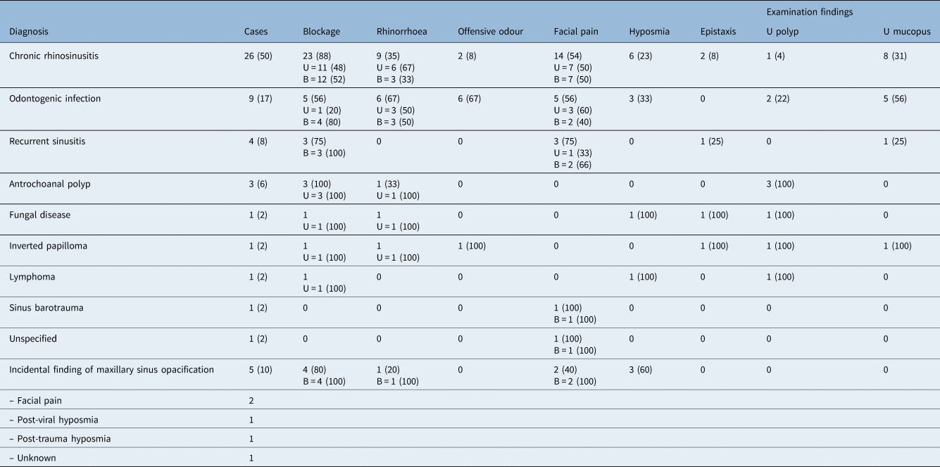
The diagnoses and clinical findings of the 52 patients are summarised in Table 1. The predominant clinical features of chronic rhinosinusitis were blockage (88 per cent) and facial pain (54 per cent). Odontogenic infection was frequently associated with rhinorrhoea (67 per cent), offensive odour (67 per cent), blockage (56 per cent), facial pain (56 per cent) and nasendoscopic signs of unilateral mucopus (56 per cent). Patients with recurrent sinusitis commonly had blockage (75 per cent) and facial pain (75 per cent). Epistaxis was seen in the fungal disease and inverted papilloma cases. All cases that had more than simple inflammatory disease, including antrochoanal polyp, fungal infection, inverted papilloma and lymphoma, had unilateral blockage, rhinorrhoea, and a unilateral polyp or mass in common.
Diagnoses and clinical findings in unilateral maxillary sinus opacification patients*

Data represent numbers (and percentages) of cases.
* Total n = 52. U = unilateral; B = bilateral
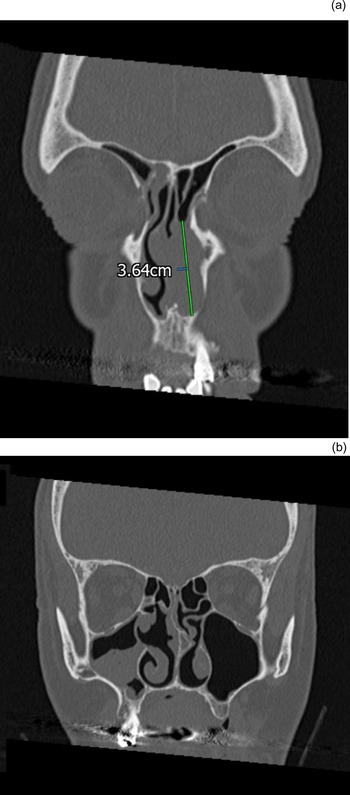
There was 1 case (2 per cent) of benign neoplasm in our cohort of 52 patients. The biopsy showed inverted papilloma. Another patient with a malignant lesion presented with a unilateral mass within the left nasal cavity, and the ipsilateral eye was proptosed. This case met our study inclusion criteria as the patient had unilateral maxillary sinus opacification of the contralateral maxillary sinus (Figure 1) that was incidental rather than representing neoplasia. A diagnosis of large B-cell lymphoma was made following endoscopic biopsy. Bony erosion was reported on the CT scans of both of these patients.
Coronal computed tomography sinus scans in bone window of a patient with primary nasal lymphoma. (a) A well-defined soft tissue mass with homogeneous attenuation within the anterior half of the left nasal cavity. (b) Incidental finding of right-sided maxillary partial opacification in the same patient.

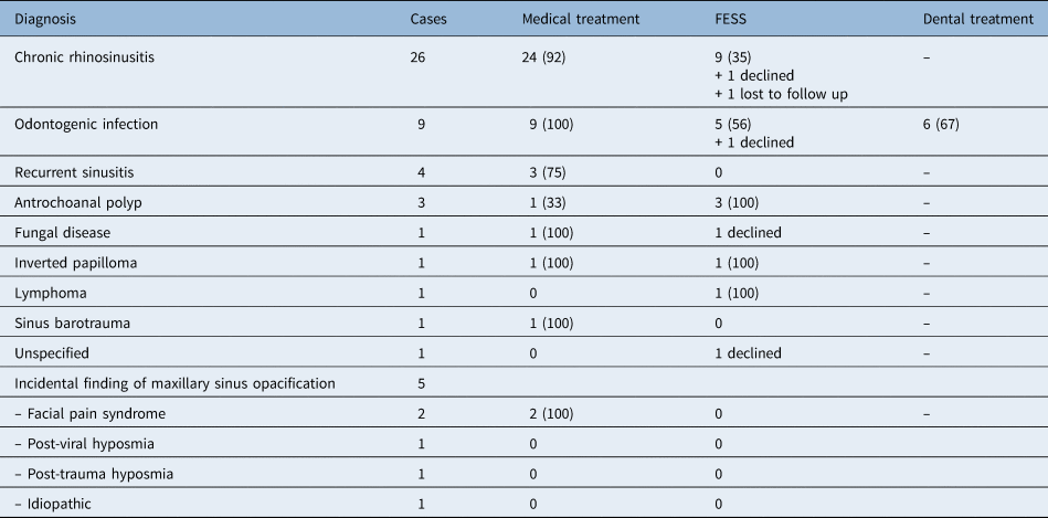
Overall, 36.5 per cent of the 52 patients in our series underwent FESS. Table 2 summarises the treatments provided to our patients. Functional endoscopic sinus surgery was performed as a diagnostic and/or therapeutic procedure in patients with: chronic rhinosinusitis (35 per cent), odontogenic infection (56 per cent), antrochoanal polyp (100 per cent), inverted papilloma (100 per cent) and lymphoma (100 per cent). The five odontogenic infection patients who underwent FESS also had dental extractions as part of their treatment.
Treatments provided to unilateral maxillary sinus opacification patients*

Data represent numbers (and percentages) of cases.
* Total n = 52. FESS = functional endoscopic sinus surgery
All patients in our series achieved clinical resolution except: one patient with chronic rhinosinusitis for whom medical and FESS treatment failed, and who was subsequently referred to a tertiary centre; two other chronic rhinosinusitis patients who were re-referred from primary care, one of which was continued on medical treatment, and the other had already undergone FESS and improved with further medical therapy; and one patient with recurrent sinusitis who was re-referred with an infective episode on the contralateral side, and who responded to medical therapy. No other instances of failed treatment or revision of diagnosis were detected over 24 months.
Discussion
We found a unilateral maxillary sinus opacification rate of 9 per cent. Higher rates of 14–23 per cent have been reported,Reference Lee4,Reference Chen, Chen, Chang and Huang5 although these studies included only patients who underwent surgery. Others reporting a lower rate of 2.5 per cent used a stricter definition of complete unilateral maxillary sinus opacification.Reference Ahsan, El-Hakim and Ah-See7 Chen et al.Reference Chen, Su and Yeow8 identified a rate of 10.7 per cent by including scans with both partial and complete opacification, as well as patients who did not undergo surgery, similar to our study methodology.
Neoplasia represented 2 per cent of patients who had unilateral maxillary sinus opacification in our series. Chen et al.Reference Chen, Su and Yeow8 found a similarly low neoplasia rate of 3.9 per cent in 76 patients. However, most other studies report considerably higher rates, ranging from 11 per cent (n = 64),Reference Kaplan and Kountakis9 15.1 per cent (n = 116),Reference Chen, Chen, Chang and Huang5 19.8 per cent (n = 121),Reference Lee4 20.2 per cent (n = 114),Reference Salami10 to 34 per cent (n = 110).Reference Eckhoff, Cox, Luk, Maidman, Wise and DelGaudio1 These studies had obtained histopathological diagnoses and therefore the samples represented study populations that had all undergone operative management. Selection bias may have caused an overestimation of the neoplasia rate. Our study mitigated this bias by including patients who did not have surgical intervention in the denominator of our rate calculation. Interestingly, two studies that also included patients without histopathological findings reported high neoplasia rates of 30–34.4 per cent.Reference Zahedi, Mohamed, Husain, Kew and Gendeh11,Reference Rudralingam, Jones and Woolford12 However, the number of CT scans requested in both of these studies was vastly lower per unit of time than in our study. This indicates that the clinician threshold for scanning may be an important confounding factor in the detection of neoplasia.
One case of lymphoma was detected in our series. This patient presented with a unilateral left-sided nasal mass and had an incidental finding of unilateral maxillary sinus opacification on the contralateral side. This case was not included in our reported rate of neoplasia as the unilateral maxillary sinus opacification itself did not represent a tumour. This case draws attention to the notion that localised pathology on one side of the nasal cavity does not protect the contralateral sinuses from an inflammatory process. This phenomenon has previously been highlighted in cases of mycetoma and mucocele.Reference Kaplan and Kountakis9
Functional endoscopic sinus surgery was carried out for chronic rhinosinusitis (35 per cent), odontogenic infection (56 per cent), and all cases of antrochoanal polyp, inverted papilloma and lymphoma. The rationale for surgery was probably therapeutic for inflammatory conditions, and diagnostic in cases where neoplasia had to be ruled out. However, we cannot be certain about the clinicians’ reasoning behind the surgery given the retrospective nature of our study.
The two-year follow-up data did not reveal any missed cases of malignancy, although our data collection methodology was within the confines of a retrospective observational study.
The patients with unilateral chronic rhinosinusitis and odontogenic infection who did not undergo FESS were diagnosed on clinical grounds. The diagnosis of unilateral chronic rhinosinusitis in patients who were managed non-operatively was based on the same criteria for chronic rhinosinusitis, according to clinical, endoscopic and CT findings, as defined in the European Position Paper on Rhinosinusitis and Nasal Polyps 2020.Reference Fokkens, Lund, Hopkins, Hellings, Kern and Reitsma3
The nine diagnoses of odontogenic infection were obtained by the following means: five were based on the CT report affirming dental pathology; one was diagnosed following recognition of dental pathology on the CT images by the otolaryngologist; two were diagnosed by general dental practitioners and referred to ENT following persistent symptoms after dental extraction; and one was diagnosed after referral to an oral and maxillofacial surgeon because of treatment failure after FESS.
Overall, an oral examination was performed in only 4 of the 52 patients (8 per cent) in our series. The CT reports commented on positive or negative dental findings in only 5 out of the 52 cases (10 per cent) of unilateral maxillary sinus opacification. This indicates that odontogenic infection was underappreciated by both otolaryngologists and radiologists. Our reported rate of odontogenic infection may be an underestimate, and it is possible that some odontogenic infection cases were mistaken for unilateral chronic rhinosinusitis. Turfe et al.Reference Turfe, Ahmad, Peterson and Craig13 also identified poor reporting of dental findings in 66 per cent of CT sinus scans in their series of odontogenic sinusitis. Collaboration between otolaryngologists and dental practitioners leads to a dramatic rise, up to 73 per cent, in the diagnosis of odontogenic infection in unilateral maxillary sinus opacification cases.Reference Matsumoto, Ikeda, Yokoi and Kohno14 A shared care model for unilateral maxillary sinus opacification should be encouraged, as recognition of odontogenic infection then allows the dental pathology to be addressed, which increases treatment success compared to FESS alone.Reference Gâta, Toader, Valean, Trombitaș and Albu15
Further research
The present body of research on neoplasia associated with unilateral maxillary sinus opacification may have overestimated its prevalence because of bias in patient selection and a lack of standardisation of CT indications. Our inclusion of patients without a histopathological diagnosis is a step closer to obtaining data on the neoplasia rate associated with unilateral maxillary sinus opacification that are not skewed towards patients who are subjected to surgery, as the decision to operate in these cases is usually underpinned by a genuine clinical concern about neoplasia. This concern is often missing in cases of unilateral maxillary sinus opacification, and diagnosis of inflammatory conditions may be reasonably reached based on clinical and CT findings with adequate follow up. Our series takes this group of patients into account in our calculation of the neoplasia rate associated with unilateral maxillary sinus opacification.
The generalisability and applicability of our findings may be improved by conducting further studies with: a prospective study design, pre-defined indications for CT scans, the inclusion of patients managed operatively and non-operatively, large sample sizes, and consideration of geographical variation.
Whilst we found that patients with unilateral symptoms and signs were likely to have more than simple inflammatory disease, warranting operative management, these findings alone were not discriminatory, as demonstrated in Table 1 by the overlap of symptoms between conditions. Bony erosion on CT was seen in the only case of inverted papilloma and lymphoma, and this can be a ‘red flag’ sign prompting further investigation. Bony erosion on CT is a worrying sign of malignant tumours,Reference Ahsan, El-Hakim and Ah-See7 as well as benign tumours.Reference Salami10 However, this feature has also been reported in mucoceles,Reference Lee4 invasive fungal diseaseReference Chen, Su and Yeow8,Reference Salami10 and in some cases of chronic rhinosinusitis.Reference Chen, Su and Yeow8 Although lacking in specificity, bony erosion does tend to be more extensive in malignancy.Reference Lee4
• Neoplasia is a potential cause of unilateral maxillary sinus opacification on computed tomography
• Current guidelines advise a low threshold for early surgical management in isolated maxillary sinus opacification cases
• The rate of unilateral maxillary sinus opacification in this retrospective observational study of 641 scans was 9 per cent
• Of 52 patients, 1 inverted papilloma case was detected; this neoplasia rate of 2 per cent is lower than previously reported (11–34 per cent)
• A unilateral nasal polyp or mass was predictive of pathology that required operative management
• Careful history and examination of nasal cavity, oral cavity and orbit are paramount in triaging patients for further investigations to exclude neoplasia
Magnetic resonance imaging (MRI) has recently shown promise in the investigation of unilateral maxillary sinus opacification, with higher specificity and positive predictive values for neoplasia compared to CT.Reference Chua, Navaratnam, St Leger, Lam, Unadkat and Weller16 Further research may be directed at evaluating the additive role of MRI in order potentially to mitigate the need for invasive biopsy in unilateral maxillary sinus opacification cases.
Conclusion
There was a high prevalence of simple inflammatory pathology causing unilateral maxillary sinus opacification on CT sinus scans. A much lower neoplasia rate of 2 per cent was observed in our study compared to the published literature. Therefore, an operative strategy for the purpose of ruling out neoplasia in all cases of unilateral maxillary sinus opacification would lead to many unnecessary procedures. A careful history and examination of the nasal cavity with a nasendoscope, oral cavity and orbit, and recognition of worrying signs on CT, are paramount in appropriately selecting patients for biopsy.
Competing interests
None declared