Pulmonary atresia and intact ventricular septum is an uncommon congenital cardiac malformation, with an incidence of from 4 to 7 cases for each 100,000 live births.Reference Ferencz, Rubin and McCarter1, Reference Freedom, Wilson, Trusler, William and Rowe2 About two-fifths of the affected children have fistulous connections, better described as ventriculo-coronary arterial communications, between the right ventricle and the coronary arteries, compromising the flow of blood in the myocardium. In the presence of such ventriculo-coronary arterial communications with proximal stenoses, parts of the myocardium are dependent on flow from the right ventricle to the coronary arteries. Although the options for treatment have developed rapidly during the last twenty years, mortality is still high.Reference Ashburn, Blackstone and Wells3–Reference Fenton, Pigula, Gandhi, Russo and Duncan6 To survive, the children usually have to undergo repeated and extensive surgery. The intended endpoints of the surgery are to achieve a biventricular repair or univentricular palliation. Myocardial pathology, including infarction, has been thoroughly described in infants with pulmonary atresia and intact ventricular septum.Reference Freedom, Wilson, Trusler, William and Rowe2, Reference Akiba and Becker7–Reference Oosthoek, Moorman, Sauer and Gittenberger-de Groot9 These investigations mainly comprised post-mortem histology in infants. There are only few studies of infarctions or myocardial pathology in older children or adults.Reference Robicsek, Bostoen and Sanger10 The aim of our study, therefore, was to explore whether perfusion scintigraphy would show deficient uptake, and if so, to compare the deficits in children with and without ventriculo-coronary arterial communications, as well as in those having biventricular repair as opposed to functionally univentricular palliation.
Material and methods
Study design
All children underwent thorough clinical examination, electrocardiography, transthoracic echocardiography, exercise test and myocardial perfusion scintigraphy at submaximal work load. The medical records were studied, focusing on anatomical defects and surgical methods. The study was approved by the local medical ethics committee and informed consent was obtained from each participant, and/or the parents.
Population
Our initial cohort comprised 18 children, 10 males and 8 females, with pulmonary atresia and intact ventricular septum who were born in the western part of Sweden between 1980 and 1994. Of these, 5 died, 4 within 30 days from surgery at ages from 3 days to 5 years, with 2 of these having ventriculo-coronary arterial communications. Another child died suddenly at home at 15 months of age, not related to surgery. He was palliated with a systemic-to-pulmonary arterial shunt, and also had ventriculo-coronary arterial communications.
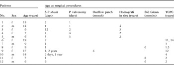
We invited all 13 surviving patients to participate in the study. Of these, 1 boy with biventricular repair without presence of ventriculo-coronary arterial communications declined for non-medical reasons. Thus, 12 accepted the invitation, 5 boys and 7 girls, having a mean age of 11 years, with a range from 6 to 19 years. The repair was biventricular in 5, with the other 7 having had functionally univentricular palliation, all by construction of a total cavopulmonary connection with an intracardiac tunnel between the inferior caval vein and the pulmonary arteries. Of these 12 children, 11 had received a systemic-to-pulmonary shunt in the neonatal period (Table 1). The period since the last operations varied from 4 to 15 years, with a median of 5.5 years. The clinical course had proved unusual in 2 children.
Demographic data.

Patients 1 through 5 had biventricular repairs, while those numbered 6 through 12 had univentricular palliations. Those numbered 9 through 12 also have ventriculo-coronary arterial communications.
Abbreviations: S-P shunt: systemic to pulmonary shunt; P valvotomy: valvotomy of the pulmonary valve; Bid. Glenn: Bidirectional Glenn (connection of the superior caval vein to the pulmonary artery); TCPC: total cavopulmonary connection (intracardiac tunnel in all cases).
*Fontan operation at four years of age.
The first, a girl now 19 years of age, with a diminutive right ventricle, was subjected to univentricular palliation at 4 years of age. A homograft had been inserted between the right atrium and the pulmonary arteries, and another homograft inserted between the inferior caval vein and the right atrium. At 11 years of age, the homografts were removed, and a total cavopulmonary connection was completed. She underwent reoperation at 14 years of age owing to a leakage in the tunnel between the inferior caval vein and the pulmonary artery.
The second was now a 17-year-old girl with ventriculo-coronary arterial communications from the right ventricle to a single left coronary artery. She was initially considered to be inoperable, but survived thanks to persistent patency of the arterial duct. At 3 months of age, she underwent reconstruction of the outflow from the right ventricle, and a systemic-to-pulmonary shunt was created at 1 year of age. The tricuspid valve, however, did not grow, so a total cavopulmonary connection was performed when she was 12 years of age.
Methods
All children performed bicycle ergometer tests on two consecutive days. The workload was increased in a stepwise manner by 10 Watts per minute. A standard 12-lead electrocardiogram was recorded at rest and during exercise. The first test was used to identify the performance ability. The test was interrupted when it was impossible to push the child further. During the second exercise test, 120–190 MBq technetium Tc-99m tetrofosmin per m2 body surface area was injected at a sub maximal work load, 10 Watts lower than the level obtained the day before. The exercise test was continued for at least one more minute after injection. The child was given ice cream in an attempt to reduce the activity in the liver, while waiting for the imaging to take place. The time period between injection and imaging was at least 30 minutes.
For acquisition, we used a triple-head gamma camera (Picker Prism 3000) fitted with low energy high resolution or low energy ultra high resolution parallel hole collimators, the machine running in a continuous mode over 360 degrees, with 3 degrees per projection, and a total acquisition time of 20 minutes. Data were reconstructed with correction for attenuation using the standard 3-dimensional post filtering algorithm.
Echocardiography was performed on the same occasion, focussing on myocardial function according to biplane Simpson’s rule, analysis of wall motion, and atrioventricular regurgitation. All investigations were performed by one investigator (B-M Ekman-Joelsson), using an Acuson 128XP machine.
Analysis of data
The scintigrams were analysed for perfusion defects in a semi-quantitative manner, classifying the perfusion as normal, scored zero, and slightly, moderately, or severely reduced, scored at 1 to 3. The location was described as the apical or basic parts of the ventricular septum or left ventricular free wall. As there are limitations in studying the right ventricular wall with perfusion scintigraphy, and as the right ventricle is often small and has a bizarre shape in this particular group of patients, it was not included in the analysis.
Wall motion was analysed on two-dimensional images of the apical four chambers, apical two chambers, parasternal long axis and parasternal short axis views, at echocardiography. The left ventricular wall was divided into 16 segments and the mural motion was scored according to a six point scale, from 1 for normokinesia, through hypokinesia, akinesia, dyskinesia, aneurysm, and 6 for paradoxical movements, according to the recommendations from the American Society of Echocardiography.Reference Schiller, Shah and Crawford11 The results for analysis were analysed blinded by two other independent investigators with experience from echocardiography performed on adults and children. When the observers were in disagreement, a mean value was reported.
Results
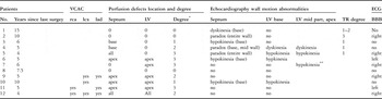
Myocardial perfusion defects, presence of ventriculo-coronary arterial communications, and findings on echocardiography and electrocardiography in each patient, are presented in Table 2. With regard to analysis of wall motion, the observers were in agreement in 6 cases with univentricular palliation, but in disagreement regarding the sixth patient in Tables 1 and 2. In all 5 patients with biventricular repair, both observers considered wall motion to be abnormal, but their grading differed slightly. Left ventricular systolic function measured as ejection fraction varied from 44% to 74%, with a median of 58%. In only one case was ejection fraction below 50%, this being the 10th patient in the tables.
Myocardial perfusion defects.

The patients are as listed in Table 1. Age means age at examination.
Perfusion defects, location, septum: interventricular septum; LV: left ventricular free wall; VCAC: ventriculocoronary arterial communications; rca: right coronary artery; lcx: left circumflex coronary artery; lad: left anterior descending coronary artery; TR: tricuspid regurgutation; ECG: electrocardiogram; BBB: bundle branch block. *0: normal perfusion; 1: slightly reduced perfusion; 2: moderately reduced perfusion; 3: severely reduced perfusion.
**Incomplete examination, as there was no 2-chamber-view.
Of the 5 children with biventricular repair, 3 had perfusion defects within the ventricular septum. All children with biventricular repair had some sort of abnormality in the ventricular septum, and paradoxical septal movements were seen in 3. There was no correlation between perfusion defects and any specific electrocardigraphic pathology, as for example, left bundle branch block. All patients had some degree of tricuspid regurgitation, which was minor in 4 patients, and more pronounced in 1.
Of the 7 children with univentricular palliation, 6 had perfusion defects, located in the left ventricular free wall in all, and also in the ventricular septum in 5. All children with ventriculo-coronary arterial communications had perfusion defects in the left ventricular free wall and in the ventricular septum, most pronounced in the apical parts. The child without perfusion defects did not have any ventriculo-coronary arterial communications.
There was no correlation between degree of perfusion defects and the presence of ventriculo-coronary arterial communications, or with younger age at surgery.
In a 9-year-old boy and a 7-year-old girl, the myocardial scintigraphy was repeated. The boy, listed as the 7th patient in the tables, had undergone univentricular palliation. At the first scintigraphy, a very large perfusion defect was found in the apical part of the left ventricle. The scintigraphy was repeated after one month, performed at rest and after exercise, verifying the perfusion finding.
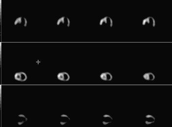
The girl, listed as the 11th patient in the tables, had undergone univentricular palliation and had ventriculo-coronary arterial communications (Fig. 1). At the first scintigraphy, she exhibited chest pain and breathing difficulties during exercise, and the scintigraphy showed perfusion defects. Findings consistent with myocardial ischaemia during activity were seen on exercise testing and Holter-electrocardiography. At subsequent catheterisation and angiocardiograhpy three ventriculo-coronary arterial communications were embolised, with definite relief of symptoms. Myocardial scintigraphy two years later, at rest and after exercise, showed the same extension and distribution of perfusion defects as the initial scintigraphy.
Myocardial perfusion abnormalities, showing views from horizontal long axis, short axis and vertical long axis in our 11th patient.

Discussion
Our main finding was the presence of perfusion defects, and their correlation with the presence of ventriculo-coronary arterial communications. We found that children undergoing biventricular repair had perfusion defects in the ventricular septum, while those undergoing functionally univentricular palliation had more extensive perfusion defects, located not only in the septum but also the left ventricular free wall.
Ventriculo-coronary arterial communications
Several studies have reported myocardial pathology and reduced function, as well as abnormalities in the motion of the walls of the left ventricle, at birth and in relation to surgery in the presence of ventriculo-coronary arterial communications.Reference Freedom, Wilson, Trusler, William and Rowe2, Reference Gentle, Colan, Giglia, Mandell, Mayer and Sanders12–Reference Hausdorf, Grävinghoff and Keck15 It is also well known that myocardial infarction may occur in this group of children after birth and in relation to surgery.Reference Freedom, Wilson, Trusler, William and Rowe2, Reference Fyfe, Edwards and Driscoll8, Reference Oosthoek, Moorman, Sauer and Gittenberger-de Groot9, Reference O’Connor, Stahr, Cottrill, Todd and Noonan16
Ventriculo-coronary arterial communications connect the hypertensive right ventricle with the coronary arteries, thus producing systolic flow with poorly oxygenated blood in the arteries, in addition to the normal diastolic flow from the aortic root, and thus compromising the supply of oxygen to the myocardium. One of our patients experienced symptomatic relief when the communications were embolized, and there was no progress of perfusion defects seen on myocardial scintigraphy performed after this procedure. There are several reports of successful ligation of ventriculo-coronary arterial communications at the time of right ventricular decompression in patients with pulmonary atresia and intact ventricular septum.Reference Hisagi, Suzuki and Nakayama17, Reference Steinberger, Berry and Bass18 In our study, all children with these communications had perfusion defects in the ventricular septum and the left ventricular wall, most pronounced in the apical parts, where the myocardium is most sensitive to impaired coronary perfusion. There was a tendency towards more severe perfusion defects in this group of patients (Table 2).
Perfusion defects in the ventricular septum
Perfusion defects in the septal parts were seen in 3 of the 5 children undergoing biventricular repair, and 5 of the 7 having functionally univentricular palliation. A false-positive ischaemic pattern in the ventricular septum is correlated to left bundle branch block, but there was no such correlation in our group.Reference Ellemann, Van Heerden, Van Heerden and Kloppwe19 There was a correlation between perfusion defects and abnormal septal motion seen on echocardiography in three of the children with biventricular repair (Table 2). Abnormal ventricular septal motion has been described in several studies of adults following valvar heart surgery in the left ventricle using cardiopulmonary bypass. It seems to be most pronounced immediately after surgery, in some reports involving nine-tenths of the patients, but tends to decrease during the months after surgery.Reference Burggraf and Craige20, Reference Vignola, Boucher and Curfman21 The isotopic investigations in our study were performed 4 to 15 years, with a median of 5.5 years, after the last operation. Decreased septal motion has been seen after right ventricular decompression in children with pulmonary atresia and intact ventricular septum.Reference Gentle, Colan, Giglia, Mandell, Mayer and Sanders12 In a recent study paradoxical septal movements, studied by tissue Doppler imaging, was reported in twothirds of such patients after biventricular repair.Reference Mi and Cheung22 We found abnormalities of mural motion in all children undergoing biventricular repair, and in 2 children with univentricular palliation. Previous pathological studies have shown disproportionate septal hypertrophy and myocytic disarray, more pronounced in pulmonary atresia and intact ventricular septum as compared with findings in other congenitally malformed hearts.Reference Bulkely, Weisfeldt and Hutchins13 This is consistent with the finding of initial myocardial pathology in the septum, regardless of right ventricular size or appearance of ventriculo-coronary arterial communications.Reference Oosthoek, Moorman, Sauer and Gittenberger-de Groot9 Oosthoek et al.Reference Oosthoek, Moorman, Sauer and Gittenberger-de Groot9 found no pathology in the left ventricular free wall, when studying post mortem myocardium from such children aged from 1 to 54 days. In our study, the perfusion defects in those undergoing biventricular repair were located only in the ventricular septum and not in the left ventricular free wall, which might support the theory that the changes in the left ventricle may appear later.
Perfusion defects in patients with univentricular palliation
Of the 7 children with univentricular palliation, 6 had perfusion defects in the left ventricular free wall. Impaired coronary perfusion has been described in the Fontan circulation with a high pressure in the atrium.Reference Ilbawi, Idriss and Muster23 All children in our study, however, had low atrial pressures at the time of the investigation. Only the sixth patient in the tables, with severe perfusion defects in all locations, had high atrial pressures, these persisting for 11 years. Only one of these children had no perfusion defects, the 8th in the tables. She had undergone an early total cavopulmonary connection operation at the age of 2 years, and she did not have ventriculo-coronary arterial communications. The question of whether there is a correlation between the reduction in volume overload at an early age and the absence of severe perfusion defects is impossible to answer with this limited study, but it is an interesting finding, and might be consistent with the hypothesis of the stress affecting the arterial ventricle.Reference Gewillig and Kalis24 We found no perfusion defects in the left ventricular free wall in children with biventricular repair. An explanation might be that the exposure to volume overload was reduced in the neonatal period, when right ventricular decompression was performed. Another explanation could be that no children in this group had ventriculo-coronary arterial connections.
Methodological considerations
Single-photon emission computed tomography with technetium Tc-99-m labelled markers is a reliable diagnostic imaging procedure to study myocardial perfusion abnormalities in adults.Reference Lavalaye, Schroeder-Tanka, Tiel-van Buul, van der Wall, Lie and van Royen25 A correlation between perfusion defects and risk of future events in patients with coronary arterial disease has been reported in large studies.Reference Hachamovitch, Hayes, Friedman, Cohen and Berman26 Coronary angiography provides excellent details of the anatomy, but adds little if any information on myocardial viability. In Kawasaki disease, when the inflammation engages the entire heart, there is discordance between angiography and technetium Tc-99m single-photon emission computed tomography, with more perfusion defects than abnormal angiographies.Reference Fu, Kao, Hwang, Jan and Chi27 Myocardial scintigraphy has been of greater prognostic value than coronary angiography in an 8-year follow-up study of 90 cases of coronary aneurysms in Kawasaki disease.Reference Miyagawa, Mochizuki and Murase28 In the study reported by Fukuda and colleaguesReference Fukuda, Ishbashi and Yokoyama29, 86 children with Kawasaki disease and a control group of 20 children without heart disease were investigated with technetium Tc-99m single-photon emission computed tomography. Hypoperfusion was detected in the group with coronary arterial stenoses, with very high precision.
Among other congenital cardiac defects, transposition is the most studied, and abnormalities in myocardial perfusion have been demonstrated, although the correlation to coronary arterial stenoses seems to be uncertain.Reference Yates, Marsden and Badawi30
To our knowledge, there are only few reported studies of myocardial scintigraphy in patients with pulmonary atresia and intact ventricular septum.Reference Björkhem, Evander, White and Lundström31, Reference Lee32 In a retrospective study no localised perfusion defects in the left ventricular free wall was found, when studied with 201Thallium in 3 children with ventriculo-coronary arterial communications.Reference Björkhem, Evander, White and Lundström31 Technetium Tc-99-m single-photon emission computed tomography, however, is a more sensitive method than 201Thallium.Reference Ellemann, Van Heerden, Van Heerden and Kloppwe19 Fluorine-18-deoxyglucose positron emission tomography is probably an even more sensitive method, reflecting myocardial viability while technetium Tc-99-m tetrofosmin uptake reflects myocardial blood flow and may underestimate viability.Reference Altehoefer, vom Dahl and Biedermann33
Artefacts are mainly seen in the apical parts of the left side of the ventricular septum. The extent of artefacts is reduced when gated single-photon emission computed tomography is used, and technetium Tc-99-m single-photon emission computed tomography is a more specific method.Reference Ellemann, Van Heerden, Van Heerden and Kloppwe19, Reference Dogruca, Kabasakal, Yapar, Nisil, Vural and Önsel34
Limitations
Limitations are derived from the small number of children, absence of a control group, and the small number of investigations in each child, owing to ethical constraints concerning radiation exposure in children. From an ethical standpoint, recruitment of age-matched control groups for any radionuclide study on children is difficult.
Clinical relevance of findings in present study
We found a correlation between perfusion defects and presence of ventriculo-coronary communications. Further examinations with scintigraphic methods are needed to explore the nature of this process. If there is an ongoing ischaemic process destroying the myocardium, the question arises whether it is possible to protect the myocardium. Ligation of the communications might be the treatment of choice, but is not always possible. In adults, it has been shown that betablockade reduces the demand for oxygen in the myocardium, which may be of value in children as well. It is urgent to identify the dynamics in this process to enable as good protection as possible during a lifetime.
Grants: The Swedish Heart Children’s Foundation. Research fund at Skaraborg Hospital.