Introduction
The protympanum is a middle-ear space anterior to the mesotympanum. It is also known as the bony portion of the Eustachian tube, and merges with the junctional then cartilaginous portions anteriorly. It joins into the epitympanum superiorly, with the superior border of the tensor tympani canal or supratubal recess (if present) marking the transition. It is confluent with the hypotympanum inferiorly with the junction marked by the protiniculum, a consistent bony ridge from the promontory to the lateral wall. The posteromedial and posterolateral borders are the tympanic branch of the glossopharyngeal (Jacobson's) nerve and the anterior annulus of the tympanic membrane respectively.Reference Jufas, Marchioni, Tarabichi and Patel1
Although the protympanum is a key anatomical region in a number of otological pathologies and operative procedures, detailed anatomy of the protympanum has often been ignored because of difficulties encountered in its complete visualisation. The increased popularity of endoscopic ear surgery has allowed objective lenses with angulation to be introduced into the middle-ear space, enabling the visualisation of regions such as the entire protympanum. In fact, quantitative analysis of the protympanum using three-dimensional modelling has demonstrated that over 90 per cent visibility is possible with endoscopes compared with 14 per cent for microscopes.Reference Bennett, Zhang, Labadie and Noble2 This improved visualisation has meant that the anatomy of and anatomical features within the protympanum can now be described and categorised in detail.
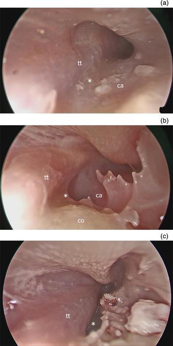
The overall protympanum conformation can be quadrangular or triangular in a two-dimensional plane perpendicular to the Eustachian tube long axis.Reference Djerić and Savić3 The demarcation between the two is based on whether the inferior wall is more or less than half of the equivalent superior wall length respectively. The lateral wall of the protympanum – from the annulus of the anterior tympanic membrane to the junctional portion of the Eustachian tube – can either be convex or concave in orientation. The protiniculum (from the Latin protinus, meaning ‘farther on’) follows from the previously described promontorial ridges of the middle ear: the ponticulus, subiculum and finiculus.Reference Marchioni, Alicandri-Ciufelli, Piccinini, Genovese and Presutti4 It has three conformations: A = ridge, B = bridge and C = absent (Figure 1).Reference Jufas, Marchioni, Tarabichi and Patel1
Examples of the three conformations of the protiniculum (asterisks): (a) type A – ridge, (b) type B – bridge and (c) type C – absent (all images are right ears). Tt = tensor tympani muscle; ca = carotid artery prominence; sb = subtensor recess; co = cochlea

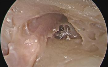
The protympanum can also variably contain several air cells, defined as discrete blind ending spaces within the protympanum. They are predominantly located posteroinferomedially, implying that they may pneumatise into the protympanum from hypotympanic air cells. Bony spicules are occasionally present in the protympanum, particularly on the carotid prominence. Occasionally, the bony spicules may coalesce into a linear formation termed the protympanic spine. They may be related to bony fusion over the carotid canal in embryological development (Figure 2).Reference Tóth, Medvegy, Moser and Patonay5
Specimen (right ear) displaying air cells, a bony spicule and a protympanic spine in the protympanum. ps = protympanic spine; pt = protiniculum; ac = air cells; ca = carotid artery prominence; bs = bony spicule; tt = tensor tympani muscle; co = cochlea

Finally, the subtensor recess is an area of variable pneumatisation inferomedial to the tensor tympani canal. The subtensor recess has three conformations: A = absent, B = shallow and C = deep.Reference Jufas, Marchioni, Tarabichi and Patel1 This is in keeping with retrotympanic classifications previously established.Reference Marchioni, Mattioli, Alicandri-Ciufelli and Presutti6 The presence of a flat tensor tympani canal necessarily means there is an absent subtensor recess. In types B and C, the tensor canal is raised, with the demarcation between the two based on whether the fundus of the subtensor recess extends superior to the midpoint of the tensor tympani canal (Figure 3).
Examples of the three conformations of subtensor recess (asterisks): (a) type A – absent, (b) type B – shallow and (c) type C – deep (all images are right ears). tt = tensor tympani muscle; ca = carotid artery prominence; co = cochlea

The primary aim of this study was to describe the anatomy of the protympanum in more detail, by ascertaining the prevalence of protympanum, protiniculum and subtensor recess conformations. The secondary aims of the study were to investigate other features, such as lateral wall conformation, and the presence of air cells, bony spicules, a protympanic spine and carotid dehiscence.
Materials and methods
A retrospective review of video recordings of endoscopic dissection and surgical procedures, carried out between October 2014 and January 2016, was conducted across four tertiary university referral centres.
All dissections were performed using either a 0-, 30- and/or 45-degree endoscope, depending on the visualisation achieved. These were 3-mm diameter, 14-cm length endoscopes, with a 3-chip full high-definition camera and full high-definition monitor (Karl Storz, Tuttlingen, Germany).
In all cases, a tympanomeatal flap was created. If necessary in cadaveric specimens, the tympanic membrane could be detached from the malleus handle to facilitate easy visualisation. This was not planned to be performed for visualisation in any of the live patients, unless required for the procedure itself.
A minimum of two authors reviewed each of the case recordings and independently classified the morphological anatomy. Any discrepancy in grading was settled by discussion amongst the authors, or, if needed, settled by a third author.
In each specimen, the following morphological anatomy was classified and tabulated. This was based on the descriptive text, diagrams and definitions in Jufas et al. (2016): protympanic conformation – quadrangular or triangular; protiniculum – ridge (type A), bridge (type B) or absent (type C); subtensor recess – ridge (type A), bridge (type B) or absent (type C); lateral wall conformation – concave or convex; air cells – present or absent; bony spicules – present or absent; and protympanic spine – present or absent.Reference Jufas, Marchioni, Tarabichi and Patel1
Both this analysis and the descriptive analysis of the data were performed with an Excel spreadsheet (Microsoft, Redmond, Washington, USA).
Results
In total, there were 97 ears used for analysis; 73 (75.3 per cent) were on human cadavers and 24 (24.7 per cent) were on live humans. Of the total number, 57 (58.8 per cent) were right sided and 40 (41.2 per cent) were left sided. None of the specimens selected needed to be excluded because of visualisation failure. None of the specimens or patients had previously been operated on or had a history of head trauma or skull base fractures. Age and gender was not recorded, as the videos had been de-identified at the point of retrospective review.
Protympanum conformation
With respect to the conformation of the protympanum, there were a total of 58 (59.8 per cent) with a quadrangular conformation and 39 (40.2 per cent) with a triangular conformation.
Protiniculum
In assessment of the protiniculum, 56 (57.7 per cent) were classified as type A (ridge), 22 (22.7 per cent) were classified as type B (bridge) and 19 (19.6 per cent) were classified as type C (absent).
Subtensor recess
Regarding the prevalence of subtensor recess conformation amongst the study group, 29 (29.9 per cent) had type A (absent), 47 (48.5 per cent) had type B (shallow) and 21 (21.6 per cent) had type C (deep).
Other protympanic features
The conformation of the lateral wall was found to be convex in 41 specimens (42.3 per cent), whilst 56 specimens (57.7 per cent) had a concave conformation.
Air cells were present in 18 specimens (18.6 per cent), bony spicules were present in 69 specimens (71.1 per cent), and 50 specimens (51.5 per cent) also had a protympanic spine.
No carotid dehiscence was noted in any specimen.
Discussion
The protympanum and lateral wall conformation appear to be predominantly quadrangular and concave respectively. There was an association noted between a triangular protympanum and convex lateral wall. This stands to reason as a prominent lateral wall that causes convexity is likely to encroach on the protympanic space and limit it inferiorly, thus meeting the definition of a triangular conformation. However, the two groups are not exclusively associated, with just over 30 per cent of specimens either having quadrangular and convex, or triangular and concave conformations.
• The protympanum is a middle-ear space anterior to the mesotympanum, also known as the bony portion of the Eustachian tube
• The protympanum has two conformations, quadrangular (seen in 60 per cent) and triangular (40 per cent)
• The protympanum is confluent with the hypotympanum inferiorly with the junction marked by the protiniculum
• The protiniculum can be type A (ridge, seen in 58 per cent), type B (bridge, 23 per cent) or type C (absent, 19 per cent)
• The subtensor recess is an area of variable pneumatisation inferomedial to the tensor tympani canal
• This area can be type A (absent, seen in 30 per cent), type B (shallow, 48 per cent) or type C (deep, 22 per cent)
Abou-Bieh et al. had described but not named a protympanic crest of variable size, direction and shape.Reference Abou-Bieh, Al-Abdulhadi, Al-Tubaikh and Haberkamp7 It has more recently been named the protiniculum in a separate study by some of the authors of this paper, and has been classified into three distinct types in keeping with other described promontorial ridges, such as the ponticulus, subiculum and finiculus.Reference Jufas, Marchioni, Tarabichi and Patel1 In the current paper, a similar proportion of distribution to the ponticulus was found. A total of 57.7 per cent of specimens were found to be type A (ridge), 22.7 per cent were type B (bridge) and 19.6 per cent were type C (absent). The ponticulus has been found to have proportions of 66 per cent ridge, 20 per cent bridge and 14 per cent absent.Reference Abou-Bieh, Al-Abdulhadi, Al-Tubaikh and Haberkamp8 In contrast, the subiculum has proportions of 87 per cent ridge, 4 per cent bridge and 9 per cent absent, and the finiculus has 81 per cent ridge, 3 per cent bridge and 16 per cent absent.Reference Marchioni, Alicandri-Ciufelli, Piccinini, Genovese and Presutti4, Reference Marchioni, Soloperto, Colleselli, Tatti, Patel and Jufas9
The subtensor recess has also been recently described. It is an area of importance given its variability and proximity to the carotid artery inferiorly and tensor tympani muscle superiorly. In approximately 20 per cent of specimens, the fundus of the subtensor recess could not be visualised because of the underlying anatomy. This conformation of the subtensor recess with a deep recess (type C) could harbour disease and cause difficulty in complete extirpation of protympanic cholesteatoma.
The carotid artery lies medial to the protympanum. Analysis of temporal bones established that the thin bone which separates it from the protympanum has a mean thickness of 1.5 mm.Reference Savić and Djerić10 A larger series of almost 1000 temporal bones reported the prevalence of medial wall dehiscence of up to 7.7 per cent.Reference Moreano, Paparella, Zelterman and Goycoolea11 Our series of 97 reported no carotid canal dehiscences in the protympanum. Whilst this result is consistent with what would be expected, it may also be an underestimation given the difficulty in assessing dehiscence in a cadaver. Alternatively, the previous estimations of prevalence may be an overestimation due to a processing artefact in formalin fixed sectioning.
Improved endoscopic visualisation has allowed the protympanum to be better defined, categorised and analysed. A detailed understanding of the anatomy of this region is a key aspect of a number of surgical procedures. For example, this could allow existing approaches, such as packing of the protympanum to avoid a cerebrospinal fluid leak following translabyrinthine cerebellopontine angle access, to be improved, given that the most common route through which this occurs is transnasally via the protympanum. This technique is performed endoscopically via a transmastoid-facial recess approach.Reference Deep and Weisskopf12 Additionally, it may allow the safe undertaking of novel approaches to procedures, such as transtympanic balloon dilatation of the Eustachian tube,Reference Jufas, Treble, Newey and Patel13, Reference Tarabichi and Najmi14 or insertion of a shim into the Eustachian tube to improve patulous Eustachian tube symptoms.Reference Oh, Lee, Goh and Kong15
This anatomical study significantly expands on knowledge of pertinent anatomical relationships in the protympanum. Additional study of the clinical relevance of the anatomical variations of the structures contained within it is an important future direction.
Competing interests
None declared.