Introduction
During field work in 2004 on Crete, the first author discovered a bizarre lichen with lirelloid apothecia growing on the bark of Pinus brutia, and subsequently showed the specimen to several colleagues. The lichen initially defied any attempt at identification, but eventually it became clear that it had affinities to the genus Elixia. In 2008 we obtained DNA from the sample and were able to test this hypothesis and confirm that the lichen is, indeed, an Elixia, only the second species known in the genus and the third species in the family Elixiaceae (after the Tasmanian Meridianelia maccarthyana; see Kantvilas & Lumbsch Reference Kantvilas and Lumbsch2009). We delayed describing the taxon as new until now, in the hopes of acquiring new or more substantial material, since all of our knowledge of this lichen is based on a single collection. However, in our view three considerations have come to outweigh our hesitation to publish: 1) the species is clear-cut morphologically and there is no overlap with related taxa, 2) DNA evidence confirms our hypothesis and 3) one may wait a long time to obtain more material of rare taxa, and in the meantime conservationists and lichenologists could make use of the data to search for more or ensure protection of known habitats. We therefore describe the taxon here as new and hope that doing so encourages lichenologists to search for and find more.
Material and Methods
Lichen specimens were investigated using material mounted in water or Lugol's iodine (I) and examined using a Zeiss Axioskop microscope fitted with an AxioCam MRc5 digital camera. Ascospore measurements are given as smallest measurement–(mean)–largest measurement.
Selected additional material studied. Elixia flexella (Ach.) Lumbsch. Austria: Kärnten: Nationalpark Hohe Tauern, 1988, Hafellner 28652 (GZU); Salzburg, Hüttwinkl Tal S von Rauris, 1978, Hafellner 2851 (GZU).—Italy: Veneto: Passo Tre Croci, 1985, Mayrhofer 8304 (GZU).—Romania: Válye Valeriaszka, Retezat, 08 viii 1872, Lojka (GZU).—USA: New Hampshire: Jefferson Notch, next to parking area, 2009, T. Spribille 31212 & V. Wagner (GZU, NY).
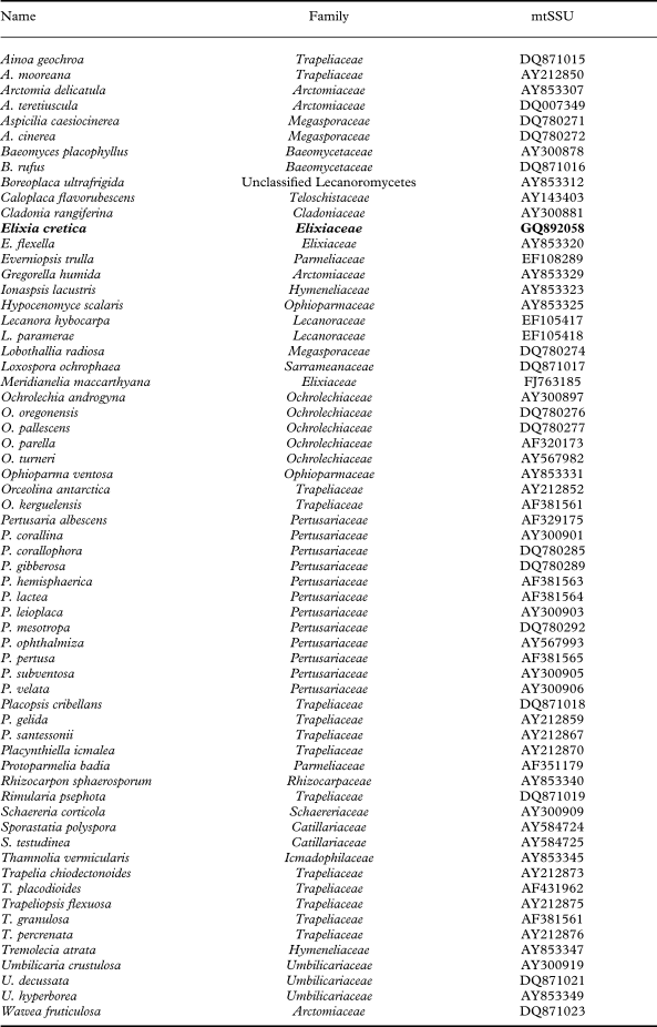
Species and GenBank sequences included in this study. Family placement follows Lumbsch & Huhndorf (Reference Lumbsch and Huhndorf2009).

Molecular Methods
Sequence data of 64 species were assembled using sequences of the mitochondrial small subunit rDNA (Table 1) with a newly obtained sequence extracted from the holotype of Elixia cretica [GenBank no: GQ892058] using apothecial material. The taxon sampling includes different clades of Lecanoromycetes and taxa that were previously believed to be close to Elixia or recently shown to be close to this genus (Lumbsch Reference Lumbsch1997; Wedin et al. Reference Wedin, Wiklund, Crewe, Döring, Ekman, Nyberg, Schmitt and Lumbsch2005; Miądlikowska et al. Reference Miądlikowska, Kauff, Hofstetter, Fraker, Grube, Hafellner, Reeb, Hodkinson, Kukwa and Lücking2006). Attempts to obtain DNA sequences of the new species for additional genes, such as ITS and nu LSU rDNA, were unsuccessful. Total DNA was extracted using the DNeasy Plant Mini Kit (Qiagen) following the instructions of the manufacturer. Dilutions (10−1 up to 10−2) of DNA were used for PCR amplifications. Primers for amplification were: mr SSU1 (Zoller et al. Reference Zoller, Scheidegger and Sperisen1999) and MSU 7 (Zhou & Stanosz Reference Zhou and Stanosz2001). The 25 μl PCR reactions contained 2·5 μl buffer, 2·5 μl dNTP mix, 1 μl of each primer (10 μM), 5 μl BSA, 2 μl Taq (0·5 U/μl), 2 μl genomic DNA extract and 9 μl distilled water. Thermal cycling parameters were: initial denaturation for 3 min at 95 °C, followed by 30 cycles of 1 min at 95 °C, 1 min at 52 °C, 1 min at 73 °C, and a final elongation for 7 min at 73 °C. Amplification products were viewed on 1% agarose gels stained with ethidium bromide and subsequently purified using the QIAquick PCR Purification Kit (Qiagen). Fragments were sequenced using the Big Dye Terminator reaction kit (ABI PRISM, Applied Biosystems). Sequencing and PCR amplifications were performed using the same sets of primers. Cycle sequencing was executed with the following program: 25 cycles of 95 °C for 30 sec, 48 °C for 15 sec, 60 °C for 4 min. Sequenced products were precipitated with 10 μl of sterile dH2O, 2 μl of 3 M NaOAc, and 50 μl of 95% EtOH before they were loaded on an ABI 3100 (Applied Biosystems) automatic sequencer. Sequence fragments obtained were assembled with SeqMan 4.03 (DNASTAR) and manually adjusted.
Sequence Alignments and Phylogenetic Analysis
Alignments were done using Clustal W (Thompson et al. Reference Thompson, Higgins and Gibson1994). Ambiguously aligned regions were removed manually. The alignments were analysed by maximum likelihood (ML) and a Bayesian approach (B/MCMC). Maximum likelihood analyses were performed using the program GARLI (Zwickl Reference Zwickl2006) employing the general time reversible model of nucleotide substitution (Rodriguez et al. Reference Rodriguez, Oliver, Martín and Medina1990) including estimation of invariant sites, assuming a discrete gamma distribution with six rate categories. Bootstrapping (Felsenstein Reference Felsenstein1985) was performed based on 2000 replicates. The B/MCMC analyses were conducted using the MrBayes 3.1.1 program (Huelsenbeck & Ronquist Reference Huelsenbeck and Ronquist2001). The analyses were performed assuming the general time reversible model of nucleotide substitution (Rodriguez et al. Reference Rodriguez, Oliver, Martín and Medina1990) including estimation of invariant sites, assuming a discrete gamma distribution with six rate categories. A run with 8 000 000 generations starting with a random tree and employing 12 simultaneous chains was executed. Every 100th tree was saved into a file. The first 400 000 generations (i.e. the first 4000 trees) were deleted as the “burn in” of the chain. We plotted the log-likelihood scores of sample points against generation time using TRACER 1.0 (http://evolve.zoo.ox.ac.uk/software.html?id=tracer) to check whether stationarity was achieved after the first 400 000 generations by checking whether the log-likelihood values of the sample points reached a stable equilibrium value (Huelsenbeck & Ronquist Reference Huelsenbeck and Ronquist2001). Additionally, we used AWTY (Nylander et al. Reference Nylander, Wilgenbusch, Warren and Swofford2007) to compare splits frequencies in the different runs and to plot cumulative split frequencies to ensure that stationarity was reached. Of the remaining 152 000 trees (76 000 from each of the parallel runs) a majority rule consensus tree with average branch lengths was calculated using the sumt option of MrBayes. Posterior probabilities were obtained for each clade. Only clades that received bootstrap support equal or above 70% under ML and posterior probabilities ≥ 0·95 were considered as strongly supported. Phylogenetic trees were visualized using the program Treeview (Page Reference Page1996).
Results and Discussion
Phylogenetic analyses
The new sequence of Elixia cretica was aligned with sequences obtained from GenBank as listed in Table 1. A matrix of 715 unambiguously aligned nucleotide position characters was produced; 315 characters in the alignment were constant. ML analysis yielded a maximum likelihood tree that did not contradict the Bayesian tree topology. In the B/MCMC analysis of the combined data set, the likelihood parameters in the sample had the following mean (Variance): LnL = −9238·206 (0·19), base frequencies π(A) = 0·332 (0·00023), π(C) =0·146 (0·00021), π(G) = 0·206 (0·00024), π(T) = 0·316 (0·00026), rate matrix r(AC) = 0·113 (0·009), r(AG) = 0·28 (0·0008), r(AT) = 0·132 (0·0004), r(CG) =0·005 (0·0001), r(CT) = 0·354 (0·0004), r(GT) = 0·007 (0·0001), the gamma shape parameter alpha = 0·773 (0·0076), and p(invar) = 0·32 (0·00151).
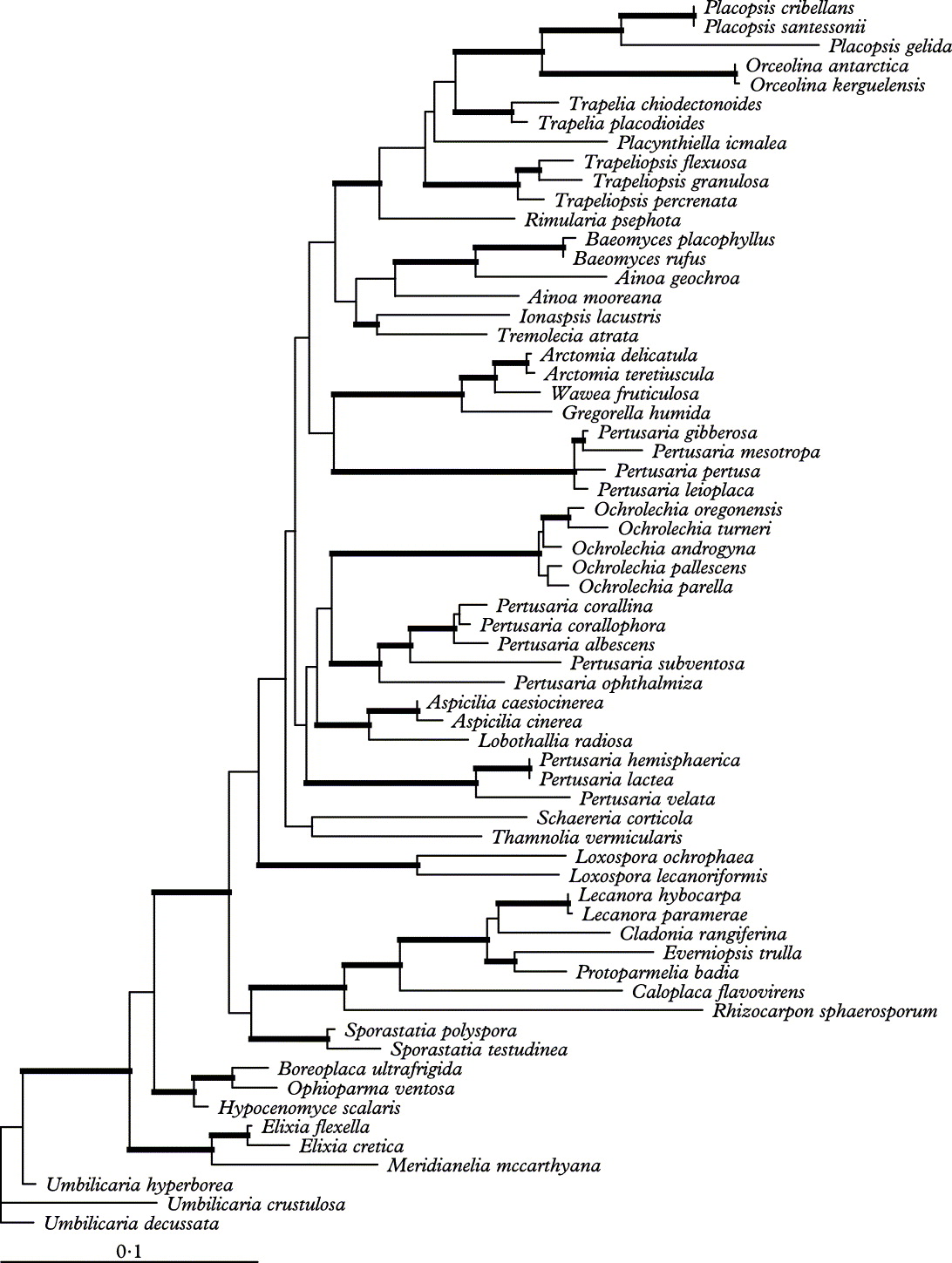
Since the topologies of the ML and B/MCMC analyses did not show any strongly supported conflicts, only the tree of the ML analysis is shown (Fig. 1). Elixia cretica clusters strongly supported with E. flexella (ML-bootstrap support 99%, B/MCMC posterior probability 1·0); the two Elixia species form a strongly supported sister-group with Meridianelia maccarthyana and this thus supports placement of the new taxon in Elixiaceae. The supported nodes in the topology of the other parts of the phylogenetic tree are in agreement with previously published phylogenies (Lumbsch et al. Reference Lumbsch, Schmitt, Lücking, Wiklund and Wedin2007a, Reference Lumbsch, Archer and Elixb) and are consequently not discussed further here.
Phylogeny of Lecanoromycetes as inferred from a mtSSU rDNA analysis to investigate the phylogenetic placement of Elixia cretica. Maximum likelihood tree obtained using the program GARLI. Branches with posterior probabilities equal to or above 0·95 and ML bootstrap support values above 70% are indicated by wide internodes.

Taxonomy
Elixia cretica T. Sprib. & Lumbsch sp. nov
Mycobank No. MB 515175
Elixiae flexellae similis sed thallo bene evoluto, apotheciis magnis, ascosporis magnis et hymeniis non-euamyloideis differt.
Typus: Greece, Kriti, Chania Prefecture, Levka Ori, just W of Agii Theodori, c. 3 km SW of Omalos, between mountains Mavri Kimite and Tourli, 35°19.5′N, 23°52.2′E, on bark of Pinus brutia near base of tree, 1125 m, 10 June 2004, T. Spribille 13340 (GZU—holotypus; B—isotypus).
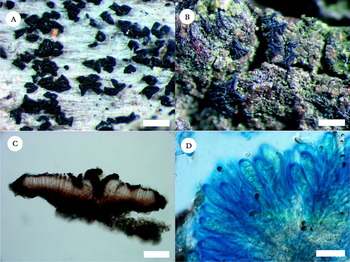
(Fig. 2B–D)
Elixia species. A, E. flexella, habit (Austria, Hafellner 28652, GZU). B–D, E. cretica (holotype); B, habit; C, section of apothecium in water; D, asci and paraphyses in Lugol's after pretreatment with KOH. Scale: A & B = 1 mm; C = 100 μm; D =10 μm.

Thallus crustose, areolate to cracked areolate or ±confluent-rimose in places, thin, not >50 μm thick; areoles 0·4–0·5(–0·6) mm, brownish to greenish in sheltered bark cracks, matt; soredia and isidia absent. Photobiont chlorococcoid, cells to 10–11 μm.
Ascomata lirelloid apothecia, young apothecia relatively simple lirellae, elongate with two to three ends, disc initially concealed, developed later into larger apothecia with exposed discs and up to seven points on star-shaped ascomatum; apothecia at their widest ranging from 0·5–1·4 mm diam.; margin jet black, matt, protruding above disc, often appearing inrolled, creases developed at apothecial corners; disc dark brown but lighter than the margin, matt. Excipulum in section 15–20 μm wide, darkly pigmented to near carbonaceous, of paraplectenchymatous cells; hypothecium occupying a narrow zone between excipulum and hymenium, of light brown or reddish brown paraplectenchymatous cells, to 10 μm thick; hymenium basically colourless, 55–60 μm deep, of conglutinated paraphyses not easily separable in water, deep rusty red in I but blue after pretreatment with KOH; paraphyses septate, unbranched, 1·5–2·0 μm wide, with strongly expanded tips to 3·0–5·0 μm, the tips with dark brown pigment inclusions that combine with diffuse brownish pigment to form an epihymenium; asci cylindrical to clavate, 34–40 × 10–13 μm, I+ blue after KOH, with light amyloid tissue throughout but darker staining regions located around the apex and in a narrow zone below the inner upper wall of the tholus (‘Elixia type’ sensu Lumbsch Reference Lumbsch1997, see Fig. 2D); ascospores 8 per ascus, ellipsoid, simple, colourless or faintly light brown in water, 7·0–(8·2)–10·0 × 3·2– (3·7)–4·5 μm.
Pycnidia not seen.
Chemistry. No substances detected by thin layer chromatography; all thallus spot tests negative.
Distribution. Currently known only from the type locality on the island of Crete (Greece).
Comments. Elixia cretica differs from the only other species known in the genus, E. flexella (Fig. 2A), in possessing a well-developed surficial thallus (Fig. 2B), which somewhat recalls that of some Hypocenomyce species. Anatomical details of the apothecia of E. flexella have been described by Lumbsch (Reference Lumbsch1997) and Coppins (Reference Coppins, Purvis, Coppins, Hawksworth, James and Moore1992, as Ptychographa) and likewise differ on several counts from our new species. Apothecia tend to be substantially larger in E. cretica than in E. flexella (0·5–1·4 mm versus 0·2–0·5 mm; no apothecia > 0·5 mm have been seen in E. flexella) and develop a free, expanded disc, whereas in many specimens of E. flexella the disc remains concealed and larger apothecia instead develop a substantial umbo or become strongly gyrose. Interestingly, the hymenium in E. cretica reacts I+ rusty red (hemiamyloid) if not pre-treated with K, whereas in E. flexella it reacts blue (euamyloid). The ascospores of E. cretica are also longer than those in E. flexella (7–10 μm in cretica versus 5–8 μm in flexella) and paraphysis tips more expanded (to 5 μm in E. cretica versus usually < 3 μm in E. flexella).
The two species of Elixia are also ecologically and geographically distinct. Elixia flexella is a temperate-boreal species; it is known from scattered records in Europe, Asia and North America (Spribille & Björk Reference Spribille and Björk2008), with the first record for eastern North America reported here (see ‘selected additional material studied’ above). It appears that all but one of these (a record from Idaho, USA.) are from wood, and many are associated with calicioid lichens such as Calicium glaucellum or C. trabinellum. The only collection of E. cretica to date was from bark of Pinus brutia, where it was closely associated with Caloplaca herbidella.
This study was supported financially by a start up fund of the Field Museum to HTL and a Field Museum scholarship to TS. The newly obtained DNA sequence was generated in the Pritzker Laboratory for Molecular Systematics and Evolution at the Field Museum. Fabian Ernemann (Chicago) assisted with laboratory work.