I. INTRODUCTION
Sol–gel-derived CaO–SiO2 bioactive glasses (BG) have generated significant interest as biomaterials for hard tissue replacement and regeneration, as they encourage the precipitation of an apatite-like surface layer when soaked in simulated body fluid (SBF),Reference Li, Clark and Hench1 promote proliferation in vitro of bone-forming cellsReference Jones, Tsigkou, Coates, Stevens, Polak and Hench2, Reference Marques, Almeida, Thiema, Wang, Falk and Jain3 and stimulate in vivo tissue growth.Reference Wang, Falk, Rashad, Saad, Marques, Almeida, Marei and Jain4 In addition, having intrinsic nanoporosity and high specific surface area, they show significant biodegradability and, therefore could be promising materials for bioscaffold in tissue engineering applications.Reference Sepulveda, Jones and Hench5, Reference Zhang, Jain, Hupa and Hupa6 On the other hand, interconnected macropores (at least 100 μm) are required for bioscaffold to allow new tissue ingrowth and blood vessel formation.Reference Karageorgiou and Kaplan7 The newly proposed nano/macro dual porous structure has been achieved by various fabrication methods for bioinert as well as bioactive glasses. For example, nano/macroporous (with dimensions ∼2–10 nm and ∼0.1–40 μm, respectively) samples can be fabricated by polymerization-induced phase separation in various silica sol–gel systems using different polymers, such as poly(ethylene oxide) (PEO), poly(ethylene glycol) (PEG), or poly(acrylic acid) (PAA)Reference Nakanishi8, Reference Yachi, Takahashi, Sato, Sodesawa, Oguma, Matsutani and Mikami9; foaming sol–gel systems where a surfactant (e.g., Teepol) is added to a sol prepared from a mixture of alkoxides, deionized water and HNO3Reference Sepulveda, Jones and Hench5, Reference Jones, Ehrenfried and Hench10; Chmelka’s method—the process, where macroporous templates such as a polystyrene foam or an oil-in-water emulsion are usedReference Maekawa, Esquena, Bishop, Solans and Chmelka11; or salt sintering process, where a glass prepared by melt quenching is crushed and mixed with sodium chloride, followed by heat treatment and final immersion into water to promote dissolution of the salt.Reference Liang and Russel12, Reference Moawad and Jain13 More recently, hierarchically three-dimensional (3D) nano/macroporous BGs were prepared using a combination of sol–gel and multipolymer templating processes,Reference Yun, Kim and Hyeon14 as well as phase separation due to spinodal decomposition.Reference Marques, Jain and Almeida15
So far, the influence of nanopores on the performance of sol–gel-derived BG has been hardly explored. To shed light on the role of pores in BG, one should be able to characterize them quantitatively (at least in number and dimensions) at different nano/macro scales first. Usually, mercury or nitrogen adsorption porosimetry methods are used for this purpose, which are limited to the open porosity of >5 and >2 nm radii, respectively.Reference Webb and Orr16–Reference Barrett, Joyner and Halenda18 However, most of the physical properties of glass, especially mechanical strength, depend not only on the amount and character of large open pores, but also on the amount and character of closed pores and/or those of smaller sizes, comparable to a few interatomic distances (vacancies, vacancy-like clusters, defects, etc.).Reference Rice19 Thus, e.g., closed porosity may help match the elastic constants of the implant to that of the bone.Reference Ondracek20
One of the best techniques capable of probing both open and closed pores at the nanoscale level is the positron annihilation lifetime (PAL) spectroscopy.Reference Krause-Rehberg and Leipner21–Reference Nakanishi, Jean, Schrader and Jean24 Its efficiency is proven for characterizing the local free volumes (closed or open-volume holes, inner pores, vacancies, and vacancy-like agglomerations) in crystals, liquids, polymers, and glasses on a subnanometer scale.Reference Krause-Rehberg and Leipner21–Reference Shpotyuk and Filipecki26 In the present article, we have shown a useful application of PAL spectroscopy to study open and closed pores in sol–gel-derived bioactive CaO–SiO2 glasses at the nanoscale (≤5 nm). The PAL results are compared with the data obtained with nitrogen adsorption porosimetry.
II. EXPERIMENTAL
Silica-based BG scaffold (disks of ∼1.5 cm in diameter) with 70 mol% SiO2–30 mol% CaO (70S30C) composition was synthesized using sol–gel-cum-spinodal decomposition technique, where interconnected macroporosity is achieved by inducing spinodal decomposition of a gelling sol, as described elsewhere.Reference Marques, Almeida, Thiema, Wang, Falk and Jain3, Reference Marques, Jain and Almeida15, Reference Vueva, Gama, Teixeira, Almeida, Wang, Falk and Jain27 First, a water soluble polymer (PEO, with an average molecular weight, M w ∼100,000) was dissolved in 0.05 N acetic acid (CH3COOH) aqueous solution, to which 9 mL of tetramethoxysilane (TMOS) were added. Calcium nitrate tetrahydrate, Ca(NO3)2•4H2O, was then added and gelation was allowed to occur at room temperature, after adding 2 mL of 2.5 vol% hydrofluoric acid (HF). The gel was aged at 40 °C for 4 days, and then dried and sintered sequentially at 700 °C.
Positron annihilation lifetime (PAL) spectra  $s(t) = \sum {(I_i /{\rm{\tau }}_i )\exp ( - t/{\rm{\tau }}_i )} $, approached by a sum of discrete exponentials with τi and Ii representing the average positron lifetime and intensity of i positron decay component, were recorded with the fast coincidence system (ORTEC) of 230 ps resolution [full width at half maximum (FWHM) of a single Gaussian, determined by measuring 60Co isotope] at the temperature, T = 22 °C and relative humidity, RH = 35%. To resolve short-lived components, each PAL spectrum was measured with a channel width of 6.15 ps (total number of channels 8000) and contained 3 × 10⁶ coincidences in total. To obtain information about long-lived components, the same samples were measured again with 61.5 ps channel width, total number of channels and coincidences being the same. Isotope 22Na (activity ∼50 kBq) was used as source of positrons (prepared from aqueous solution of 22NaCl, wrapped with Kapton® foil (DuPontTM, Circleville, OH) of 12 μm thickness and sealed), which was sandwiched between two identical samples.
All the PAL spectra of the investigated samples were decomposed into five discrete exponentials (i = 5) using standard LT 9.0 program,Reference Kansy28 where additional peaks into the envelope of fitted curve were added only if they significantly improved goodness of the fit. The uncertainties in the determination of lifetimes (τi) and corresponding intensities (Ii) are ±0.005–0.5 ns (increasing with increasing τi) and ±0.5%, respectively. Initial PAL measurements were performed on the samples dried at 120 °C for 4 h in vacuum. To reveal interconnectivity of pore structure, the same samples were soaked in water vapor (RH = 100%) during 8 h at 22 °C and then measured again. The same samples were then dried by heating in a vacuum for 4 h at 120 °C and measured once more to check the reversibility of physisorption of the water molecules at this particular temperature.
The volume fraction of porosity, as well as the size of interconnected pores in the prepared scaffolds, were measured by nitrogen adsorption (ASAP 2020, Micromeritics, Norcross, GA) Brunauer–Emmett–Teller (BET)Reference Brunauer, Emmett and Teller17 and Barrett–Joyner–Halenda (BJH) methods,Reference Barrett, Joyner and Halenda18 using adsorption and desorption curves, respectively. For these experiments, accurately weighed specimens (∼200 mg for each test) were evacuated and heated at 150 °C for at least 5 h to remove moisture and potential surface contaminants before analysis.
III. RESULTS AND DISCUSSION
The main potential of PAL spectroscopy method lies in its ability to characterize the local free volumes (including both open and closed pores) in materials on a subnanometer scale.Reference Krause-Rehberg and Leipner21–Reference Dlubek, Sen Gupta, Pionteck, Hassler, Krause-Rehberg, Kaspar and Lochhaas25 The PAL method is particularly effective when positronium (Ps) is formed, because energetic and geometric characteristics of this electron–positron bound state (hydrogen-like atom) are well known, which allows relatively easy quantification of the free volume.Reference Krause-Rehberg and Leipner21–Reference Dlubek, Sen Gupta, Pionteck, Hassler, Krause-Rehberg, Kaspar and Lochhaas25 In disordered solids Ps is usually formed in two ground states—para-positronium (p-Ps) and ortho-positronium (o-Ps)—and localized in the pores of the free volume.Reference Krause-Rehberg and Leipner21–Reference Dlubek, Sen Gupta, Pionteck, Hassler, Krause-Rehberg, Kaspar and Lochhaas25 Usually, quantification is based on the analysis of o-Ps lifetime, which has a characteristic value of 142 ns (average lifetime inside an infinite hole in vacuum). It decreases significantly due to a pick-off process (i.e., annihilation of positron with a strange electron picked from the surrounding medium and having antiparallel spin orientation), occurring as a result of collisions with the walls of pores.Reference Krause-Rehberg and Leipner21–Reference Dlubek, Sen Gupta, Pionteck, Hassler, Krause-Rehberg, Kaspar and Lochhaas25 This decrease, which depends on the size (and shape) of holes, gives important information on the void structure of the materials.Reference Krause-Rehberg and Leipner21–Reference Dlubek, Sen Gupta, Pionteck, Hassler, Krause-Rehberg, Kaspar and Lochhaas25, Reference Tao29, Reference Eldrup, Lightbody and Sherwood30 Thus, it is possible to estimate the average hole size from the o-Ps lifetime in a given material.Reference Tao29–Reference Djourelov, Suzuki, Shantarovich and Kondo32 Assuming approximately spherical shape of the free volume, the o-Ps lifetime (τo) can be related to the average radius of holes (R) by a semiempirical Tao–Eldrup equationReference Tao29, Reference Eldrup, Lightbody and Sherwood30:
 $${\rm{\tau }}_o =\! \left[ {2\left( {1\! - {R \over {R + \Delta R}}}}+ {1 \over {2{\rm{\pi }}}}\sin \left( {{{2{\rm{\pi }}R} \over {R + \Delta R}}} \right)} \!\right)}\!\! + 0.007} \right]^{ - 1} ,$$
where ΔR is the empirically determined parameter (in the classical case ΔR ≈ 0.1656 nm), describing effective thickness of the electron layer responsible for the pick-off annihilation of o-Ps in the hole.Reference Tao29, Reference Eldrup, Lightbody and Sherwood30
Although the pores in silicate gel systems, like the BG scaffold in this study, are not necessarily spherical, we idealize the pores as spherical voids with R as “effective” or “equivalent” pore radius, since such practice is common for analyzing similar irregularly shaped free volumes in other materials (e.g., polymers, zeolites, etc.) and has been demonstrated to be quite useful.Reference Dlubek, Sen Gupta, Pionteck, Hassler, Krause-Rehberg, Kaspar and Lochhaas25, Reference Goworek, Ciesieslski, Jasinska and Wawryszczuk31–Reference Misheva, Djourelov, Margaca and Miranda Salvado33
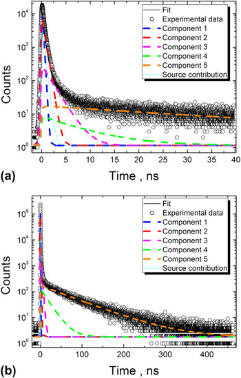
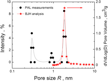
Typical PAL spectrum obtained for a sol–gel-derived 70S30C BG and its best fit are shown in Fig. 1. Five discrete exponentially decaying components (fit parameters are given in Table I) can be distinguished in this spectrum, using iterative curve fitting procedure of the LT program.Reference Kansy28, Reference Klym, Ingram, Shpotyuk, Filipecki and Hadzaman34 The first component (τ1, I 1) includes free annihilation, p-Ps decay and is related also to the positrons’ bulk lifetime (τb) in the sample. The second component (τ2, I 2) usually is caused by positrons that are trapped before annihilation in the free volume devoid of significant electron density in the glass structure, where formation of Ps is impossible for any other reason (geometric, energetic, inhibition of Ps formation, etc.).Reference Krause-Rehberg and Leipner21, Reference Shpotyuk and Filipecki26 Typical examples of such effective positron traps can be found in crystals and some amorphous materials where Ps is not formed, e.g., grain boundaries in some ceramics and glass–ceramics, shallow positron traps in chalcogenide glasses, precipitates and inclusions, etc.Reference Krause-Rehberg and Leipner21, Reference Shpotyuk and Filipecki26 This component may also convolute with greater bulk lifetime component of another phase, if it is present in the material. All the remaining three components (τ3, I 3), (τ4, I 4), and (τ5, I 5) can be associated with o-Ps formation (Table I). Contrary to the one or two o-Ps PAL components observed in silica–gel systems,Reference Goworek, Ciesieslski, Jasinska and Wawryszczuk31–Reference Misheva, Djourelov, Margaca and Miranda Salvado33 we have observed three distinct o-Ps components in the PAL spectra of the present sol–gel-derived 70S30C BG. Each of these lifetimes can be related to corresponding pores via Tao–Eldrup relation [Eq. (1)]. However, Eq. (1) describes quite accurately the relation between o-Ps lifetime and pore radius only when Ps is in the ground (often denoted as “1s”) state, which is usually satisfied at low temperatures and for relatively small pores. At higher temperatures, in very porous materials like silica gels the o-Ps in large pores can exist not only in the ground state, but also in the excited states.Reference Goworek, Ciesieslski, Jasinska and Wawryszczuk31 The probability to find o-Ps in the excited state increases with increasing temperature (lifetime  $\tau }}_o \sim1/{\rm{\sqrt T $). Thus, in the large pores of silica gels besides the ground state at room temperatures the contribution from the first excited state of o-Ps is also expected, for which pick-off annihilation lifetime is estimated at the level of ∼40 ns.Reference Goworek, Ciesieslski, Jasinska and Wawryszczuk31 If present, it should result in a separate fit component in the experimental PAL spectrum under the condition that average dwelling time of o-Ps in this excited state is much longer than actual o-Ps lifetime.Reference Goworek, Ciesieslski, Jasinska and Wawryszczuk31 No such component is seen in the experimental PAL spectra of the investigated BG (Table I). So, we can conclude that in accordance with Goworek et al.Reference Goworek, Ciesieslski, Jasinska and Wawryszczuk31 the dwelling time of o-Ps in the excited state is much shorter than the o-Ps lifetime. Therefore, the observed o-Ps lifetime ∼70 ns is a value averaged over all engaged states, the distribution weight being closer to pick-off annihilation of o-Ps in the ground state (∼80 ns).Reference Goworek, Ciesieslski, Jasinska and Wawryszczuk31 Taking this into account, we still can use classical Tao–Eldrup Eq. (1) to estimate pore sizes in the investigated sol–gel-derived 70S30C BG scaffold within a good approximation. The τo value of around ∼70 ns (τ5 in Table I) is typical for silica gel systemsReference Goworek, Ciesieslski, Jasinska and Wawryszczuk31, Reference Djourelov, Suzuki, Shantarovich and Kondo32 and corresponds to nanopores with radius (R) distribution centered near ∼1.8 nm (Fig. 2). These results agree well with BET porosimetry measurements (Fig. 2). Most probably, these pores correspond to the empty volumes associated with mismatches in the packing of extended atomic groups (clusters, fractals, building blocks, etc.). The lifetime similar to τ3 ≈ 2.1 ns was also observed in various silica gelsReference Djourelov, Suzuki, Shantarovich and Kondo32, Reference Dutta, Chatterjee, Pujari and Ganguly35 and its origin was associated with fine pores of R ∼ 0.3 nm within the oxide building blocks (individual coordination polyhedra) of silica network.
Fitting of PAL spectra recorded for dry 70S30C BG with (a) 6.15 ps and (b) 61.5 ps channel width.

Pore size distribution according to PAL data (Tao–Eldrup equation) and nitrogen adsorption porosimetry results in dry sol–gel derived 70S30C BG. Each single value of R calculated according to Eq. (1) was artificially broadened at full width at half maximum by ΔR variation between 0.16 and 0.18 nm values (frequently used along with classic 0.1656 nm) and at zero by dispersion of corresponding τo values from LT program to visualize possible distribution of R values. The lines are drawn for better visualization.

Fitting parameters for PAL spectra of sol–gel derived 70S30C BG. Average lifetime τav is calculated as  ${\rm{\tau }}_{{\rm{av}}} = \sum\limits_i {I_i {\rm{\tau }}_i } $, where τi and Ii denote lifetime and intensity of the corresponding fitting components in units of ns and %, respectively. The experimental uncertainty in various I values is ±0.5%.

The observed o-Ps lifetime of τ4 ≈ 18 ns seems to be unique to the investigated bioglass. Following Goworek–Gidley approach of Ps formation in porous materials,Reference Goworek, Ciesieslski, Jasinska and Wawryszczuk31, Reference Gidley, Frieze, Dull, Yee, Ryen and Ho36 its origin can be associated with the existence of nanopores of R ∼ 0.9 nm. Because corresponding PAL component is not resolved in PAL spectra of pure silica-gel systems,Reference Goworek, Ciesieslski, Jasinska and Wawryszczuk31–Reference Misheva, Djourelov, Margaca and Miranda Salvado33, Reference Dutta, Chatterjee, Pujari and Ganguly35 it is likely to be caused by the presence of Ca2+ modifier cations in the network of the investigated bioglasses, which make the pore structure somewhat different from that of silica gels. While it is well-known that introduction of Ca atoms into a silica network produces nonbridging oxygen and increases the density of glass,Reference Varshneya37 the above lifetime component of o-Ps can be associated with pick-off annihilation in the nanopores created by neighboring coordination polyhedra and modified by the presence of Ca2+ cations and nonbridging oxygen atoms. Fraction of nanopores associated with each o-Ps lifetime can be estimated by the intensities of corresponding long-lived PAL components (I 3, I 4, I 5). However, contrary to the short-lived components, annihilating almost entirely via two-quantum annihilation, substantial part of o-Ps annihilates also via three-quantum process, which is a compete process to pick-off annihilation.Reference Krause-Rehberg and Leipner21–Reference Nakanishi, Jean, Schrader and Jean24, Reference Goworek, Ciesieslski, Jasinska and Wawryszczuk31 Different efficiency of the registration for two- and three-quantum processes can distort the proportion between observed o-Ps annihilation intensities (I 3, I 4, I 5), introducing uncertainty into the estimation of the number of pores.
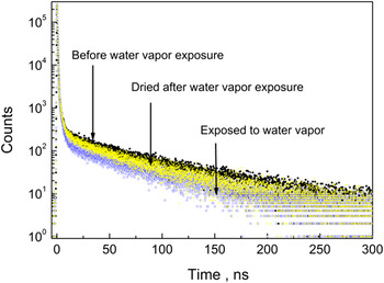
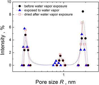
PAL spectra of sol–gel-derived 70S30C BG exposed to water vapor reveal well-expressed changes in the long-lived components associated with o-Ps formation. PAL spectra of the samples before and after exposure to water vapor, followed by drying in vacuum are shown in Fig. 3 (corresponding fitting parameters are given in Table I). A number of interesting features can be noticed in the samples exposed to water vapor. Three-modal pore distribution remains, but the number of large pores (∼0.9 and ∼1.8 nm), as estimated from the intensity of corresponding lifetime components in the fit of PAL spectra (I 4, I 5), is reduced drastically (Table I, Fig. 4). Respective τ4 and τ5 values decrease after water exposure, indicating also a decrease in the radius of the pores according to Eq. (1). Thus, the water molecules appear to enter these pores (some of them being fully occupied with water, others filled only partially), changing the o-Ps lifetime characteristics. Therefore, these pores should have access to the environment and should be interconnected at the nanoscale level. It is also noteworthy to think that part of them should exist as closed pores, since the intensities of corresponded lifetime components (I 4, I 5) do not drop to zero completely (Fig. 4). The presence of closed pores in the present samples has been observed at the micrometer level by scanning electron microscopy.Reference Marques, Almeida, Thiema, Wang, Falk and Jain3, Reference Marques, Jain, Kiely, Song, Kiely and Almeida38 It results from a secondary, nucleation-growth type phase separation during the fabrication of samples. In principle, this process must be beginning at the nm scale, but it was not possible to observe closed nanoporosity directly by electron microscopy. The PAL technique has provided the evidence of the early stages of phase separation that produces closed porosity.
Comparison of PAL spectra of dry (dark circle), exposed to water vapor (blue circle) and dried after the exposure to water vapor (yellow circle) 70S30C BG. Total number of counts 3 × 10⁶ is the same for each spectrum.

Pore size distribution derived from PAL data (Tao–Eldrup equation) in sol–gel derived 70S30C BG samples. Each single value of R calculated according to Eq. (1) was artificially broadened at full width at half maximum by ΔR variation between 0.16 and 0.18 nm values (frequently used along with classic 0.1656 nm) and at zero by dispersion of corresponded τo values from LT program to visualize possible distribution of R values. The lines are drawn for better visualization.

After drying in vacuum at 120 °C for 4 h of the samples previously exposed to the water vapor, the initial pore distribution tends to be restored (Table I, Fig. 3). However, it does not recover fully to the initial state (Fig. 4), suggesting that a fraction of the water molecules remains adsorbed. This result is consistent with other investigators’ observation that physisorbed water is not fully eliminated until 170 °C.Reference Hench and West39
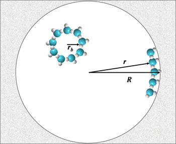
The most significant permanent changes produced by the water sorption process are observed for the largest pores of ∼1.8 nm radius (Fig. 4). The intensity of the corresponding PAL component is restored only ∼50% after the drying procedure, while τ5 remains almost unchanged (Table I). Regaining of I 4 and I 5 values after drying can be simply understood as the removal of water from the pores. At the same time, the behaviors of τ5 value and (τ3, I 3) component need additional consideration. The decreased τ5 value for the samples dried after the water vapor exposure (Table I) can be explained as the development of thin layers of water molecules covering the walls of big pores, which are not completely removed after annealing at 120 °C. Thus, the effective pore radius decreases from R to r value, which is a characteristic of the pores covered by a layer of water molecules (Fig. 5). It leads to a decrease in τ5 value according to Eq. (1). The presence of liquid water in some of the pores (even to some extent after the annealing procedure) is a reason for the increase in I 3 intensity and slight decrease in τ3 value (Table I). Note that the lifetime of o-Ps in water “bubbles” with radius rb ≈ 0.3 nm is around 1.8 ns,Reference Magalhaes, Abbe and Duplatre40 which could be easily convoluted with the observed (τ3, I 3) component.
Schematic diagram showing possible changes in pore radius after water vapor exposure: initial pore volume is decreased by physisorbed water molecules at the wall of the pore; in the case of pore fully filled with water, the o-Ps annihilates through a “bubble” (rb ≈ 0.3 nm) mechanism within liquid water.Reference Magalhaes, Abbe and Duplatre40

The present investigation of BG pore structure with PAL technique can be considered as complementary to gas adsorption porosimetry studies, having certain advantages and disadvantages at the same time. Thus, BET/BJH method, which is based on the adsorption-desorption of nitrogen molecules at critical pressure and at boiling temperature (77 K), gives slightly different values for pore sizes and distribution if calculated from adsorption or desorption branches of isotherm, as regards the hysteresis between them.Reference Brunauer, Emmett and Teller17, Reference Barrett, Joyner and Halenda18, Reference Dutta, Chatterjee, Pujari and Ganguly35 Besides, there is a critical lower value of relative pressure for nitrogen, which limits the detection of smaller nanopores. Actual pore size may not be correctly estimated at the lower limit of the nanopore region because of the changes in surface tension of capillary liquid with changing curvature of its meniscus. Finally, gas adsorption porosimetry is limited to open pores, which should have an access to the environment to be detected. On the other hand, PAL spectroscopy can probe both open and closed pores of sizes ranging from atomic scale to several tens of nanometers. The disadvantages are associated with different efficiency for registration of two and three-quantum annihilation events and applicability of various models relating o-Ps lifetime and pore size (like Tao–Eldrup equation). In spite of the above limitations of PAL spectroscopy and BET/BJH analysis, the dimensions of pores obtained in present studies from PAL and BET/BJH analysis are in excellent agreement with each other in the region where these techniques overlap.
IV. CONCLUSIONS
In conclusion, the usefulness of positron annihilation technique to study nanopore size distribution in sol–gel-derived 70S30C BG is demonstrated. Three-modal distribution of o-Ps lifetimes is derived from the fitting of the experimental PAL data. The observed o-Ps lifetimes ∼2, ∼18, and ∼70 ns are related to the pores with radius of ∼0.3, ∼0.9, and ∼1.8 nm, respectively, based on classic Tao–Eldrup equation. The latter data were confirmed by nitrogen adsorption porosimetry using BJH method. Three-modal pore distribution remains when the investigated glasses are exposed to water vapor. However, the number of large pores (∼0.9 and ∼1.8 nm), as estimated by the intensities of corresponded lifetime components in the fit of PAL spectra, is reduced substantially. The redrying in the vacuum at 120 °C of the samples previously exposed to water vapor does not restore the initial pore distribution fully, testifying to the sensitivity of PAL method to various amount of water molecules physisorbed in the pores. Finally, closed nanopores have been identified by PAL spectroscopy, indicating the early stages of phase separation by nucleation and growth in the present sol–gel system.
ACKNOWLEDGMENTS
The authors thank U.S. National Science Foundation through IMI-NFG (Grant No. DMR-0844014), for initiating the international collaboration and providing partial financial support for this work. SW and HJ thank the National Science Foundation for support via the Materials World Network (DMR-0602975) program.