Introduction
Echinococcus granulosus and its genotypic variants are the aetiologic agents of cystic hydatid or echinococcal disease, which occurs widely in the American continent from Alaska and northern Canada to Tierra del Fuego (Moro & Schantz, Reference Moro and Schantz2006) and is caused by the metacestode stage of the tapeworm. Metacestodes are fluid-filled cysts with cellular and acellular compartments. The outer, acellular, layer of the metacestode is formed by a laminated membrane which becomes surrounded by a very prominent, host-derived fibrous capsule or adventitial layer (Hemphill et al., Reference Hemphill, Spicher, Stadelmann, Mueller, Naguleswaran, Gottstein and Walker2007). Spontaneous cure of hydatidosis occurs sometimes; it starts with a process of calcification of the laminated membrane. Bisphosphonates are synthetic, non-hydrolysable analogues of pyrophosphates, which block bone degradation by a complex mechanism that involves calcium deposition and inhibition of calcium crystal dissolution; their affinities for bone are inversely related to pH. Bisphosphonate induces increases in cell permeability to ions such as NH4+ and H+ (Sato et al., Reference Sato, Grasser, Endo, Akins, Simmons, Thompson, Golub and Rodan1991). Modulation of cytosolic calcium levels in osteosarcoma cells has also been described with olpadronate (OPD) and ibandronate (IB) (Vazquez et al., Reference Vazquez, Santillan, Boland, Roldán and Pérez-Lloret2003; Drake et al., Reference Drake, Clarke and Khosla2008). A group of bisphosphonates has been shown to be active both in vitro and in vivo against protists and they have been proposed as potential antiparasitic agents. Hence, alternative mechanisms of action have been postulated – for instance, the preferential accumulation of bisphosphonates in certain parasites due to the presence of the acidocalcisome, a calcium and pyrophosphate rich organelle found in trypanosomes, and their interference with phosphate metabolism (Ortiz-Gómez et al., Reference Ortiz-Gómez, Jiménez, Estévez, Carrero-Lérida, Ruiz-Pérez and González-Pacanowska2006; Sanz-Rodríguez et al., Reference Sanz-Rodriguez, Concepción, Pekerar, Oldfield and Urbina2007). Calcareous corpuscles in protoscoleces contain 142.7 mg/g calcium, 41.3 mg/g magnesium and 18.0 mg/g inorganic phosphate on a dry weight basis, as assessed by energy-dispersive X-ray microanalysis. The predominant anion is carbonate, as X-ray diffraction patterns reveal a poorly crystalline material including calcite. The X-ray absorption near-edge spectrum of corpuscles taken at the phosphorus K edge resembles brushite (CaHPO4.2H2O) and suggests that phosphate within the corpuscles is present in an amorphous hydrated form that could be mobilized for the metabolic processes of the parasite (Smith & Richards, Reference Smith and Richards1993). Hence, E. granulosus has many potential targets for drugs with high affinity for minerals, such as bisphosphonates.
Five different bisphosphonates were tested alone or in combination with other compounds able to modify calcium intracellular accumulation in a cell culture model derived from bovine E. granulosus protoscoleces (EGPE). The model was developed earlier in this laboratory and the established cell line can form cystic colonies in agarose (Echeverría et al., Reference Echeverría, Isolabella, Prieto, Leonardelli, Prada, Perrone and Fuchs2010). The effect of bisphosphonates on EGPE cells was confirmed with this three-dimensional model and the interaction of bisphosphonates with different compounds was assessed. The calcium storage variation induced by bisphosphonates was linked with the cellular metabolic energy level of cyst colony formation in vitro. Some of the advantages of using this model over fresh protoscoleces were that it circumvented the difficult recruitment of viable protoscoleces from bovine cysts due to the differential viability of the embryonic cells, and eliminated the risk of contamination from the slaughterhouse (Daryani et al., Reference Daryani, Sharif, Amouei and Nasrolahei2009). The EGPE cells proliferate in axenic cultures, this characteristic, shared by cultured cell lines, enabled the in vitro observation of the primary effects of a series of drugs and metabolic antagonists without the influence of host tissues. By studying simultaneously a number of conditions, consisting of variable compound combinations and controls, EGPE cells presented an opportunity to compare mechanisms of drug action.
The effects of bisphosphonates on EGPE cell growth, on cystic colony formation and on intracellular calcium levels are reported here. The examination of the changes in intracellular calcium level that resulted from the combined action of bisphosphonates and amiloride, vitamin D3 or proline, and in the respective cellular adenosine triphosphate (ATP) production of treated cells, supports the conclusion that the decrease of EGPE cell growth caused by bisphosphonates is associated with a pharmacological effect on calcium storage. However, understanding how proline modulates calcium metabolism with bisphosphonates will require further studies.
Materials and methods
Cell cultures of the EGPE cell line and bisphosphonates
The cell line was obtained from bovine E. granulosus protoscoleces (EGPE) (Echeverría et al., Reference Echeverría, Isolabella, Prieto, Leonardelli, Prada, Perrone and Fuchs2010). Cells were maintained with two passages per month and, once a week, replacing half of the culture medium for fresh. Bisphosphonates were synthesized and provided by Gador SA (Buenos Aires, Argentina). Stock solutions of bisphosphonates were prepared in phosphate buffered saline (PBS) (Hyclone Laboratories Inc., Logan, Utah, USA) at 10 mm and pH was adjusted to 7.2 with NaOH (Sigma, Stockholm, Sweden). Cells were treated with sodium or disodium etidronate salts (EHDP), pamidronate (APD), alendronate (ABP), ibandronate (IB) and olpadronate (OPD) as appropriate. Control cells received PBS instead of bisphosphonates. In the reported experiments, frozen cells from passage 10 were used up to passage 30. The effects of the above-mentioned bisphosphonates on cells were tested in vitro using two different culture methods and three different treatments under 5% CO2 /95% air, 37°C (Sanyo incubator, MCO 17AC, Gunma-Kan, Japan). Cells grown in liquid were seeded in 24-well culture plates (Falcon, Oxnard, California, USA) at a density of 104/well in medium 199 containing 1% inactivated fetal bovine serum (Internegocios SA, Mercedes, Argentina) and 0.6 mm CaCl2 (Mallinckrodt, Hazelwood, Missouri, USA). Bisphosphonates were added on seeding (T0) at concentrations of 10, 30 and 90 μm and cells were counted every 2 or 3 days. PBS instead of bisphosphonates was added to control cells. The cells were cultured for 3 or 7 days and then analysed. Cell viability was determined with trypan blue (Sigma, St. Louis, Missouri, USA). In the three-dimensional culture model, cystic colonies developed in 2% agarose (Merk, Darmstadt, Germany). Briefly, 103 cells were seeded in 2% agarose prepared in 5 mm 4-(2-hydroxyethyl)-1-piperazineethanesulphonic acid (MP Biomedicals Inc., Illkirch, France)/5 mm phosphate (JT Baker, Hazelwood, Missouri, USA) buffer, pH 7.4 (Echeverría et al., Reference Echeverría, Isolabella, Prieto, Leonardelli, Prada, Perrone and Fuchs2010). The semisolid phase was covered with culture medium containing 10% of inactivated fetal bovine serum. Bisphosphonates at 30 μm were mixed in both phases. The residual effect of the tested bisphosphonates was analysed in another set of experiments. Accordingly, 102 viable cells previously treated with bisphosphonates in liquid medium were seeded in agarose as in the three-dimensional culture model and incubated without bisphosphonates for seven additional days. In all three-dimensional assays, colonies were observed and quantified at 400 × magnification under an inverted microscope (Arcano, Beijing, China). Ten fields per well were examined. The reported cell/cyst counts correspond to the mean of triplicate samples per assay; unless otherwise stated, every experiment was repeated at least twice.
Bisphosphonates and calcium storage in cells
Given that deposition of calcium is the most important physiological mechanism in cyst rejection, the ability of bisphosphonates to affect calcification in the EGPE cell cyst colony model was studied by localizing first calcium deposits in EGPE cells using a specific staining technique (see below). Stock solutions were prepared of amiloride hydrochloride hydrate (Sigma, St. Gallen, Switzerland) at 1 mm in dimethyl sulphoxide (Mallinckrodt); of vitamin D3 (Sigma-Aldrich Chemie GmbH, Poznari, Poland) at 1 mm in ethanol (Merk); and of proline (Sigma, Tokyo, Japan) at 10 mm in water. The effect of each of these was tested on EGPE cells in association with 30 μm of bisphosphonates: EHDP, OPD or IB. Controls consisted in cells treated only with bisphosphonates, proline, vitamin D3 or amiloride at 30 μm. The negative controls received PBS instead of bisphosphonates. Cell growth, colony formation and calcium concentration were then assessed.
Cell morphology and histochemical localization of calcium were assayed in cell colonies fixed with neutral formaldehyde (Merk), dehydrated, embedded in paraffin and cut into 5 μm slices. They were then stained with haematoxylin–eosin or 5% AgNO3 (E Van Rossum & Cia, SRL, Buenos Aires, Argentina) following von Kossa's method and counterstained with fast red. Slides were observed at 400 × magnification and recorded digitally (Motic, Beijing, China).
The intracellular calcium concentration in treated cells was quantified using a colorimetric method. The pellets obtained after cell centrifugation at 4000 rpm were digested with 0.1 m HCl (Cicarelli, San Lorenzo, Sante Fe, Argentina). Then, the pH was neutralized with the colorimetric test kit buffer (Ca-Color AA kit, Weiner Lab, Rosario, Argentina). Samples were read at 570 nm (Biowave II, Biochrom Ltd, Cambridge, England). Protein content in each pellet was determined using the Bradford method (BioRad, Hercules, California, USA). Results were expressed in μg of calcium/μg of protein.
Adenosine triphosphate production in treated cells
Adenosine triphosphate (ATP) content was measured in 2 × 10⁵ cells cultured in RPMI medium which does not contain nucleotides. Cells were treated with 30 μm bisphosphonates, proline or EHDP plus proline, and were incubated for 24 h. ATP was measured with CellTiter-Glo® Luminescent Cell Viability Assay (Promega, Madison, Wisconsin, USA) after 5 min of incubation using a Glomax Multidetection System (Promega).
Data analysis
Results were analysed using Student's t-test and the Kruskal–Wallis test. Differences with P< 0.05 were considered as statistically significant. For some experiments, data from treatments which were not significantly different were clustered for further analysis by grouping the data from two concentrations of the same compound.
Results
Dose responses to bisphosphonates and cystic colony formation
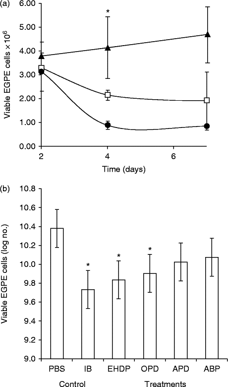
The dose–response assay results for the EGPE cells cultured in liquid medium are shown in fig. 1a. The plots indicate that 30 μm EHDP had a maximum antiproliferative effect that was significant by day 4 and persisted until the treatment ended (day 7). Figure 1b shows IB and OPD also had significant antiproliferative effects on EGPE cells, but treatments with APD and ABP did not lead to any significant changes in cell growth. No further antiproliferative effects were observed between treatments with 30 and 90 μm bisphosphonates (data not shown). Dose-clustered data analysed using the Kruskal–Wallis test showed P< 0.001 differences among all groups studied, and P< 0.001 intra-group differences in cells exposed to EHDP, IB and OPD. The inhibitory effect ranked in the order IB > EHDP > OPD.
Viability of cells (mean ± SD) derived from a bovine protoscoleces. (a) Dose responses of EGPE cells treated with 10 μm (white squares) and 30 μm (black dots) etidronate (EHDP) for up to 7 days with an antiproliferative effect on day 4, versus PBS control (black triangles). (b) Cells exposed to ibandronate (IB), etidronate (EHDP) and olpadronate (OPD), n= 9, pamidronate (APD) and alendronate (ABP), n= 4, clustered concentrations 30 and 90 μm for 4–7 days with PBS control, n= 8. *, Significant at P< 0.05.

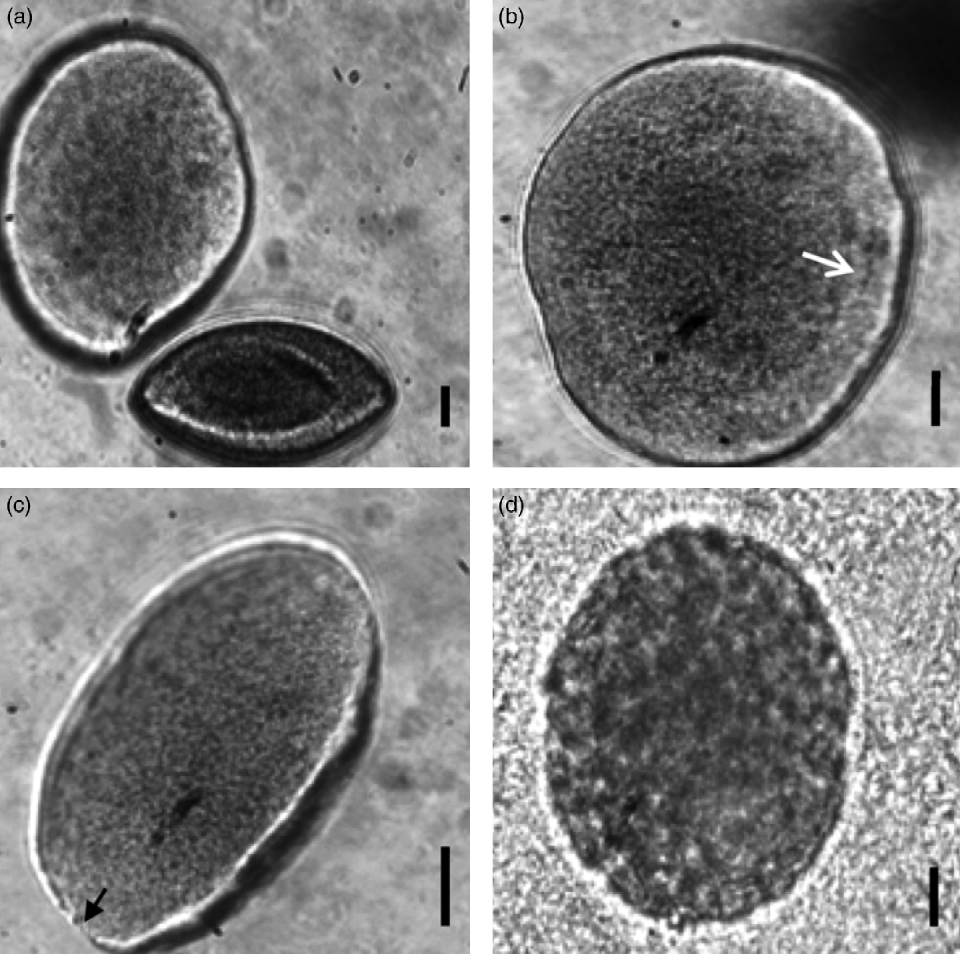
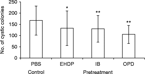
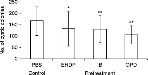
In the three-dimensional model, cells treated with 30 μm EHDP, IB and OPD showed a decrease (P< 0.05) in colony numbers. Colony counts were 111 ± 15.8 (n= 4) for controls and 66.3 ± 9.2 (n= 4) for EHDP-treated cells. In another set of experiments, 125.9 ± 17.1 (n= 3) colonies were observed in the control sample, 92.6 ± 12.3 (n= 3) and 89.5 ± 19.8 (n= 3) in cultures treated with IB and OPD, respectively. Figure 2 shows the typical colony morphology of control and EHDP-treated cultures. Colonies exposed to EHDP for a week showed flocculent contents with thinner and more irregular borders than control colonies. Seven independent experiments were performed to study the residual effect of bisphosphonates. Briefly, EGPE cell duplicates were cultured in liquid medium for 4 days and treated with 30 μm EHDP, IB and OPD, respectively. The cells seeded and grown in agarose without any further treatment showed the persisting antiproliferative effects of EHDP, IB and OPD exposure (fig. 3).
The effect of etidronate (EHDP) on the cystic colony morphology in agarose: (a) EHDP-free agarose control with an entire turgid cyst typically full of fluid, limited by a thick, non-cellular wall with concentric rings; (b) cyst contents with the cells (arrowed) in contact with the cystic membrane and with a central homogeneous region; (c) cyst becoming flaccid after 4 days in agarose with 30 μm EHDP, with a thinner perforated wall (arrow); (d) cyst after 7 days in agarose with vacuolated core material and flocculent cellular residues. Scale bars = 20 μm.
Persistent effect in cells exposed to 30 μm bisphosphonates (BPs): etidronate (EHDP), ibandronate (IB) and olpadronate (OPD), transferred into BP-free agarose for comparison with untreated PBS control cells after 7 days; PBS-control single-tailed (*) and two-tailed (**) test with significance at P< 0.05.
Changes in intracellular calcium and ATP production
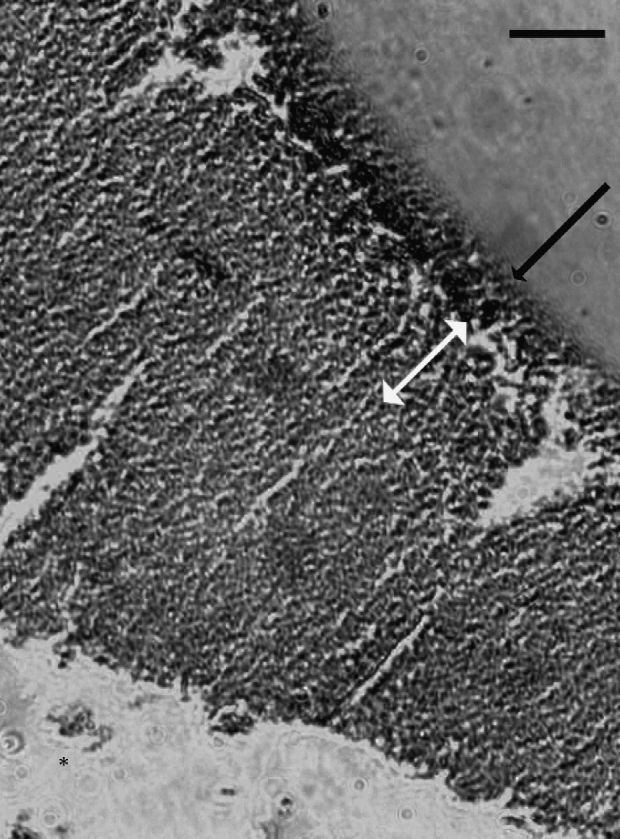
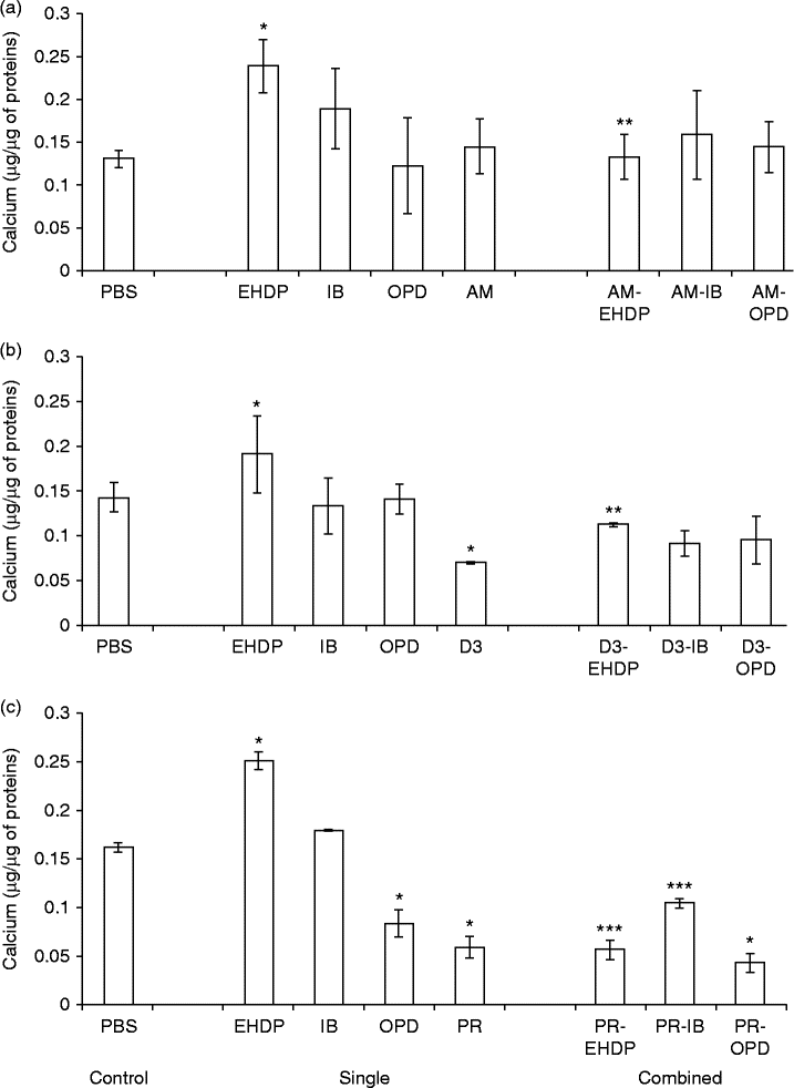
Calcium deposits in EGPE colonies were mostly observed in the external layers and scattered calcium-positive cells were found near the colony centre (fig. 4). A change in the intracellular calcium concentration was observed in cells treated with EHDP; this was the only bisphosphonate that increased the calcium concentration in cells compared to the control (P< 0.05) (fig. 5a, b and c). However, the combined treatment of EHDP with 1.3 μm amiloride abolished the effect of EHDP on calcium concentration (fig. 5a). A decrease in calcium concentration was measured with 13 μm vitamin D3 compared to the untreated controls (P< 0.05) but no changes were observed when D3 was combined with bisphosphonates (n= 2) (Fig. 5b). Proline provoked a considerable decrease in calcium concentration (P< 0.05) in bisphosphonate-treated or untreated cells (fig. 5c). The decrease in calcium concentration seen with the proline treatment correlated with the increase in cystic colony formation. Cells treated with either EHDP or IB with proline in liquid medium and seeded in agarose without bisphosphonates and proline showed increased colony counts (P< 0.05; n= 3): EHPP 16.67 ± 0.88 compared with EHDP-proline: 23.23 ± 1.79 and IB 20 ± 3.33 compared with IB-proline 38.33 ± 7.78 colonies. Moreover, cells previously treated only with proline did not alter the number of cell colonies when compared with controls incubated with PBS (data not shown). The association of bisphosphonates with either amiloride or vitamin D3, however, did not have any significant effect on colony formation. Nevertheless, treatment of EGPE cells with 13 μm vitamin D3 alone showed an increase in colony formation that was not statistically significant: the counts were 20.12 ± 2.12 colonies (n= 2) in the control and 33.64 ± 6.16 colonies (n= 2) for vitamin D3-treated cells.
Localization of calcium in cells cultured in agarose with the staining of calcium in the external layer ( ) and neighbouring cells (
 )
 . Scale bar = 10 μm.
The concentration of calcium (mean ± SD) in cells in response to single treatments with 30 μm etidronate (EHDP), ibandronate (IB), olpadronate (OPD), 1.3 μm amiloride (AM), 13 μm 1,25-dihydroxycholecalciferol (D3) and 10 μm proline (PR) and combined treatments: (a) each EHDP, IB and OPD with AM, n= 3; (b) each EHDP, IB and OPD, with D3, n= 3; (c) each EHDP, IB and OPD with PR, n= 2. PBS used as control, with P< 0.05 for treated cells (*); combined treatment compared to single bisphosphonate treatment data (**); combined treatment compared to grouped single bisphosphonate treatment and PBS control data (***).
The effect of bisphosphonates on ATP production was measured in EGPE cells cultured in 96-well plates and treated for 24 h with bisphosphonates, proline and with EHDP-proline. Control cells produced 41,966 ± 2091 luminescent arbitrary units (lau) ATP (100% reference). Presence of bisphosphonates provoked a significant decrease in ATP production (P< 0.05), with OPD, APD, ABP and IB eliciting ATP readings between 85 and 85.76% (n= 6). The decays in ATP production in cells treated with EHDP were 35,466 ± 3387 lau (84.57%), while cells treated only with proline gave control readings: 42,243 ± 4793 lau. Interestingly, proline reversed the effect of EHDP on ATP production, since the cells incubated with the combined EHDP-proline showed 96% of the ATP values found in control cells (40,677 ± 3825 lau); the difference with EHDP alone being significant P= 0.05 (n= 4).
Discussion
Different strategies are currently available to prevent or treat hydatidosis in humans, while many are being developed (Giorgio et al., Reference Giorgio, Tarantino, Francica, Mariniello, Aloisio, Soscia and Pierri1992; Hemphill et al., Reference Hemphill, Spicher, Stadelmann, Mueller, Naguleswaran, Gottstein and Walker2007). The ideal strategy would be to prevent the human infections by vaccinating grazing animals that are the natural intermediate hosts of the parasite in the field. Vaccines for veterinary use have been developed in Australia and in Argentina (Heath et al., Reference Heath, Jensen and Lightowlers2003). However, government programmes for the control and prevention of animal hydatidosis and private investment in animal vaccination and treatment are often insufficient. Consequently, other approaches and more efficient and affordable human therapies are needed.
The process of laminal layer calcification of the cyst, which may lead to cyst degeneration and sterilization, is a potential therapeutic target. Flubendazole, for example, interferes with cell homeostasis and promotes an increase in free cytosolic calcium (Cumino et al., Reference Cumino, Elisondo and Denegri2009), but adverse reactions have been reported with long-term benzimidazole chemotherapies. Consideration of biphosphonates, a family of calcification-promoting drugs, as anti-hydatidic agents was encouraged by the intrinsic ion distribution and calcium crystal formation in the cyst (Smith & Richards, Reference Smith and Richards1993). Indeed, potassium, magnesium and calcium levels are higher in protoscoleces than in cystic fluid (Frahya & Haddad, Reference Frayha and Haddad1980). Other authors found levels similar to those in plasma for some ions but potassium and calcium were slightly higher in cyst fluid than in plasma. Additionally, phosphate concentration in cyst fluid was reported to be ten times lower than in plasma (Vidor et al., Reference Vidor, Piens, Abbas and Petavy1986). Calcium and magnesium concentrations have been described to decrease as the cyst size increases; in thin-walled cysts, an increase in magnesium concentration was followed by calcium deposition in the laminar layer of the cysts (Chowdhury & Singh, Reference Chowdhury and Singh1993). One of the molecules involved in calcium storage is myo-inositol hexakisphosphate, which forms microcrystalline solids in the laminated layers and is also found inside membrane vesicles of the germinal layer cells. Each crystal comprises around 200 myo-inositol hexakisphosphate molecules (300 mm) and contains 74% calcium and 6–9% magnesium. The phosphorus content in these crystals accounts for 92% of the total phosphorus in the hydatid cyst wall (Irigoin et al., Reference Irigoin, Casaravilla, Iborra, Sim, Ferreira and Díaz2004; Casaravilla et al., Reference Casaravilla, Brearley, Soulé, Fontana, Veiga, Bessio, Ferreira, Kremer and Díaz2006). Other macromolecules involved in calcium storage in cestodes have also been described, such as a 260 kDa antigenic protein (Zurabian et al., Reference Zurabian, Carrero, Rodríguez-Contreras, Willms and Laclette2005) and calreticulin (Cabezón et al., Reference Cabezón, Cabrera, Paredes, Ferreira and Galanti2008).
Certain E. granulosus cyst cells have been reported to accumulate calcium in their cytoplasm in the form of corpuscles. It has been postulated that such calcified cells would be unable to divide and are involved in calcium equilibrium in the cyst. Cation channels have been identified in protoscoleces: a Ca2+ channel blocked by Ba2+ and another that displays cation affinities in the order K+> Na+> Li+> Ca2+. The hyperpolarizing effect of amiloride, a Na+ blocker, on the cell membrane potential has already been characterized in fresh protoscoleces (Ibarra & Reisin, Reference Ibarra and Reisin1994; Grosman & Reisin, Reference Grosman and Reisin1995). A non-selective mode-shifting cation channel showing permeability for Ca2+, Ba2+ and tetraethylammonium was identified in E. granulosus tapeworms (Grosman & Reisin, Reference Grosman and Reisin1997). Recently, a rapamycin-binding protein-mediated Ca2+ channel implicated in protoscolex viability has been described (Cumino et al., Reference Cumino, Lamenza and Denegri2010).
The present studies showed that bisphosphonates decrease EGPE cell growth rate in liquid medium and the cell's clonogenic growth in agarose. A dose–response relationship was observed in the 10–30 μm bisphosphonate range but not at higher concentrations, such as 90 μm. Moreover, cells that were exposed to bisphosphonates in liquid medium, washed and seeded in agarose still showed significant decreases in colony formation rate, which are clear indicators of residual bisphosphonate action. The residual effect was observed after treatment with EHDP, a first-generation bisphosphonate with the lowest impact on human tissues other than bone. Bisphosphonate binds specifically by chemical adsorption to the calcified structures of living tissues, physically affecting the properties of mineralized structures of bones. EHDP was the only bisphosphonate that led to a significant calcium accumulation in this model. In bone, EHDP reacts with calcium phosphate and forms crystals (Cukrowski et al., Reference Cukrowski, Popović, Barnard, Paul, van Rooyen and Liles2007). The kind of crystals EHDP forms at increasing cell calcium concentrations in the EGPE cells was not investigated. However, some light was shed on the role of calcium storage, on in vitro cell proliferation and on cyst colony development. Amiloride did not affect calcium concentration but prevented the EHDP-induced increase in calcium concentration in EGPE cell cultures. In normal rat kidney fibroblasts, K+ accumulation stimulated by amiloride modified the calcium-clearing activity mediated by a slow endothelial reticulum calcium channel and inositol-3-phosphate (Kusters et al., Reference Kusters, Dernison, van Meerwijk, Ypey, Theuvenet and Gielen2005). In the Xenopus oocyte model, 0.2–2 mm ABP and risedronate (a bisphosphonate not included in this study) inhibited [Na+]out or [K+]out currents without affecting membrane dielectric properties (Shao et al., Reference Shao, Orlando and Awayda2005).
Vitamin D3 decreased calcium concentration in untreated EGPE cells, but only caused a minor, non-significant reduction in calcium levels in cells that had been previously exposed to bisphosphonates. EHDP is known to impair intestinal absorption of Ca2+ (Lidor et al., Reference Lidor, Meyer, Wasserman and Edelstein1987). The combined treatment with vitamin D3 and EHDP in humans increased bone mineral density (Masud et al., Reference Masud, Mulcahy, Thompson, Donnelly, Keen, Doyle and Spector1998) but vitamin D3 action entailed a different mechanism (Farach-Carson & Ridall, Reference Farach-Carson and Ridall1998). Moreover, vitamin D3 could have discrete effects in combination with calcium (Panda et al., Reference Panda, Miao, Bolivar, Li, Huo, Hendy and Goltzman2004). Vitamin D3 impaired the increase in cellular calcium concentrations caused by EHDP in EGPE cells. Recently, an antifibrotic effect of vitamin D3, achieved by suppressing transforming growth factor (TGF)-β3 induced fibrosis in human uterine leiomyoma cells was reported (Halder et al., Reference Halder, Goodwin and Al-Hendy2011), and a molecular family that responds to transforming growth factor stimulation has been described in Echinococcus multilocularis (Epping & Brehm, Reference Epping and Brehm2011). Since another effect of vitamin D3 alone observed in this study was a slight increase in colony formation, it prompted us to investigate the role of the extracellular compartment in the clonogenic potential of EGPE cells.
Studies in mice have shown that EHDP decreased the incorporation of proline into collagen (Guenther et al., Reference Guenther, Guenther and Fleisch1981) while collagen (Mahmoud & el-Garhy, Reference Mahmoud and el-Garhy2002) and the activity of lysyl–prolyl hydroxylase (Guerret et al., Reference Guerret, Vuitton, Liance, Pater and Carbillet1998) were described in the hydatid cyst wall. Bovine E. granulosus protoscoleces cells treated with proline showed the most significant decrease in calcium level for both bisphosphonate-treated and non-treated cells. Colony-formation rates were higher when EHDP was combined with proline, suggesting at least one of proline's effects is exerted on the extracellular structures. Cyst walls were affected by bisphosphonates but cells treated with bisphosphonates + proline did not show any visible abnormalities. The five bisphosphonates of this study decreased ATP levels, and proline counteracted this effect in cells treated with EHDP. Proline has been observed to provide energy to cells (Phang et al., Reference Phang, Liu, Hancock and Christian2012) and could be modulating the metabolism of bisphosphonate-treated EGPE cells. Calcium deposition could be a consequence of a loss of cell viability caused by bisphosphonates. However, there may be mechanistic differences among the studied compounds as all bisphosphonates decreased ATP levels but only three of them reduced colony formation and only EHDP increased intracellular calcium concentration measured by the colorimetric method. In fact, bisphosphonates could inhibit pyrroline-5-carboxylate reductase I, the enzyme catalysing the committed step for proline synthesis (Phang et al., Reference Phang, Liu, Hancock and Christian2012), but this enzyme, or another with similar metabolic activity, has not been described in E. granulosus. Moreover, the effects of bisphosphonates on these cells is more likely associated with calcium storage, colony formation and morphology than to other mechanistic hypotheses.
Bisphosphonates interfere with the mevalonate pathway by inhibiting farnesyl pyrophosphate synthase, thereby preventing prenylation of small GTPases such Rho, Rac and Rabs (Kavanagh et al., Reference Kavanagh, Guo, Dunford, Wu, Knapp, Ebetino, Rogers, Russell and Oppermann2006). A Ral (Ras-like) GTPase homologue which binds GTP has been found in E. multilocularis, with 53% sequence homology with human RalA and 54% homology with RalB (Spillotis & Brehm, Reference Spillotis and Brehm2004), the end-product in the cholesterol synthesis pathway in E. granulosus and cysticerci of Taenia hydatigena being 2-cis,6-trans farnesol (Frayha & Haddad, Reference Frayha and Haddad1980).These mechanisms were seen to be affected mostly by nitrogen-containing bisphosphonates such as APD, ABP, IB and OPD used in this study, which may have thus exerted additional metabolic effects which were not included in the scope of this study.
The fact that the bisphosphonate concentrations used in the in vitro analysis are potentially achievable in human treatment is relevant as they are lower or comparable to those approved for pharmaceutical use. The antihydatidic effect of the five compounds shown here does not parallel their known skeletal effects and potency, and would not impact bone metabolism at the described concentrations. Indeed, EHDP is the least potent bone resorption inhibitor and likely the safest agent in a novel extraskeletal indication. In humans, EHDP toxicity appears after months of accumulation, a schedule which may not be necessary in its re-profiled antiparasitic use.
In conclusion, calcium storage is related to EGPE cell line growth, and the more effective bisphosphonates are in the order: EHDP > IB > OPD in terms of EGPE cell growth inhibition in the liquid medium and the three-dimensional colony model. Calcium deposition in these cells may result from steady-state mechanisms of the extracellular compartments. Calcium influx in these cells is partially dependent on cationic channel blockage by amiloride. The preliminary results suggest that bisphosphonates possess antiparasitic qualities to be probed further in in vivo therapies alone, or in combination with other antihelminthic drugs, at their lowest active concentration or for a short period of time. EHDP has the advantage of being safe, with fewer potential side-effects, in humans than current approved therapeutics, and adequate antiparasitic clinical doses deserve exploring, in spite of the proline antagonism.
Acknowledgements
This work used the in vitro model for pharmacological studies in Echinococcus, applied patent P090102320 Argentina, by A.G. Fuchs, Universidad Abierta Interamericana. The work was supported by Universidad Abierta Interamericana grants. We thank Dr Gabriela Canziani for careful reading of the manuscript. The authors declare no conflicts of interest.