

Introduction
It is well documented that olfactory dysfunction is a common symptom in patients with neuro-degenerative disorders (Alves, Petrosyan, & Magalhães, Reference Alves, Petrosyan and Magalhães2014; Kovács, Reference Kovács2013; Ottaviano, Frasson, Nardello, & Martini, Reference Ottaviano, Frasson, Nardello and Martini2016; Wilson et al. Reference Wilson, Xu, Sadrian, Courtiol, Cohen and Barnes2014). Recent research on aging indicates that lifespan development and aging, age-associated cognitive decline, as well as the development of Alzheimer's disease (AD) and other neuro-degenerative diseases, are all closely connected with olfactory impairment (Doty & Kamath, Reference Doty and Kamath2014; Hüttenbrink, Hummel, Berg, Gasser, & Hähner, Reference Hüttenbrink, Hummel, Berg, Gasser and Hähner2013; Katotomichelakis et al., Reference Katotomichelakis, Balatsouras, Tripsianis, Tsaroucha, Homsioglou and Danielides2007; Kovács, Reference Kovács2004; Rawson et al., Reference Rawson, Gomez, Cowart, Kriete, Pribitkin and Restrepo2012). Olfactory senescence begins around the age of 36 years in both genders and accelerates with advancing age, but the trajectory of this decline seems to be non-linear (Hawkes, Reference Hawkes2006; Kouklidou, Sfakianaki, Moraitou, Papantoniou, & Masoura, Reference Kouklidou, Sfakianaki, Moraitou, Papantoniou and Masoura2015). Research shows that over 50% of people from 65 to 80 years old, and almost 80% of people over 80 years of age, show signs of serious olfactory impairment (Attems, Walker, & Jellinger, Reference Attems, Walker and Jellinger2015; Doty, Shaman, & Dann, Reference Doty, Shaman and Dann1984). Moreover, olfactory dysfunction is estimated to have an even more remarkable prevalence (90–100%) in Parkinson disease (PD) and AD (Alves et al., Reference Alves, Petrosyan and Magalhães2014).
Some of the variables that have been systematically associated with olfactory functioning are age, gender, smoking, and cognitive functioning (Doty, Petersen, Mensah, & Christensen, Reference Doty, Petersen, Mensah and Christensen2011; Velayudhan, Pritchard, Powell, Proitsi, & Lovestone, Reference Velayudhan, Pritchard, Powell, Proitsi and Lovestone2013). According to the recent findings within the field of cognitive neuro-psychology, impairment in olfactory function is associated with low-processing speed (Dulay, Gesteland, Shear, Ritchey, & Frank, Reference Dulay, Gesteland, Shear, Ritchey and Frank2008), impaired attention, and impaired working memory capacity (Hulshoff-Pol, Hijman, Baare, van Eekelen, & van Ree, Reference Hulshoff-Pol, Hijman, Baare, van Eekelen and van Ree2000). Cognitive decline is associated with poor performance in olfactory tests of patients with AD and mild cognitive impairment (MCI) (Djordjevic, Jones-Gotman, Sousa, & Chertkow, Reference Djordjevic, Jones-Gotman, Sousa and Chertkow2008), as well as patients with other neuro-degenerative disorders (Baba et al., Reference Baba, Takeda, Kikuchi, Nishio, Hosokai and Itoyama2011; Doty, Reference Doty2012; Kovács, Reference Kovács2013). The main explanation of these associations is given at a structural level, in terms of the complex connections of the olfactory bulb to frontal brain areas, as well as the involvement of medial temporal lobe structures in both olfactory functioning as odour identification and memory processes (Conti et al., Reference Conti, Vicini-Chilovi, Riva, Zanetti, Liberini, Padovani and Rozzini2013).
The most common methods to examine olfactory dysfunction in humans are the psychophysical tests. Almost all of them require a conscious response from the examinee. They usually consist of procedures that test the ability to detect an odour, the ability to identify an odour, or/and the ability to discriminate one odour from another or different concentrations of the same odour. Odour detection threshold is the minimum concentration at which an odor can be detected without any requirements to recognise the stimulus, while odour identification threshold is the minimum concentration at which an odorant can be identified or recognised. For the assessment of odour discrimination abilities, the detection of similarities and differences between odorants or different concentrations of a given odorant is required (Kovács, Reference Kovács2004; Schofield, Finnie, & Yong, Reference Schofield, Finnie and Yong2014). According to Doty and Kamath (Reference Doty and Kamath2014), the results of these tests are positively associated with each other (see also Doty, Reference Doty2015), and this is possibly indicative of the existence of a ‘general olfactory acuity’ factor of olfaction such as the ‘g’ factor of intelligence. Nevertheless, besides the well-established overlapping to some extent, there is also evidence that odour detection threshold tests represent a relatively discrete measure from odour identification and odour discrimination tests. This could be explained on the basis of the involvement of higher-level cognitive processes in the latter, while odour detection is claimed to be rather relying on lower-level perceptual processes (see Rahayel, Frasnelli, & Joubert, Reference Rahayel, Frasnelli and Joubert2012). Moreover, Xu et al. (Reference Xu, Lopez-Guzman, Schoen, Fitzgerald, Lauer, Nixon, Levy and Wilson2014), based on their results regarding an animal model of AD, suggest that odour identification requires linking the odour percept to associated labels in regions upstream of the primary olfactory cortex (POC), and thus it differs from the other two procedures that seem to require basic odour processing.
In the same vein, some differences in odour detection, identification, and discrimination abilities of the patients diagnosed with MCI, AD and other neuro-denenerative diseases have been found in recent studies. According to the meta-analysis of Mesholam et al. (see Djordjevic et al., Reference Djordjevic, Jones-Gotman, Sousa and Chertkow2008), odour detection and identification abilities significantly worsen in patients diagnosed with AD, although this decline appears to be greater in odour identification, compared to odour detection (Djordjevic et al., Reference Djordjevic, Jones-Gotman, Sousa and Chertkow2008; Kovács, Reference Kovács2004; Wilson, Arnold, Schneider, Tang, & Bennett, Reference Wilson, Arnold, Schneider, Tang and Bennett2007). A broader systematic meta-analysis of 81 studies on the effects of AD and PD on olfaction (Rahayel et al., Reference Rahayel, Frasnelli and Joubert2012) showed that, indeed, AD and PD patients are more impaired on odour identification tasks than on odour detection ones. However, PD patients are more impaired on threshold detection, compared to AD patients, and this indicates that the aetiology of olfactory dysfunction in these diseases is probably different: In PD, lower-level problems, such as the altered sniffing pattern, may play a role in the olfactory decrements, along with the presence of Lewy bodies in specific neurons, as well as the presence of dopaminergic and cholinergic deficits (see also Zhang, Xiao, & Le, Reference Zhang, Xiao and Le2015). Olfactory dysfunction has been reported in frontotemporal dementia (FTD) too: The few existing studies showed, in general, that odour identification is impaired in FTD patients (similarly to AD), while odour discrimination is preserved. No differences were found in odour identification in different variants of FTD (Alves et al., Reference Alves, Petrosyan and Magalhães2014; Ottaviano et al., Reference Ottaviano, Frasson, Nardello and Martini2016). Beyond the differences, which could also be attributed to methodological reasons to some extend (Doty, Reference Doty2015; Doty & Kamath, Reference Doty and Kamath2014), the well-established olfactory dysfunction in aging and dementia has led recent research on the field to the examination of olfactory function in MCI.
A series of empirical – mainly cross-sectional – studies showed that patients diagnosed with MCI display lower performance in odour identification tests, compared to their cognitively healthy counterparts (Bahar-Fuchs, Moss, Rowe, & Savage, Reference Bahar-Fuchs, Moss, Rowe and Savage2010; Devanand et al., Reference Devanand, Michaels-Marston, Liu, Pelton, Padilla, Marder and Mayeux2000; Eibenstein et al., Reference Eibenstein, Fioretti, Simaskou, Sucapane, Mearelli, Mina and Fusetti2005; Laakso et al., Reference Laakso, Tervo, Hänninen, Vanhanen, Hallikainen and Soininen2009; Wang et al., Reference Wang, Tian, Huang, Qin, He and Zhou2002). In a next step, the longitudinal work of Conti et al. (Reference Conti, Vicini-Chilovi, Riva, Zanetti, Liberini, Padovani and Rozzini2013) indicated that odour identification assessment may be useful for identifying MCI patients with high risk of developing dementia. Eighty eight participants diagnosed with MCI were divided into two groups based on the performance in odour identification (40% with normal smell and 60% with moderate deficits in smell). Within 2 years of follow up, 47% of the MCI patients displaying olfactory deficits and 11% of the MCI patients with normal smell developed dementia. Recently, Roberts et al. (Reference Roberts, Christianson, Kremers, Mielke, Machulda, Vassilaki and Petersen2016) examined the association between odour identification ability and MCI subtypes and progression from MCI to AD, in a sample of 1430 participants who were enrolled in the population-based, prospective Mayo Clinic Study of Aging between 2004 and 2010, and were cognitively normal at baseline. Based on their findings, they suggest that odour identification tests have potential utility for screening for MCI and amnestic MCI conversion to AD. Another longitudinal study aimed at assessing whether odour identification and neuro-psychological testing can distinguish MCI patients to those who will probably develop Dementia with Lewy bodies (MCI-DLB), those who develop AD, and non-converters (Yoon, Kim, Moon, Yong, & Hong, Reference Yoon, Kim, Moon, Yong and Hong2015), showed that MCI due to Dementia with Lewy bodies patients had a lower odour identification ability as well as worse executive functions and visuospatial ability, compared to the other groups. The pathology of Lewy bodies was considered a possible reason for their greater olfactory dysfunction, compared to AD patients.
As regards the association between odour identification ability and cognitive functioning, research has clearly indicated that impaired odour identification sensitivity is consistent with the impairment of semantic memory, and this association seems to reflect the progressive loss of knowledge about odours. Impaired odour identification ability seems thus to function as a precursor of verbal learning decline, memory decline, and general intelligence decline (Djordjevic et al., Reference Djordjevic, Jones-Gotman, Sousa and Chertkow2008; Huart et al., Reference Huart, Rombaux, Gerard, Hanseeuw, Lhommel, Quenon and Mouraux2015). In this research direction, Vyhnalek et al. (Reference Vyhnalek, Magerova, Andel, Nikolai, Kadlecova, Laczo and Hort2015) examined odour identification and its potential cognitive correlates in amnestic and non-amnestic MCI. They found that odour identification is impaired in both groups but is proportional to memory decline only in amnestic MCI. The sharing of a common neuro-anatomical substrate, which suffers from AD pathology and primarily, medial temporal lobes’ involvement in both odour identification and memory function, was considered as a strong possible reason for this association in amnestic MCI (see Kovács, Reference Kovács2013). Kjelvik et al. (Reference Kjelvik, Saltvedt, White, Stenumgård, Sletvold, Engedal and Håberg2014) also suggest odour identification as an early indicator of medial temporal lobe degeneration, based on their findings according to which odour identification is associated with hippocampal volume in amnestic MCI and AD.
At this point, despite the advantages and the broad use of odour identification measures, it should be mentioned that the extant research has shown that they are culture- and language-dependent, while odour discrimination performance appears to be associated with semantic memory as well. On the other hand, it has been suggested that odour detection tests rather represent measures of basic olfactory sensitivity which are relatively more independent from socio-cultural factors (Sorokowska et al., Reference Sorokowska, Sorokowski and Hummel2014).
In this line of research, the study of Stamps, Bartoshuk and Heilman (Reference Stamps, Bartoshuk and Heilman2013) is probably the most publicised. Stamps et al. (Reference Stamps, Bartoshuk and Heilman2013) compared amnestic MCI, AD patients, patients with other types of dementia, and healthy controls in odour detection ability, using only the odour of peanut butter and following a unirhinal examination in which the distance from the naris (either of the pair of openings of the nose or nasal cavity) was measured. They found that left nostril performance in odour detection was significantly lower in AD patients. According to Stamps et al. (Reference Stamps, Bartoshuk and Heilman2013), this can be explained by the fact that the olfactory epithelium projects mainly to the ipsilateral olfactory cortex (left) which is usually more damaged in the AD, compared to the right olfactory cortex.
However, studies that tried to replicate and expand the work of Stamps et al. (Reference Stamps, Bartoshuk and Heilman2013), failed to find similar evidence: no meaningful left:right asymmetry in odor detection was apparent (Doty et al., Reference Doty2014). According to Doty et al. (Reference Doty2014), left-side lateralised pathology in MCI and AD is contradictory, and such a lateralisation can potentially result from sample selection on the basis of neuro-psychological testing that is heavily weighted towards processes mainly supported by the left hemisphere. Nevertheless, a recent study that aimed to examine olfactory cortex degeneration in AD and MCI, via measuring total structural volumes and fMRI activation volumes of the POC and the hippocampus (Vasavada et al., Reference Vasavada, Wang, Eslinger, Gill, Sun, Karunanayaka and Yang2015), partially supported lateralisation in terms of an earlier and quicker left POC atrophy. This was on the basis of the finding that the right POC was larger than the left POC in MCI and AD participants, while no such difference was found in the normal controls. The same study showed that unlike lateralisation of atrophy of the POC, POC lateralisation at the functional level (activation) was not found. Besides the POC, this study and one other neuro-imaging study have found almost the same type of lateralisation effects of MCI or AD in the hippocampus volume, together with a high association between the loss of left hippocampal volume and participants’ performance in odour recognition tasks (Vasavada et al., Reference Vasavada, Wang, Eslinger, Gill, Sun, Karunanayaka and Yang2015; Zou, Lu, Liu, Zhang, & Zhou, Reference Zou, Lu, Liu, Zhang and Zhou2016).
There is also some evidence of left:right lateralisation concerning other structures and functions that are involved in olfactory processing (see Attems et al., Reference Attems, Walker and Jellinger2015; Doty & Kamath, Reference Doty and Kamath2014). Moreover, a marked asymmetry of olfactory function –including odour detection – in MCI patients, compared to age-matched controls (Huart et al., Reference Huart, Rombaux, Gerard, Hanseeuw, Lhommel, Quenon and Mouraux2015), and in AD patients compared to amnestic MCI (Bahar-Fuchs et al., Reference Bahar-Fuchs, Moss, Rowe and Savage2010) was found, when best/worst nostril performance was measured. This suggests that unirhinal examination should be included as an additional procedure to the classic bilateral one, to assess olfactory function in very mild cognitive impairment. In sum, recent literature suggests that the assessment of olfactory function can be used as a tool for early diagnosis of cognitive decline related to dementia and other neuro-cognitive disorders (Albers, Tabert, & Devanand, Reference Albers, Tabert and Devanand2006; Djordjevic et al., Reference Djordjevic, Jones-Gotman, Sousa and Chertkow2008; Velayudha et al., Reference Velayudhan, Pritchard, Powell, Proitsi and Lovestone2013; Wang et al., Reference Wang, Eslinger, Doty, Zimmerman, Grunfeld, Sun and Yang2010; Wesson, Levy, Nixon, & Wilson, Reference Wesson, Levy, Nixon and Wilson2010).
In this light, the present study refers to the development of a new brief odour detection test. The rationale for such a test is that smell function can be easily measured due to the fact that injury to central neural structures similarly affects networks that code or transmit information from many classes of receptor cells (Doty, Reference Doty2015). Hence, an accurate assessment of olfactory functioning at the psychophysical level can be achieved via the careful selection and use of only a few odours. The aim of the study was to examine whether this test could differentiate community dwelling older adults with very mild cognitive impairment from their cognitively healthy counterparts. The following two hypotheses were formulated: older adults with very mild cognitive impairment would display significantly lower performance in the two conditions of the Brief Odor Detection Test (B-ODT) in terms of both odour detection and identification, compared to cognitively healthy older adults (Hypothesis 1); older adults with very mild cognitive impairment would display significantly lower odor-detection sensitivity of the left nostril, compared to cognitively healthy older adults (Hypothesis 2).
Methods
Participants
Thirty four community-dwelling older adults, free of a diagnosis related to cognitive impairment, participated in the study. It was a convenience sample recruited from Open Care Centers for Older Adults (OCCOA) in Thessaloniki, Greece. The Social Worker – responsible for every OCCOA department accessed by the authors – permitted the access after approving the design of the study. Participants were divided into two groups according to their performance in the Montreal Cognitive Assessment – MoCA test – designed to assist health professionals for cognitive screening and detection of MCI – as adapted in Greek (MoCA, 2006; Nasreddine, Reference Nasreddine, Chertkow, Phillips, Whitehead, Collin and Cummings2004). The group with probable mild cognitive impairment (pMCI group) consisted of 16 older adults who achieved a MoCA total score from ‘24’ to ‘26’. The group of cognitively healthy older adults (CH group) consisted of 18 participants who achieved a MoCA total score from ‘27’ to ‘30’.
There had been a series of exclusionary criteria for participation in the study: (a) a MoCA total score < 24 indicating cognitive impairment of a higher level than the level of interest in this study; (b) a Geriatric Depression Scale-15 score > 6 (GDS-15; Fountoulakis et al., Reference Fountoulakis, Tsolaki, Iacovides, Yesavage, O'Hara, Kazis and Ierodiakonou1999; Yesavagr et al., Reference Yesavage, Brink, Rose, Lum, Huang, Adey and Leirer1982–Reference Yesavage, Brink, Rose, Lum, Huang, Adey and Leirer1983) indicating probable depression; (c) to be smoker or former smoker who had stopped smoking less than one year ago; (d) to have history of olfactory problems (for example, asthma, rhinitis, sinusitis, chronic polyps, and so on); (e) to have history including surgery in the nasal area; and (f) to have a diagnosis related to cognitive impairment and/or dementia. For the aforementioned reasons, 54 possible participants were excluded from the study.
Ethical Standards
The authors assert that all procedures contributing to this work comply with the ethical standards of the relevant national and institutional committees on human experimentation and with the Helsinki Declaration of 1975, as revised in 2008. The Ethics Committee of the School of Psychology of the Aristotle University of Thessaloniki, after reviewing the research protocol, confirmed that all ethical guidelines for research on human subjects were followed. The participants in the study were active members of the Open Care Centers for Older Adults and participated in the study voluntarily. They were informed about the procedure and the aim of the study, and their written informed consent was taken. The same procedure was followed as regards the students of the Military School of Corps Officers who participated in the pilot studies.
Main Instrument
Olfactory measure: the B-ODT. The B-ODT tests both odour detection and odour identification but not odour discrimination. Odour detection was measured via vanillin solutions that were created and renewed for each administration in the laboratory of the Department of Chemistry of the Aristotle University of Thessaloniki (AUTH). Vanillin is the primary component of the extract from the vanilla bean. The criterion of vanillin's selection was the fact that it is a widely used, pure odorant substance that activates mainly the olfactory nerve. Moreover, it is a fairly common odour in the Greek population. The decision about solutions’ concentrations was made after a pilot examination for threshold detection, which was conducted in a sample of 20 students of the Military School of Corps Officers. According to the literature (Ruth, Reference Ruth1986), the detection of the threshold of pure vanillin rises to 2×10⁻⁷ mg/m3. The department of Chemistry of AUTH assisted in the production of these solutions. Certain changes were made by the chemists due to the fact that the above amount is extremely low and it would be very difficult to be produced as well as to be detected. Thus, they created four solutions with the following concentrations of vanillin: 30 mg/L, 30 μg/L, 30 ng/L, and 30 pg/L. Next, these solutions were administered in a convenience sample of 20 students: Due to inability of detecting the weakest solutions, the chemists changed the concentrations to 150 mg/L, 30 mg/L, 15 mg/L, 3 mg/L, and .03mg/L.
The first condition of the B-ODT was developed based on the study of Djordjevic et al. (Reference Djordjevic, Jones-Gotman, Sousa and Chertkow2008), and specifically on their procedure of a scale solution administration which they used to determine the threshold of detection. For the administration of the concentrations of vanillin in B-ODT Condition 1, five pairs of transparent glass bottles are used. Each pair comprises of one bottle with transparent vanillin solution and one with water. The experimenter presents the pair of bottles to each participant, who has to close their mouth according to experimenter's guidelines, and decide which of the stimuli ‘smells stronger’. The assessment starts with the lowest density concentration. In case of failure to detect the odour, the process continues using the next solution in terms of density (the solution of pure vanillin with label ‘1’ had the highest concentration and with label ‘5’ had the lowest concentration).
Once participants have detected the odour (at whichever strength), the second step involves asking participants to then identify the odour, starting at the level at which the odour was detected. If they cannot identify the odor at that level, they are then presented with the next strongest solution and so on. The identification is based on information retrieval from semantic memory, without auxiliary hint. However, if the participant reaches the final level (level 1) before detecting the odour, for the identification step, the experimenter presents the participant with a list with four possible answers (aniseed, rose, vanilla, and lemon) and asks the participant to choose the correct answer (identification based on semantic memory with auxiliary hint).
As regards the scoring procedure, higher scores represent better performance. Odour detection is scored on a scale (5–1), as per the level at which the odour is first detected. The score for identification starts from the detection score: For example, if one detects the third solution in terms of density (detection score = 3), they proceed to odour identification. The identification score starts from ‘3’ in case they identify the odour of this solution, but can result to ‘1’ in case they can only identify the odour of the solution with the higher density. The identification with auxiliary hint can only score ‘1’, that is, the same as the identification without auxiliary hint for the same solution (1).
The second condition of the B-ODT was developed based on the procedure of Stamps et al. (Reference Stamps, Bartoshuk and Heilman2013). B-ODT Condition 2 examines the change in air odour and it requires vanillin solution of 30 mg/L and a 30-cm metric ruler. At this point it must be noted that the specific solution was selected due to the fact that solution concentration of 150 mg/L appeared to have quite strong smell and could be detected even from a 30-cm distance. As for the rest of solutions, they were rejected for this condition due to difficulty to detect them in a pilot administration to 20 undergraduate students of the Military School of Corps Officers.
The Experimenter gives the following instructions: ‘Once you detect a change in the air smell, report it’ and, after detection, ‘Can you identify what it is?’ In case they do not manage to identify the odour without auxiliary hint, four possible answers are given at the end, from which the participant must choose the correct one. The solution is initially placed 30 cm below participant's nostril (the solution of pure vanillin was moved on a 30-cm ruler – where ‘30 cm’ was the largest distance from the nostril and ‘0 cm’ was the nearest point under the nostril) and it rises 1 cm at a time (that is 1 cm upon each exhale), until the participant detects the odour. The process does not stop but continues, until the participant identifies the odour. The procedure is performed first on the one nostril and after a 90-s delay on the other, and each time, detection and identification distance is recorded. The scores from this component of the test are the distance in centimetres at which detection or identification is made.
In summary, the six variables measured by the B-ODT are: (a) odour detection 1-scale measuring (O-DETECT-SCALE), (b) odour identification 1-scale measuring (O-IDENT-SCALE), (c) odour detection 2a-distance measuring-left nostril performance (O-DETECT-DISTANCE-L), (d) odour detection 2b-distance measuring-right nostril performance (O-DETECT-DISTANCE-R), (e) odour identification 2a-distance measuring-left nostril performance (O-IDENT-DISTANCE-L), (f) odour identification 2b-distance measuring-right nostril performance (O-IDENT-DISTANCE-R).
Procedure
All participants were tested individually in a quiet and clean context which was free of strong smells. Both participants and researchers did not wear any perfumes. The entire session took about 40–60 minutes. Each participant was informed that participation was voluntary and they could leave the process at any time. The first task was to fill in a questionnaire concerning health status, olfactory aptitude, and individual –demographics information. Next, MoCA and GDS-15 were administered to possible participants. Following successful completion of these tests, the B-ODT was administered. The order of administration of the two B-ODT conditions to every participant was randomly changed. During the unirhinal examination (Condition 2), the order of nostril administration was alternated from participant to participant as follows: L–R, R–L, L–R, and R–L.
Data Analysis
Data analysis was performed in SPSS v. 21.0 (IBM Corp., 2012). Specifically, analyses were conducted in three stages. In order to reveal the relationships between the discrete measures, dimensions of olfaction that are estimated with the B-ODT, Pearson correlations were computed. The strength of the correlation coefficients was estimated on the basis of Cohen's d: d = .20, .50, and .80 for small, medium, and large effects, respectively (cf. Brown, Reference Brown2006, p. 314). Taking into account the small sample size, to test the effects of the participants’ grouping, according to their general cognitive function, on each one of the specific olfactory measures of the B-ODT, a series of one-way ANOVAs was subsequently performed. Then, for the scores showing significant group-level differences, Receiver Operating Characteristic (ROC) curves were constructed to evaluate their ability to distinguish early cognitive decline from healthy cognition at the individual level.
Results
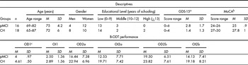
Table 1 shows the demographic characteristics of the two groups of older adult participants, their MoCA and GDS-15 score as well as the mean and standard deviation for all variables of olfactory functioning measured via the B-ODT (Table 1). At this point it should be noted that the two groups did not differ significantly in age [F (1, 33) = 2.77, p > .05], gender [χ 2 (1, N = 34) = 1.42, p > .05], and educational level [χ 2 (2, N = 34) = 2.89, p > .05]. As regards the GDS-15 mean score, the two groups differed significantly [F(1, 33) = 7.04, p = .01]: The pMCI group displayed a higher mean score, compared to the CH group. However, as already mentioned, all the participants had scored lower than the cut-off value for diagnosing probable depression with the GDS-15.
Descriptives and Performance in the Brief Odor Detection Test (B-ODT) of Probable Mild Cognitive Impairment (pMCI) and Cognitively Healthy (CH) Older Adult Groups

a GDS-15 = Geriatric Depression Scale-15.
b MoCA = Montreal Cognitive Assessment.
c OD1 = O-DETECT-SCALE, OI1 = O-IDENT-SCALE, OD2a = O-DETECT-DISTANCE-L, OI2a = O-IDENT-DISTANCE-L, OD2b = O-DETECT-DISTANCE-R, OI2b = O-IDENT-DISTANCE-R.
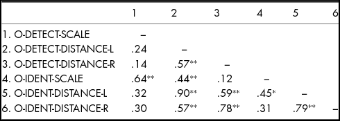
Next, Pearson correlations were computed between the variables related to olfactory functioning. As shown in Table 2, the results showed a positive statistically significant correlation of a medium level between the two variables of the first condition of the B-ODT (O-DETECT-SCALE, O-IDENT-SCALE). However, O-DETECT-SCALE had not been found to display any other significant correlation, while O-IDENT-SCALE has also a very limited number of significant correlations with other variables. On the other hand, all the variables of the second condition of the B-ODT were found to relate with each other, with O-DETECT-DISTANCE-L and O-IDENT-DISTANCE-L displaying a very high positive correlation with each other.
Pearson Correlations between Odour Detection and Odour Identification Measures of the Brief Odor Detection Test (B-ODT)

*p < .05.
**p < .01.
Subsequently, to examine the relationships between general cognitive functioning as measured via the MoCA test, and odour detection and identification as measured via the B-ODT, a series of one-way ANOVAs were performed. The first of them, in which the group of participants according to cognitive functioning was entered as the independent variable, and the performance in the O-DETECT-SCALE variable (Condition 1) as the dependent variable, showed a statistically significant difference in odour detection between the group of older adults with probable MCI (pMCI) and the group of cognitively healthy older adults (CH): The CH group displayed a significantly higher performance, compared to the pMCI group, F (1, 32) = 5.54, p < .05, η2 = .15 (Figure 1).
Odour detection ability of probable MCI (pMCI) and cognitively healthy (CH) older adult groups: birhinal examination using different concentrations of vanillin.

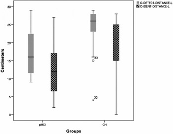
In the same vein, there was found a statistically significant higher performance of the CH group, compared to the pMCI group, in odour detection, when detection was measured as threshold distance from the left nostril (Condition 2: O-DETECT-DISTANCE-L), F (1, 32) = 6.99, p = .01, η2 = .18 (Figure 2). As expected, given the high inter-correlation between odour detection and identification when measured via distance from the left nostril, the CH group presented a statistically significant higher performance in odour identification (O-IDENT-DISTANCE-L) too, compared to pMCI group, F (1, 30) = 7.17, p = .01, η2 = .19 (Figure 2). However, the participants in the pMCI group did not differ from the CH group in O-DETECT-DISTANCE-R. Moreover, they did not differ in odour identification performance either via scale measuring or via distance (from the right nostril) measuring. Finally, as shown in Figure 2, the box plot from the Condition 2a displays two outliers in the first column (O-DETECT-DISTANCE-L) for the CH group.
Odour detection and identification ability of probable MCI (pMCI) and cognitively healthy (CH) older adult groups: unirhinal examination measuring vanillin's distance from the left nostril.

For the two odour detection scores showing significant group-level differences, ROC curves were constructed to evaluate their ability to distinguish pMCI and CH groups at individual level. Given the presence of the outliers, the ROC analyses were conducted both with their inclusion, and then recalculated with the two outliers excluded. The area under the ROC curve (AUC) was used as an index of discrimination performance. For each measure, the ability to distinguish different groups was assessed by examining whether AUC was significantly different from 0. Only the O-DETECT-DISTANCE-L score was able to discriminate pMCI and CH older adults (AUC = .76, p = .009, 95% CI: .60–.93; sensitivity: 75%, specificity: 72%, including outliers; AUC = .86, p = .001, 95% CI: .72–.99; sensitivity: 75%, specificity: 87%, without outliers). The value of ‘22’ was considered an optimal cut-off value as regards this measure of the B-ODT (Figure 3).
Receiver Operating Characteristics (ROC) curves computed to assess the ability of the score in the B-ODT condition of ‘odour detection from the left nostril’ to discriminate between cognitively healthy controls (CH) and older adults with probable mild cognitive impairment (pMCI). The first figure refers to the variable of ‘O-DETECT-DISTANCE-L’ including outliers, while the second refers to the same variable without the outliers shown in Figure 2.

Discussion
The aim of this study was to describe a new brief instrument, namely the B-ODT, developed to measure odour detection sensitivity in older adults, in order to differentiate persons with very mild cognitive impairment from their counterparts who maintain a relatively robust level of cognitive functioning. The findings primarily support the claim that functional asymmetries in odour detection sensitivity, as estimated via unirhinal assessment, may assist in this differentiation.
As regards odour detection measurement using two different methods, the preliminary results showed that older adults seem to recruit different cognitive processes, in order to perform each one of them. Even if it is suggested that threshold determination is rather driven by lower-level perceptual functions, and correlates with the volume of the olfactory bulb that is related to peripheral sensory input (Haehner, Rodewald, Gerber, & Hummel, Reference Haehner, Rodewald, Gerber and Hummel2008; Rahayel et al., Reference Rahayel, Frasnelli and Joubert2012), the pattern of the correlations between the different variables related to odor detection in this study, showed that besides the recruitment of the same lower-level functions, the two conditions of the B-ODT may involve different types of higher-order cognitive functions.
In relation to odour detection as measured by the ordinal scale, it is obvious that the process entailed some kind of discrimination between stimuli (i.e., the odour from the water). Although the discrimination is based primarily on sensory properties of the stimuli, it can be suggested that executive functions, such as inhibitory control, updating, and switching, may also be prevalent in threshold assessments of this type (Hedner, Larsson, Arnold, Zucco, & Hummel, Reference Hedner, Larsson, Arnold, Zucco and Hummel2010). In other words, odour detection ability as measured by the first condition compared to the second condition may be found to differ in that the former poses higher-level demands on cognitive functioning.
At the methodological level, it should be mentioned that the unidirectional procedure of stimuli presentation, that is followed in the first condition of the B-ODT, has been considered a less reliable way than a procedure in which the concentration of an odorant is increased and subsequently decreased, following trials where failure and correct detection occur, respectively, and an average score is used to estimate the threshold (Doty, Reference Doty2015). Moreover, a plausible explanation for the limited number of correlations between the measures of the first condition of the B-ODT and the other measures is that a relatively non-sensitive scale (ordinal, 5-point scale) used in this condition, compared to the second one.
Involvement of cognitive functions in odour detection measures of the B-ODT appears to be corroborated by the findings regarding the differentiation of older adults with pMCI from the CH participants (as they were divided according to their performance in the MoCA) in odour detection sensitivity. The MoCA assesses several cognitive domains. Among these, multiple aspects of executive functions are assessed using an alternation task adapted from the trail-making B task, a phonemic fluency task, and a verbal abstraction task. Attention, concentration, and working memory are evaluated using a sustained attention task, a serial subtraction task, and digits forward and backward. Language, short-term memory recall and delayed recall, visuospatial abilities, and orientation to time and place are also assessed (MoCA, 2006; Nasreddine et al., Reference Nasreddine, Chertkow, Phillips, Whitehead, Collin and Cummings2004). On the basis of the findings, there seems to be a cerebral substrate connecting general cognitive function – including executive functions – and olfactory sensitivity.
It has been proposed that among the brain regions that could constitute such a substrate should be included the amygdala, which is responsible for emotional processing and uncertainty estimation, the olfactory tubercle involved in motivated behaviour, the entorhinal cortex – hippocampus network that supports episodic and working memory, as well as the orbitofrontal cortex (OFC) involved in higher-order flexible cognitive behaviour such as decision making and expectations (Wilson et al., Reference Wilson, Xu, Sadrian, Courtiol, Cohen and Barnes2014).
In fact, the connections among these regions are reciprocal: for instance, the piriform cortex which is responsible for basic odour quality discrimination displays heavy reciprocal connections with the entorhinal cortex and the OFC. In this vein, the pathology of a connected region could be reflected not only on the behaviour that this specific region supports, but also on the behaviour supported by other regions of the network (Doty & Kamath, Reference Doty and Kamath2014). Thus, for example, the vulnerability of the Layer II neurons of the entorhinal cortex to AD pathology (Braak & Braak, Reference Braak and Braak1995) could explain to some extent the olfactory deficits in AD (see also Rahayel et al., Reference Rahayel, Frasnelli and Joubert2012).
Recently, the mediodorsal thalamic nucleus (MDT) has been indicated as an important region that is involved in both higher-order cognition and olfaction (Courtiol & Wilson, Reference Courtiol and Wilson2015): The MDT has dense reciprocal connections with prefrontal regions supporting executive functions and its role in cognition is well established. Several studies have indicated the role of MDT in working memory, goal-directed behaviour, and cognitive flexibility (Courtiol & Wilson, Reference Courtiol and Wilson2015). In addition, the MDT is bidirectionally connected with primary and secondary olfactory areas. Hence, it can be viewed as a higher-order olfactory thalamus, the dense connections of which may be involved in both complex cognition and olfactory processing including odour detection (Courtiol & Wilson, Reference Courtiol and Wilson2015). Significant neuro-chemical changes, that are likely present prior to the established onset of a neuro-cognitive disorder and can be region specific, may also explain the association between cognition and olfaction (Doty & Kamath, Reference Doty and Kamath2014; Larsson et al., Reference Larsson, Farde, Hummel, Witt, Erixon Lindroth and Backman2009; Miwa et al., Reference Miwa, Watanabe, Mitsumoto, Furukawa, Fukushima and Moriizumi2004).
In sum, Hypothesis 1, namely that older adults with very mild cognitive impairment would display significantly lower performance in the two conditions of the B-ODT in terms of both odour detection and identification, compared to cognitively healthy older adults, was partially confirmed. Clearly, the performance in the B-ODT can differentiate older adults with pMCI and CH persons, in terms of odour detection sensitivity measured as odour detection distance from the left nostril, while, based on the strength of the respective correlation coefficient, odour identification distance from the left nostril appeared to be a simultaneous and highly inter-connected to detection process. Such a finding verifies Doty's (Reference Doty2015) claim that there possibly exists a ‘general olfactory acuity’ factor.
As regards left-side lateralisation (Hypothesis 2), the findings of the present study appear to confirm studies, suggesting that to differentiate older adults with and without neuro-degenerative disorders affecting olfactory function, unirhinal assessments might be more informative than birhinal ones (Bahar-Fuchs et al., Reference Bahar-Fuchs, Moss, Rowe and Savage2010; Huart et al., Reference Huart, Rombaux, Gerard, Hanseeuw, Lhommel, Quenon and Mouraux2015; Stamps et al., Reference Stamps, Bartoshuk and Heilman2013). However, at this point it should be noted that most of the extant research that supports asymmetrical progression of AD-related neuro-pathology does not necessary suggest a predominant involvement of a specific hemisphere (Bahar-Fuchs et al., Reference Bahar-Fuchs, Moss, Rowe and Savage2010; Bottino et al., Reference Bottino, Castro, Gomes, Buchpiguel, Marchetti and Neto2002; Huart et al., Reference Huart, Rombaux, Gerard, Hanseeuw, Lhommel, Quenon and Mouraux2015; Pantel, Kratz, Essig, & Schroder, Reference Pantel, Kratz, Essig and Schroder2003). Nevertheless, the findings of this preliminary study support left-side lateralisation.
In addition to an earlier and quicker left POC and hippocampal volume atrophy found in the study of Vasavada et al. (Reference Vasavada, Wang, Eslinger, Gill, Sun, Karunanayaka and Yang2015), recent voxel-based morphometric studies of grey matter volume loss, consistently find significantly greater left than right mesial temporal lobe atrophy in MCI or healthy older adults who were progressed to AD (see, for a review, Ferreira, Diniz, Forlenza, Busatto, & Zanetti, Reference Ferreira, Diniz, Forlenza, Busatto and Zanetti2011). From this standpoint, the significant differentiation of the two groups of older adult participants in the present study, in odour detection from the left nostril, seems to ‘capture’ asymmetrical atrophy such as the aforementioned one, and in this way it fully confirms Hypothesis 2.
Limitations
The preliminary ‘nature’ of the study is the major limitation. The study was based on a small sample of participants in each group, and therefore the findings are essentially tentative. Replication of these findings in larger and pathological samples of older adult participants and the opportunity for follow-up analyses are needed to increase confidence in these findings. Various and more objective criteria for cognitive impairment should also be used, besides common neuro-psychological tests of general cognitive functioning. More accurate ways of measuring odour detection sensitivity should be developed, to improve the validity of the B-ODT. In this vein, although the order of administration of the two conditions of the B-ODT is changed, there still remains an issue concerning potential effects of previous exposure to vanillin on the performance in the condition that follows. More research is needed to examine such effects. However, given that based on the preliminary findings the most promising tool for detection of early cognitive decline seems to be the second condition of the B-ODT, future research should examine the possibility to use only this condition, compared to the administration of both B-ODT conditions.
Conclusions
The results of the present study support the suggestion that olfactory functioning measures, mainly in the form of odour detection sensitivity tests, could predict very mild cognitive impairment in older adults. Specifically, the findings support the claim that functional asymmetries in olfactory performance measured as odour detection sensitivity may assist in differentiating cognitively healthy older adults from persons with probable MCI, and that these asymmetries can be detected through unirhinal assessment. On the basis of these results, the B-ODT seems a promising instrument for very early cognitive impairment screening in older adult population.
Financial Support
This research received no specific grant from any funding agency, commercial or not-for-profit sectors.
Conflict of Interest
The authors have no conflicts of interest to declare.
Ethical Standards
The authors assert that all procedures contributing to this work comply with the ethical standards of the relevant national and institutional committees on human experimentation and with the Helsinki Declaration of 1975, as revised in 2008.