




No CrossRef data available.
Article contents
Ventricular preexcitation associated with dilated cardiomyopathy: a causal relationship?
Published online by Cambridge University Press: 21 January 2005
Abstract
Over a six year period, we identified four children with dilated cardiomyopathy associated with ventricular preexcitation, three in the absence of any documented tachyarrhythmias, and one with but a solitary episode of tachyarrhythmia. None of them had another identifiable aetiology for the cardiomyopathy. Based on the 12 lead electrocardiogram, and/or invasive electrophysiologic studies, we localised the accessory pathways to the right atrioventricular groove in all patients. We commenced antifailure medications in two of the four patients, but did not produce any measurable improvement in ventricular function. Catheter ablation was performed in two patients, with resolution of the cardiomyopathy. The ventricular preexcitation disappeared spontaneously in one child during follow-up, again with resolution of cardiomyopathy. The youngest patient continues to receive antifailure medications, albeit without improvement. We suggest, therefore, that dilated cardiomyopathy of reversible nature is associated with manifest ventricular preexcitation, even in the absence of sustained arrhythmias.
Information
- Type
- Original Article
- Information
- Copyright
- © 2004 Cambridge University Press
The association between dilated cardiomyopathy and the Wolff–Parkinson–White variant of ventricular preexcitation with associated reentrant supraventricular tachycardia is well recognised, with dilated cardiomyopathy usually appearing as the sequel to the recurrent and sustained tachyarrhythmia.1, 2 In this report, we describe four young patients, all with Wolff–Parkinson–White syndrome associated with dilated cardiomyopathy, but in the absence of any documented arrhythmia in three of the patients, and with only a single self-limiting episode of reentrant supraventricular tachycardia in the other patient. In two of them, catheter ablation of the accessory pathway resulted in resolution of the cardiomyopathy. The third patient had spontaneous loss of preexcitation during follow-up, which was again associated with resolution of the cardiomyopathy. Our final patient, the youngest child in our series, continues to take antifailure medications.
Patients
The four patients, 3 girls and 1 boy, presented to one of the institutions between July 1997 and June 2003. The presenting complaints were failure to thrive with an associated systolic murmur in two, one of whom also had tiredness with decreased exercise tolerance, a solitary documented episode of reentrant supraventricular tachycardia in one, and an isolated systolic murmur in the other. The age at presentation ranged between 12 months and 13 years. All patients had in common ventricular preexcitation as seen on the 12 lead electrocardiogram, diagnostic of the Wolff–Parkinson–White syndrome (Figs 1–4), with the configuration of the delta wave suggesting a right-sided pathway. On echocardiography, all patients had evidence of cardiomyopathy, with left ventricular end-diastolic diameters well above the 97th centile for weight (see Table 1), and impaired shortening fractions of less than 30%. Of the four patients, three have undergone annual ambulatory Holter recordings for periods of up to 4 weeks, without any tachyarrhythmia being recorded. For comparison, an average of between 40 and 60 patients with Wolff–Parkinson–White syndrome have been seen annually at each of our institutions between 1997 and 2003, and dilated cardiomyopathy has not been observed in any of these in the absence of recurrent tachyarrhythmia.

Figure 1. The 12 lead electrocardiogram from our first patient at initial presentation (a), confirming ventricular preexcitation. There was spontaneous loss of preexcitation during follow-up (b), which was associated with recovery of ventricular function.

Figure 2. The 12 lead electrocardiogram from our second patient (a), who had had one documented episode of reentrant supraventricular tachycardia. We recorded surface and intracardiac electrocardiographic recordings in this patient catheter ablation (b). The surface leads I, aVF and V5 are shown. The intracardiac leads are high right atrium (HRA), coronary sinus (CS), right ventricle (RV) and the mapping and ablation catheter (MAP). The first beats demonstrate reentrant tachycardia, with earliest retrograde atrial activation in the right atrium (large arrows), confirming that the pathway is right sided. The third ventricular complex is not followed by retrograde atrial activation, demonstrating loss of retrograde conduction to the atriums and termination of tachyarrhythmia. Thereafter, the following beat (4th beat) arises from the sinus node, with earliest activation in the right atrium (small arrow) and a normal sequence of atrioventricular activation.

Figure 3. Electrograms are shown before (a) and after (b) ablation in our third patient, showing the loss of preexcitation following ablation.

Figure 4. The electrogram from our fourth and youngest patient, who has not yet undergone catheter ablation.
Table 1. Patient characteristics data, and investigations and management.

Other investigations, such as family history, metabolic studies and viral screening, offered no additional clue to the aetiology of the cardiomyopathy. We commenced antifailure medication with digoxin, diuretics, and inhibitors of angiotensin converting enzyme in two of the four patients. Digoxin was also used in these patients, despite its potential for shortening the atrial refractory period and enhancing atrioventricular conduction, as neither of the two patients had had a previously documented tachyarrhythmia. In view of their young age, preexcited atrial fibrillation was also considered to be unlikely. Our only patient with a documented tachyarrhythmia, the second patient in the Table, received propafenone prior to catheter ablation.
Follow-up
During a follow-up ranging from three to 66 months, two of the four patients have undergone diagnostic cardiac catheterisation and right ventricular biopsy. The left ventricular end diastolic pressure was mildly elevated in both, at 10 and 13 millimetres of mercury, respectively. Biopsies from multiple sites were negative for active myocarditis, viral cultures, fibrosis, or fatty changes.
At the request of the parents, our second and third patients have undergone invasive electrophysiologic studies and catheter ablation. The accessory pathway was localised paraseptally in to the right atrioventricular groove,3 and nonsustained reentrant supraventricular tachycardia could be induced using standard extrastimulation techniques. Delivery of radiofrequency current at this site resulted in abolition of conduction across the accessory pathway, with disappearance of the delta wave. Echocardiographic examination repeated at follow-up two weeks later confirmed normalization of myocardial function. Preexcitation disappeared spontaneously from the surface electrocardiogram of our first patient at follow-up, 54 months subsequent to the initial diagnosis of cardiomyopathy. This was associated with normalization of left ventricular function compared with the findings from 6 months previously, although her presenting symptoms remained unchanged. Our fourth and youngest patient, who presented with failure to thrive, continues to receive antifailure medications, albeit with no improvement in ventricular function.
Discussion
In the setting of Wolff–Parkinson–White syndrome, cardiomyopathy is known to be the result of recurrent and sustained episodes of tachyarrhythmia. Apart from a single documented episode in one of our patients, none of the others had had symptoms suggestive of tachyarrhythmia, or any documented tachyarrhythmias. The cardiomyopathy resolved completely in three of the four patients, with a definite temporal relation between the disappearance of preexcitation and resolution of the cardiomyopathy. Only one of the patients had symptoms attributable to the cardiomyopathy, and these symptoms persisted despite spontaneous loss of conduction across the accessory pathway and normalization of left ventricular function.
A possible mechanism for the cardiomyopathy could be ventricular dysfunction secondary to nonphysiologic activation of ventricular myocardium via the accessory pathway, as has been reported in patients undergoing permanent implantation of pacemakers.4, 5 This form of cardiomyopathy related to the site of pacing, however, is associated with structural changes in the ventricular myocardium.4, 6 In the two patients in whom diagnostic cardiac catheterisation was performed, multiple right ventricular biopsies failed to reveal structural changes. In addition, cardiomyopathy associated with structural alterations in the myocardium would be unlikely to reverse rapidly. Another possibility is that eccentric ventricular activation via the accessory pathway might mimic cardiomyopathy, due to asynchronous spread of the depolarising current. Wolff–Parkinson–White syndrome, nonetheless, is relatively common, and it may be expected that dilated cardiomyopathy in asymptomatic patients would have been observed more frequently if it was entirely the result of asynchronous ventricular activation. A genetic basis for this association is unlikely, as none of the patients had a positive family history, and the cardiomyopathy was reversed with abolition of preexcitation. A potentially significant observation may be that the accessory pathway was right sided in all four patients.
Regardless of the mechanism of cardiomyopathy, the important question remains as to what we should recommend for the asymptomatic child with dilated cardiomyopathy associated with ventricular preexcitation? Catheter ablation in young patients is not without risks, although the rates of success at the present time are in excess of 90%. If screening studies to determine the aetiology of dilated cardiomyopathy are negative, and the cardiomyopathy persists at follow-up, our limited results suggest that ablation produces complete recovery of ventricular function. This option should, therefore, be considered in the management of such patients.
References
Fenelon G, Wijns W, Andries E. Tachycardiomyopathy: mechanisms and clinical implications. Pacing Clin Electrophysiol 1996; 19: 95–106.Google Scholar
Horenstein MS, Saarel E, Dick M, Karpawich PP. Reversible symptomatic dilated cardiomyopathy in older children and young adolescents due to primary non-sinus supraventricular tachyarrhythmias. Pediatr Cardiol 2003; 24: 274–279.Google Scholar
Cosio FG, Anderson RH, Kuck K-H, et al. Living anatomy of the atrioventricular junctions. A guide to electrophysiologic mapping. A consensus statement from the Cardiac Nomenclature Study Group, Working Group of Arrhythmias, European Society of Cardiology, and the Task Force on Cardiac Nomenclature from NASPE. Circulation 1999; 100: e31–e37.Google Scholar
Karpawich PP, Rabah R, Haas JE. Altered cardiac histology following apical right ventricular pacing in patients with congenital atrioventricular block. Pacing Clin Electrophysiol 1999; 22: 1372–1377.Google Scholar
Tantengco MV, Thomas RL, Karpawich PP. Left ventricular dysfunction after long-term right ventricular apical pacing in the young. J Am Coll Cardiol 2001; 37: 2093–2100.Google Scholar
Rosenqvist M, Isaaz K, Botvinnick EH, et al. Relative importance of activation sequence compared to atrioventricular synchrony in left ventricular function. Am J Cardiol 1991; 67: 148–156.Google Scholar

The 12 lead electrocardiogram from our first patient at initial presentation (a), confirming ventricular preexcitation. There was spontaneous loss of preexcitation during follow-up (b), which was associated with recovery of ventricular function.

The 12 lead electrocardiogram from our second patient (a), who had had one documented episode of reentrant supraventricular tachycardia. We recorded surface and intracardiac electrocardiographic recordings in this patient catheter ablation (b). The surface leads I, aVF and V5 are shown. The intracardiac leads are high right atrium (HRA), coronary sinus (CS), right ventricle (RV) and the mapping and ablation catheter (MAP). The first beats demonstrate reentrant tachycardia, with earliest retrograde atrial activation in the right atrium (large arrows), confirming that the pathway is right sided. The third ventricular complex is not followed by retrograde atrial activation, demonstrating loss of retrograde conduction to the atriums and termination of tachyarrhythmia. Thereafter, the following beat (4th beat) arises from the sinus node, with earliest activation in the right atrium (small arrow) and a normal sequence of atrioventricular activation.

Electrograms are shown before (a) and after (b) ablation in our third patient, showing the loss of preexcitation following ablation.

The electrogram from our fourth and youngest patient, who has not yet undergone catheter ablation.
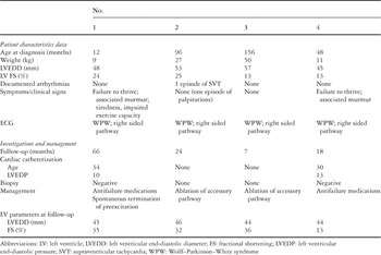
Table 1.
 You have
Access
- 63
- Cited by