Introduction
Supernumerary teeth occur in 0.1–1 per cent of the general population.Reference Thawley and LaFerriere1 The most common type encountered by dentists and oral and maxillofacial surgeons is the mesiodens, which occurs in the midline between the two central incisors. However, supernumerary nasal teeth are very rare.Reference Kirmeier, Truschnegg, Payer, Malyk, Daghighi and Jakse2
The cause of such supernumerary nasal teeth is unclear. Thawley and LaFierriereReference Thawley and LaFerriere1 advocate that supernumerary nasal teeth develop either from a third tooth bed that arises from the dental lamina near the permanent tooth bud, or possibly from splitting of the permanent bud itself. PrimoschReference Primosch3 does not accept the latter theory because the majority of supernumerary nasal teeth present as ectopic and single. Some cases of eruption in the nasal cavity are associated with aetiological factors such as genetic pre-disposition, being offset by dental trauma, local infection, cysts or cleft palate. In rare cases, osteomyelitis and congenital syphilis may also be related. Some authors believe that supernumerary nasal teeth are inverted mesiodens that erupted in the nasal cavity or in the presence of a mechanical barrier that diverts the eruption of a supernumerary nasal tooth to the nasal cavity.Reference Kim, Kim, Chae, Hwang, Lee and Lee4
Supernumerary nasal teeth are asymptomatic in the early stages. However, they may prevent and delay the eruption of normal teeth, leading to their malalignment in later life.Reference Dhingra and Gulati5 Supernumerary nasal teeth generally present with symptoms, such as nasal discharge, epistaxis, unilateral nasal obstruction, headaches, nasal discharge, abscess, and crusting of nasal mucosa with growth. The symptoms are most likely related to breaching of the nasal mucosa and partial obstruction of the nasal cavity by the erupting tooth.Reference Janardhan, Kumar, Reddy and Kumar6
Supernumerary nasal teeth are usually misdiagnosed, not only because of their rarity, but also because the supernumerary nasal tooth is embedded in hypertrophic nasal mucosa or granulation tissue.Reference Primosch3 This report presents the clinical case records of four patients with symptomatic supernumerary nasal teeth.
Materials and methods
This study was reviewed and approved by the Institutional Ethical Review Board of Yiwu Central Hospital, Wenzhou Medical University, Zhejiang. Informed consent was obtained from all participants prior to enrolment.
The clinical records of patients with supernumerary nasal teeth who presented to the Otolaryngology Outpatient Clinic at Yiwu Central Hospital, Wenzhou Medical University, between August 2005 and August 2018, were accessed through the records department of the hospital. Age, sex, clinical symptoms and signs within the nasal cavity, and treatment details were analysed for each patient.
In addition, a brief review of the literature was conducted, and a subsequent overview provided, in relation to the aetiology, symptomatology, diagnosis and treatment of intranasal teeth.
Results
All four of the patients identified were male and had a supernumerary nasal tooth in the right nasal cavity. The patients ranged in age from 16 to 28 years. Three of the four patients had previously been misdiagnosed.
All supernumerary nasal teeth were surrounded by granulation tissue or hypertrophic nasal mucosa, and were subsequently confirmed by computed tomography (CT) and endoscopy. The granulation tissue or hypertrophic nasal mucosa was removed using microwave ablation, and each supernumerary nasal tooth was successfully removed by endoscopy.
Case reports
Case one
A 22-year-old male was referred to our otolaryngology department on 30 August 2005 with a history of recurrent right-sided, blood-stained discharge. The patient had nasal obstruction, headache, chronic purulent rhinorrhoea, facial swelling and hyposmia. There was no history of trauma, allergy or smoking.
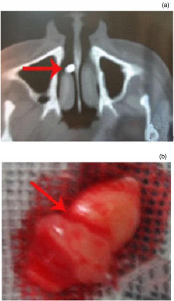
On examination, he was found to have granulation tissue between the inferior nasal conchae and the right side of his nasal septum, and mild epistaxis; in addition, hard tissue was found. Computed tomography of the paranasal sinuses revealed a radiopaque mass resembling an ectopic tooth in the right nasal floor (Figure 1).
(a) Axial computed tomography scan of the paranasal sinuses confirmed the presence of a white mass in the floor of the nasal cavity. (b) The supernumerary nasal tooth was removed. (Red arrows indicate the supernumerary nasal tooth.)

Pledgets of cotton soaked in 1 per cent Dicaine with adrenaline were placed in the right nasal cavity to improve visibility and prevent bleeding. The granulation tissue in the common meatus was removed by endoscopic microwave ablation. Endoscopy confirmed the presence of a tooth in the floor of the nasal cavity, which was surrounded by granulation tissue. The number of teeth in the arch was normal. The tooth with a conical shape was subsequently removed endoscopically, and all the granulation tissue was coagulated by microwave ablation. A diagnosis of supernumerary nasal tooth was made.
Case two
A 28-year-old male was referred to our otolaryngology department on 17 November 2005 for evaluation, following intermittent episodes of right-sided epistaxis and mild nasal obstruction for 2 months. The patient had visited two other hospitals, and had been diagnosed with a ‘rhinopolyp’ and ‘nasal cavity angioma’.
Clinical examination with a nasal speculum showed red granulation tissue in the right nostril with some blood-stained discharge. A CT scan of the paranasal sinuses revealed a well-defined radiopaque mass between the inferior nasal conchae and septum, suggestive of a supernumerary tooth erupting into the nasal cavity on the right side. The number of teeth in the arch was normal.
The granulation tissue was removed by endoscopic microwave ablation. A hard white mass was exposed, and the presence of a tooth was confirmed. The tooth with a conical shape was removed endoscopically, and all granulation tissue was coagulated by microwave ablation. A diagnosis of supernumerary nasal tooth accompanied by hyperplasia of granulation tissue was made.
Case three
A 16-year-old male was referred to our otolaryngology department on 17 March 2006 with a history of right-sided nasal obstruction and rhinorrhoea for 3 years. The patient had blood-stained discharge, headache, facial swelling and hyposmia. The patient had repeatedly visited other hospitals, and received diagnoses of ‘chronic rhinitis’ and ‘hypertrophic rhinitis’.
Clinical examination with a nasal speculum showed hypertrophic inferior nasal conchae closing to the septum. Pledgets of cotton soaked in 1 per cent Dicaine with adrenaline were placed in the right nasal cavity. After 10–15 minutes, the cotton was removed. A hard white mass resembling an ectopic tooth was found in the right nasal floor. A CT scan of the paranasal sinuses confirmed the presence of a tooth in the floor of the nasal cavity. The root of the tooth was firmly embedded in the septum and alveolar process of the maxilla, and was surrounded by hypertrophic nasal mucosa. The root of the tooth was separated from the septum with a detacher, and the tooth with a conical shape was removed by endoscopy. The number of teeth in the arch was normal.
Case four
A 21-year-old male was referred to our otolaryngology department on 25 January 2018 with a history of right-sided nasal obstruction and some recurrent blood-stained discharge for 2 years. The patient had repeatedly visited other hospitals, and had been diagnosed with ‘chronic rhinitis’ and ‘neoplasm of the nasal cavity’.
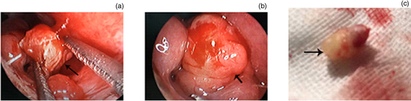
Clinical examination with a nasal speculum showed an irregular mass on the common meatus. The inferior nasal conchae and septum were extruded by the mass, thus resulting in septum deviation and smaller inferior nasal conchae (Figure 2). A CT scan of the paranasal sinuses confirmed the presence of a tooth in the floor of the nasal cavity. The tooth was surrounded by hypertrophic nasal mucosa. The number of teeth in the arch was normal. The tooth with a conical shape was removed by endoscopy under general anaesthesia.
(a) The mass was seen between the inferior nasal conchae and septum. (b & c) The supernumerary nasal tooth was removed. (Black arrows indicate the supernumerary nasal tooth.)

Discussion
The presence of a tooth in the nasal cavity or maxillary sinus is unusual. The cause of such supernumerary nasal teeth is unclear. One theory suggests that the supernumerary nasal tooth is created either from a thin tooth bud that arises from the dental lamina near the permanent tooth bud, or the tooth germ undergoes a dichotomy.Reference Dhingra and Gulati5 Another theory is that their development is a reversion to the dentition of extinct primates, which had three pairs of incisors.Reference Dhingra and Gulati5,Reference Liu7 The hyperactivity theory suggests that supernumerary nasal teeth are formed as a result of local, independent hyperactivity of the dental lamina.Reference Liu7 The role of epithelial mesenchymal interaction has also been suggested.Reference Ray, Singh, Das and Roy8
The formation of supernumerary nasal teeth can also result from: the presence of any obstruction at the time of tooth eruption due to a crowded dentition, persistent deciduous teeth or exceptionally dense bone; developmental disturbances, such as a cleft palate; rhinogenic or odontogenic infection; and displacement as a result of trauma or cysts.Reference Moreano, Zich, Goree and Graham9 Rege et al.Reference Rege, Shal and Marfati10 described osteomyelitis of the maxilla as a cause of nasal teeth and reported three ectopic nasal teeth following osteomyelitis of the maxilla.
None of the four patients in the present study had a history of odontogenic infection, or osteomyelitis of the maxilla or cleft palate, and the number of teeth in the arch was normal. However, they did have a history of repeated rhinogenic infection. We speculate that supernumerary nasal teeth could be related to a rhinogenic infection.
Supernumerary nasal teeth are usually misdiagnosed because they are asymptomatic or the symptoms are atypical. Here, we presented four patients with supernumerary nasal teeth, three of whom had previously been misdiagnosed. Supernumerary nasal teeth are commonly misdiagnosed as rhinopolyps, nasal cavity angioma, chronic rhinitis, hypertrophic rhinitis, epistaxis and so forth. This is due not only to the inexperience of the clinician, but also to the features of the supernumerary nasal tooth.
The number of teeth in the arch was normal in all four patients in the present study. In addition, the supernumerary nasal tooth was completely covered by nasal mucosa, and surrounded by granulation tissue and necrotic debris. The supernumerary nasal tooth stimulated the adjacent nasal mucosa or injured the nasal mucosa, thereby resulting in hypertrophy and proliferation of granulation tissue as it gradually increased in size. The inferior nasal conchae and septum were also extruded by the supernumerary nasal tooth, resulting in deviation of the septum. Thus, hyperactivity of supernumerary nasal teeth may cause a series of clinical symptoms. When a supernumerary nasal tooth as an ivory white mass is not covered or is only partly covered by the hypertrophic nasal mucosa or granulation tissue, a misdiagnosis of a foreign body, rhinolith, osteoma, calcifying odontogenic cyst, or malignant tumour, such as chondrosarcoma and osteosarcoma, may be made.Reference Mittal and Sultan11,Reference Kumar, Namdev, Bakshi and Dutta12
The diagnosis of supernumerary nasal teeth mainly depends on signs within the nasal cavity and radiological examination findings. Dental panoramic tomographs would be helpful in outlining the exact relationship and orientation of the supernumerary or ectopic tooth with respect to the existing dentition. However, some authors have suggested that conventional radiological examination may not be able to confirm supernumerary nasal teeth precisely, as panoramic radiographs only provide information on the state of dentition and help determine if a tooth is supernumerary, which may not be sufficient to identify intranasal teeth off the midline.Reference Mittal and Sultan11,Reference Kumar, Namdev, Bakshi and Dutta12
Nevertheless, CT is useful for identifying the exact position of the supernumerary nasal tooth, and will help in deciding whether the tooth can be removed through an intraoral or transnasal approach. Although the differential diagnosis of an intranasal white mass includes a foreign body, odontoma, osteoma, calcifying odontogenic cyst, and malignant tumours, such as chondrosarcoma and osteosarcoma, the CT findings of tooth-equivalent attenuation and a centrally located cavity are highly discriminating features.Reference Iwai, Aoki, Yamashita, Omura, Matsui and Maegawa13–Reference Vinothkumar, Kandaswamy, Arathi, Ramkumar and Felsypremila15
• Supernumerary nasal teeth are rare and usually misdiagnosed in rhinology clinics
• These teeth may be surrounded by hypertrophic nasal mucosa or granulation tissue
• Supernumerary nasal teeth should be confirmed by computed tomography (CT) and endoscopy
• Paranasal sinus CT revealed a white well-defined radiopaque mass in the nasal cavity
In order to avoid misdiagnosis of supernumerary nasal teeth, clinicians need to increase their awareness of supernumerary nasal teeth; in addition, conventional CT and dental panoramic tomographs should be performed in patients with neoformation of the nasal cavity. Treatment is not difficult, and the supernumerary nasal tooth can be removed under endoscopic guidance.
Competing interests
None declared