Introduction
Calcium is an essential mineral for snail survival, as one of the main inorganic elements making up these animals' shells, giving them the resistance necessary for protection. This ion has been described as a limiting factor in the distribution and adaptation of adult snails, as well as in their egg-laying capacity and the development of eggs and embryos (Tunholi et al., Reference Tunholi, Lustrino, Tunholi-Alves, Mello-Silva, Maldonado, Rodrigues and Pinheiro2011a). Therefore, this element is fundamental to the colonization of these organisms in the environment. Other studies have also indicated the importance of calcium as an enzyme cofactor in redox reactions, and for its involvement in the metabolic process related to acid–base balance and in the immune response of snails to parasites (Sminia et al., Reference Sminia, de Witt, Bos, van Nieuwmegen, Witter and Wondergem1977; de Witt & Sminia, 1980).
Calcium is not only essential for snail survival, it is also an important element manipulated by parasites during their development in the intermediate host to maintain their life cycle. Several studies have investigated the influence of parasite infection on the level of calcium in host snails. An increase in the calcium content of the shell of Lymnaea stagnalis infected with Trichobilharzia ocellata was demonstrated by McCleland & Bourns (Reference McCleland and Bourns1969). However, Tunholi et al. (Reference Tunholi, Lustrino, Tunholi-Alves, Garcia, Mello-Silva, Maldonado, Rodrigues and Pinheiro2011c) observed a significant increase in the haemolymph calcium concentration accompanied by a reduction in shell reserves of Biomphalaria glabrata when infected by different miracidial doses of Echinostoma paraensei. The same effect was reported by Stewart et al. (Reference Stewart, Ubelaker and Curtis1985) for the haemolymph calcium levels of B. glabrata infected by Angiostrongylus costaricensis. Therefore, the calcium metabolism pattern of snails infected by the larval stages of helminths is highly variable, influenced by intrinsic factors, including variations at the species and individual levels of both host and parasite (Zbikowska, Reference Zbikowska2003; Pinheiro et al., Reference Pinheiro, Maldonado and Lanfredi2009), and extrinsic factors such as the concentration of CaCO3 available in water (Magalhães et al., Reference Magalhães, Pinheiro and Mello-Silva2011).
Among the different species of Metastrongylidae, Angiostrongylus cantonensis stands out for its zoonotic importance, because it can accidentally infect humans, being considered as the main aetiological agent of eosinophilic meningoencephalitis (Thiengo et al., Reference Thiengo, Maldonado, Mota, Torres, Caldeira, Carvalho, Oliveira, Simões, Fernandez and Lanfredi2010). Studies indicate that different snail species can act as intermediate hosts of A. cantonensis, including both terrestrial and aquatic species, such as B. glabrata (Caldeira et al., Reference Caldeira, Mendonça, Goveia, Lenzi, Graeff-Teixeira, Lima, Mota, Pecora, Medeiros and Carvalho2007; Tunholi-Alves et al., Reference Tunholi-Alves, Tunholi, Lustrino, Amaral, Thiengo and Pinheiro2011, Reference Tunholi-Alves, Tunholi, Pinheiro and Thiengo2012). This dependence makes the snail an essential element for development and dispersal of parasites, enabling them to reach the larval stage which can infect the definitive hosts. Recently our group characterized changes in the reproductive and metabolic profile of B. glabrata experimentally infected by A. cantonensis (Tunholi-Alves et al., Reference Tunholi-Alves, Tunholi, Lustrino, Amaral, Thiengo and Pinheiro2011, Reference Tunholi-Alves, Tunholi, Pinheiro and Thiengo2012). However, no information is available about the changes in mineral metabolism during this interaction.
Therefore, because of the importance of this ion to the adaptive success of snails and the lack of information on its participation in the snail/nematode interaction, the present study evaluated, for the first time, the effects of experimental infection by A. cantonensis on the calcium content of the haemolymph and its reserves in the shell of B. glabrata, during 3 weeks of infection, which correspond to the pre-patent period of the intramolluscan larval development of this helminth (Tunholi-Alves et al., Reference Tunholi-Alves, Tunholi, Lustrino, Amaral, Thiengo and Pinheiro2011, Reference Tunholi-Alves, Tunholi, Pinheiro and Thiengo2012). Additionally, histochemical analyses were conducted to determine possible tissue changes resulting from infection.
Materials and methods
Maintenance of snails
Snails were kept in aquaria containing 1500 ml of dechlorinated water, to which 0.5 g of CaCO3 was added, in the Laboratório de Biologia e Parasitologia de Mamíferos Silvestres Reservatórios, Instituto Oswaldo Cruz, Fiocruz (Tunholi et al., Reference Tunholi, Lustrino, Tunholi-Alves, Mello-Silva, Maldonado, Rodrigues and Pinheiro2011a, Reference Tunholi, Lustrino, Tunholi-Alves, Mello-Silva, Maldonado, Pinheiro and Rodriguesb). The water was replaced once a week. Six groups were formed: three control groups (uninfected) and three infected groups. Each aquarium contained ten snails, reared in the laboratory from hatching, to be certain of their age and freedom from infection by other parasites. The entire experiment was conducted in duplicate, using a total of 90 snails, 45 snails in the control groups (uninfected) and 45 snails in the infected groups. The aquaria were kept in a room with controlled temperature of 25°C throughout the experiment. The study was conducted for 3 weeks, a period that corresponds to the pre-patent development of A. cantonensis in B. glabrata (Tunholi-Alves et al., Reference Tunholi-Alves, Tunholi, Lustrino, Amaral, Thiengo and Pinheiro2011). Snails were fed with dehydrated lettuce (Lactuca sativa L.) ad libitum. Lettuce leaves were changed every day to prevent their fermentation, at which time the number of dead snails was also noted.
Parasitological procedures
Third-stage larvae (L3) of A. cantonensis, obtained from Achatina fulica collected in the municipality of Olinda, Pernanbuco, Brazil in 2008, in the area surrounding the home of a patient diagnosed with eosinophilic meningoencephalitis, were inoculated in Rattus norvegicus in the Laboratório of Patologia do Instituto Oswaldo Cruz (Fiocruz), where the cycle was maintained. First-stage larvae (L1) utilized in this study were obtained from this experimental cycle maintained in the same laboratory Young snails (8-12 mm) were individually placed in each hole of 24-hole plates in which 1200 first-stage larvae (L1) per snail had been placed previously (Tunholi-Alves et al., Reference Tunholi-Alves, Tunholi, Pinheiro and Thiengo2012). After 24 h the snails were removed from the plates and transferred to aquaria.
Weekly for 3 weeks post-exposure, the haemolymph was collected by cardiac puncture from randomly chosen snails from each group (n= 10), after which they were dissected and the shell removed, washed and left to dry at room temperature. The haemolymph was maintained in an ice bath during collection and stored at − 10°C.
Calcium determination and histochemical analyses
Calcium content in the haemolymph of B. glabrata was determined by using commercial kits (Doles, Brazil) and the results were expressed as mg calcium/dl of haemolymph. Calcium content in the shell was determined according Pinheiro & Amato (Reference Pinheiro and Amato1995) and Soido et al. (Reference Soido, Vasconcellos, Diniz and Pinheiro2009). Calcium carbonate mass was calculated and expressed as mg CaCO3/g of ash.
Snails from each period of infection were dissected and transferred to Duboscq-Brasil fixative (Fernandes, Reference Fernandes1949). Soft tissues were processed according to routine histological techniques (Humason, Reference Humason1979). Sections (5 μm) were stained using von Kossa's method (Humason, 1979) and observed under a Zeiss Axioplan light microscope. Images were captured by an MRc5 AxioCam digital camera and processed with the Axiovision software (Carl Zeiss do Brasil Ltda, Brasil).
Data analysis
The results obtained were expressed as mean ± standard deviation and the Tukey test and analysis of variance (ANOVA) were used to compare means. A polynomial regression was calculated to analyse the relation between the values obtained and infection time and for the parasite load to which snails were exposed (α = 5%) (InStat, GraphPad, v.4.00, Prism, GraphPad, v.3.02, Prism, Inc., San Diego, California, USA).
Results
Significant changes in the haemolymph calcium concentration and its reserves in the shell of B. glabrata infected experimentally by A. cantonensis larvae were observed during the study period, when the intermediate host was able to normalize the concentrations of this mineral in both sites.
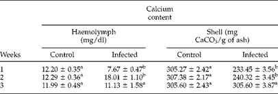
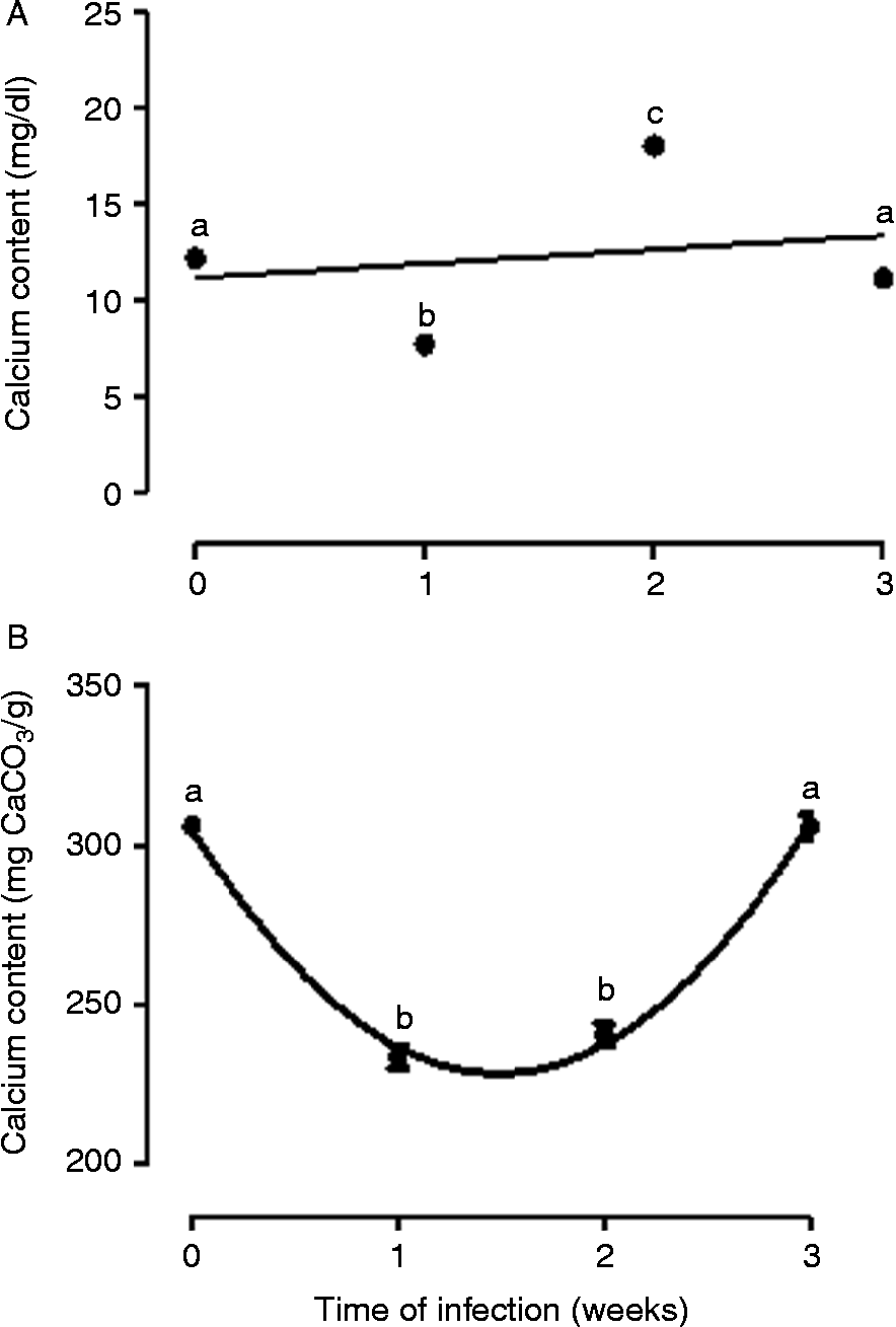
The highest levels of calcium in the haemolymph were detected 2 weeks after exposure (18.01 ± 1.10 mg/dl), an increase of 39.61% in comparison with the respective control group (12.29 ± 0.36 mg/dl) (table 1). However, a significant decrease in this ion was observed 1 week after infection in relation to the control group (7.67 ± 0.47 versus 12.20 ± 0.35 mg/dl, a decline of 37.13% (table 1). Although in both cases these values differed significantly in relation to the average of the control groups, the linear regression analysis did not reveal a pattern during the study period (fig. 1A).
Mean values of calcium content in the haemolymph (mg/dl) and shell (mg CaCO3/g of ash) of Biomphalaria glabrata infected by Angiostrongylus cantonensis, up to 3 weeks post-infection.
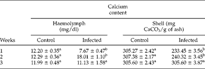
a,b Means followed by different letters differ significantly from their respective control group at each experimental week, P< 0.05.
The relationship between calcium concentration in the haemolymph (mg calcium/dl) (A) and in the shell (mg CaCO3/g of shell) (B) of Biomphalaria glabrata infected with Angiostrongylus cantonensis, at different times of infection (in weeks). Time zero (0) represents the average value of the control groups after 3 weeks of observation. a,b,c: Means followed by different letters differ significantly from the average value of the control groups at each experimental week, P< 0.05. The linear regression analysis showed the following relationships: haemolymph (A), y= 0.7250 − 9.161x; r 2= 0.05; and shell (B), y= 305 − 102.9x+34.48x 2; r 2= 0.99.

There was intense hypocalcification of the shell 1 and 2 weeks after exposure (233.45 ± 3.56 and 240.32 ± 3.45 mg CaCO3/g ash), significantly different in relation to the respective control groups (305.27 ± 2.42 and 307.38 ± 2.17 mg CaCO3/g ash, respectively), representing reductions of 23.53 and 21.81%, respectively (table 1). In contrast, there was no variation in the third week of exposure, when the calcium concentrations of the shells of infected snails fluctuated near the average value of the respective control group (306.08 mg of CaCO3/g of shell). The second-order polynomial regression revealed a negative parabolic variation pattern, with reductions in intermediate periods of analysis and a tendency to normalization at the end of the pre-patent period (fig. 1B).
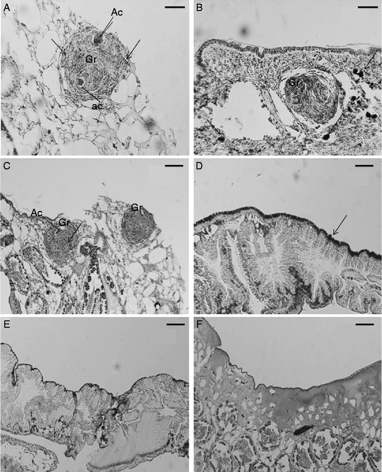
The histochemical results revealed the presence of larval stages of A. cantonensis in the digestive gland of the infected snails, as well as the occurrence of calcified areas assisting the encapsulation process of the parasite (fig. 2A–D). In the control snails there were no observed larval stages of the parasite in sections of digestive gland tissue, the integrity and functioning of which were preserved (fig. 2E and F).
The digestive gland of Biomphalaria glabrata (A– C) infected with Angiostrongylus cantonensis (Ac), showing granulomata (Gr) with concentrations of haemocytes around the larvae of A. cantonensis, producing a spherical nodular appearance, and maximum deposition of calcium ions around the granulomata (arrow). (D) Increasing deposition of calcium ions (arrow) in the epithelium of the digestive gland; (E, F) control B. glabrata to show the intact digestive gland and the absence of granulomata. Scale bars: (A) and (B) = 40 μm; (C–F) = 70 μm.
Discussion
Brockelman & Sithithavorn (Reference Brockelman and Sithithavorn1980), studying the A. fulica/A. cantonensis relationship, found a decline of the levels of carbohydrates circulating in the haemolymph and of the reserves in the tissues in the initial phase of infection. According to these authors, this period is characterized by an intense larval development process, resulting in greater competition for nutrients between the parasite and host, leading to a negative energy balance in the latter. Our results indicate that infection by A. cantonensis induced a significant reduction in the calcium concentration in the haemolymph of B. glabrata in the first week of infection, after which this concentration returned to normal as the infection progressed. These results can be partly explained by the greater need for this ion, by both the parasite and host, confirming that this is the period of greatest energy need (adenosine triphosphate), which is obtained predominantly through oxidative reactions that are catalysed by enzymes that use calcium as a cofactor. Paschoal & Amato (Reference Paschoal and Amato1996), studying the interface Bradybaena similaris/Eurytrema coelomaticum, observed deposition of calcium in the shell and digestive gland of infected snails. According to these authors, this deposition is mainly due to acceleration of the glycolytic pathway, satisfying the snail energy demand during infection (Tunholi et al., Reference Tunholi, Lustrino, Tunholi-Alves, Mello-Silva, Maldonado, Pinheiro and Rodrigues2011b). Such involvement of the calcium ion has also been observed in other snail species, indicating the participation of this element as an enzyme cofactor in cells of Pomacea sp., Lanistes libycus and B. glabrata (Adebayo-Tayo et al., Reference Adebayo-Tayo, Onilude and Etuk2011; Tunholi et al., Reference Tunholi, Lustrino, Tunholi-Alves, Garcia, Mello-Silva, Maldonado, Rodrigues and Pinheiro2011c). In addition, in the present study the significant reduction of the calcium levels in both sites analysed during this period could be a consequence of extensive damage caused during the penetration of infective stages of A. cantonensis, resulting in inflammatory reactions in B. glabrata. This has also been observed by Mostafa (Reference Mostafa2007), studying the effect of Schistosoma mansoni infection on calcium content in B. glabrata. That author attributed the decrease in haemolymph calcium content to the deposition of this ion in host tissues in response to the inflammatory reactions caused by the parasite. This mechanism can impair the snail's movement, reducing its food ingestion and lowering its calcium levels.
In contrast, in the second week of infection there was an increase in the calcium concentration in the haemolymph and a reduction of calcium in the shell. This situation has been confirmed in other parasite/snail associations (Tunholi et al., Reference Tunholi, Lustrino, Tunholi-Alves, Garcia, Mello-Silva, Maldonado, Rodrigues and Pinheiro2011c). Infection by larval stages of helminths is frequently associated with depletion of carbohydrate stocks in the intermediate host. Therefore, snails start to use proteins preferentially as an alternative substrate for production of energy, altering the haemolymph pH and resulting in an intense amino acid deamination process (Pinheiro et al., Reference Pinheiro, Maldonado and Lanfredi2009; Tunholi et al., Reference Tunholi, Lustrino, Tunholi-Alves, Mello-Silva, Maldonado, Pinheiro and Rodrigues2011b). Under these circumstances, calcium carbonate is mobilized from shell to haemolymph, helping to maintain the acid–base balance by release of carbon dioxide (CO2), an important component of the bicarbonate buffering system. This process can explain the hypocalcification observed in this study (de Witt & Sminia, Reference de Witt and Sminia1980; Pinheiro & Amato, Reference Pinheiro and Amato1995).
Other studies have demonstrated that the immune response of snails is calcium dependent. According to Couto (Reference Couto2001), the activities of lectins in the plasma of snails during the process of recognizing antigenic substrates are directly related to the calcium concentration in the haemolymph. Therefore, the hypercalcaemia observed in the second week of infection sheds light on the participation of calcium in strengthening the snail immune response when infected, by contributing to cell recruitment during this period. Additionally, the increase of calcium levels observed in the present study establishes the existence of a favourable biochemical condition for sequestration of this ion in the haemolymph and its deposition in tissues, in response to injuries caused by the presence of A. cantonensis larvae in B. glabrata, helping to encapsulate the parasite.
The process of encapsulation in B. glabrata during experimental infection by A. cantonensis was reported by Harris & Cheng (Reference Harris and Cheng1975). According to these authors, this process initially involves the participation of basophilic haemolymph cells, followed by formation of fibrous nodular aggregates. The results obtained in the present study corroborate in part those of Harris & Cheng (Reference Harris and Cheng1975), indicating the occurrence of areas of metastatic calcification strictly located in visceral organs, as a complementary reaction during encapsulation of A. cantonensis. Harris & Cheng (Reference Harris and Cheng1975) also observed the occurrence of the encapsulation process in various tissues, including the host cephalopedal mass, in the eighth week of infection. This discrepancy can be explained partly by the different isolates utilized (parasite and host) and the exposure time of the host to the parasite, since in the present study we only analysed the changes in the pre-patent period, while Harris & Cheng (Reference Harris and Cheng1975) also investigated the changes in the patent period.
The calcaemia returned to normal in the third week of infection, with calcium levels near those of the control group. According to some authors, this period is characterized by formation of the L3 stages, which are located mainly in the host muscle tissue rather than in the visceral organs such as the digestive gland (Tunholi-Alves et al., Reference Tunholi-Alves, Tunholi, Pinheiro and Thiengo2012). Therefore, despite the morphological changes found in the digestive gland of infected snails, these changes are restricted to specific points of the organ and do not impair its functionality, thus enabling the host to respond to the infection, since the digestive gland is considered to be the main metabolic site and storage location of the main energy reserves. Besides this, because A. cantonensis has a very short pre-patent period in B. glabrata, at the start of the third week the larvae of this nematode no longer needed such high quantities of calcium for their development and maintenance of their metabolic activities, many of them already having migrated through the host tissues and reached the L3 stage.
Finally, although many studies have been conducted on the metabolic aspects of trematode-infected snails, little is known about this aspect with regard to nematodes. Thus, this is the first study to show the effects of infection by A. cantonensis on calcium metabolism in B. glabrata. Our results indicate the existence of precise physiological mechanisms developed by the host, enabling it to re-establish its acid–base balance, as well as to strengthen its immune response, to minimize the harmful effects of infection. In addition, the histological/histochemical changes confirm the participation of calcium ions as an important element in the process of parasite encapsulation, resulting in the formation of calcification areas, especially in the digestive gland. This information can be used in developing measures to control host snails and consequently human angiostrongyliasis.
Acknowledgements
This study was supported in part by Conselho Nacional para o Desenvolvimento Científico e Tecnológico (CNPq) and Fundação Carlos Chagas Filho de Amparo à Pesquisa do Estado do Rio de Janeiro (FAPERJ).