INTRODUCTION
Within the Mollusca, the Solenogastres (neomeniomorphs) represent one of the two aplacophoran clades (the second being the Caudofoveata or chaetodermomorphs). They are externally characterized by a mantle that produces a chitinous cuticle throughout as well as unicellularly formed aragonitic sclerites. At present, the purely marine Solenogastres include about 250 nominal species (between 0.8 mm and 300 mm in length), with records between 1 m and 6850 m depth (mostly below 50 m).
Though more intensive research over the last 40 years has distinctly enlarged our general knowledge of the Solenogastres (summarized in Salvini-Plawen, Reference Salvini-Plawen1985b; Scheltema et al., Reference Scheltema, Tscherkassky and Kuzirian1994, García-Álvarez & Salvini-Plawen, Reference García-Álvarez and Salvini-Plawen2007), their presently known range by no means includes the true biodiversity. This holds true even for the West-European Atlantic (cf. Salvini-Plawen, Reference Salvini-Plawen1997).
Solenogastres apparently live fairly individually and in general only one or few specimens are sampled at once, which makes them rare representatives. Accordingly, the known geographical distribution of most species is spotty and based on occasional findings. Moreover, the poor condition of specimens or their immaturity often makes them difficult for accurate determination, though a principal biodiversity is documented. The present contribution reiterates this situation with respect to the Irish Sea; only in case of large-scaled surveys (see Hartley, Reference Hartley1979; Mackie et al., Reference Mackie, Oliver and Rees1995, for the Irish and Celtic Seas) have the specific and geographical diversity been more thoroughly elucidated. The two present specimens, both juvenile/immature, are described due to the poor knowledge of the Solenogastres biodiversity and fauna in the western Irish Sea.
MATERIALS AND METHODS
Two single specimens of Solenogastres were collected during the UK Strategic Environmental Assessment in region 6 (SEA 6) of the western Irish Sea. They belong to the National Museum of Scotand, NMSZ, and the two ‘?Pruvotina sp.’ were sent to the author for determination and publication. After examination of the sclerites, ribbons of semithin serial sections (cs 1–2 µm) of both specimens were made with glass knives and stained with toluidine-blue. Histological examination and reconstruction were done under a Reichert Biovar microscope.
TYPE MATERIAL
Holotype: juvenile specimen in serial sections on 12 slides, slide with mantle sclerites.
SEA 6, Station 10#3/20.10.2004 (Ref. 34936), Irish Sea, 53.48460°N 05.25727°W, 78 m.
NMSZ: 2006.013.0001.
DIAGNOSIS
Gephyroherpia(?) species of at least 2.4 mm length, body high-triangular in cross-section, dorsal region internally delimited from main body cavity. Cuticle moderately thick, spicules in several layers, with hook-shaped sclerites, without epidermal papillae. One pedal fold extending to mantle cavity. Atrial sense organ posteriorly trifid, central portion as blind pouch exceeding lateral ones. With separate, narrow oral tube; distichous radula delicate; no unpaired-dorsal papillous foregut glandular organ, ventral foregut glandular organs (type A) with long ducts; with distinct post-radular foregut (oesophagus), midgut caecum frontally paired, midgut sacculations scarcely correlated to the dorso-ventral muscle bundles. Longitudinal body wall musculature, distinctly strengthened ventrally. Genital apparatus not yet differentiated.
DESCRIPTION
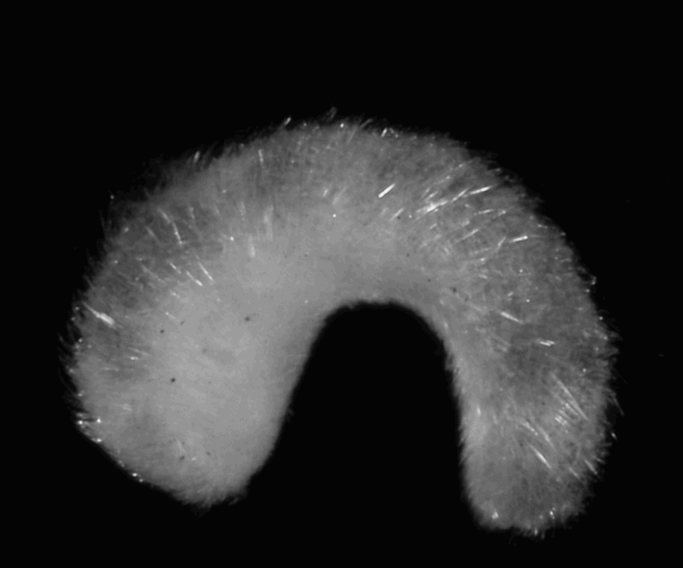
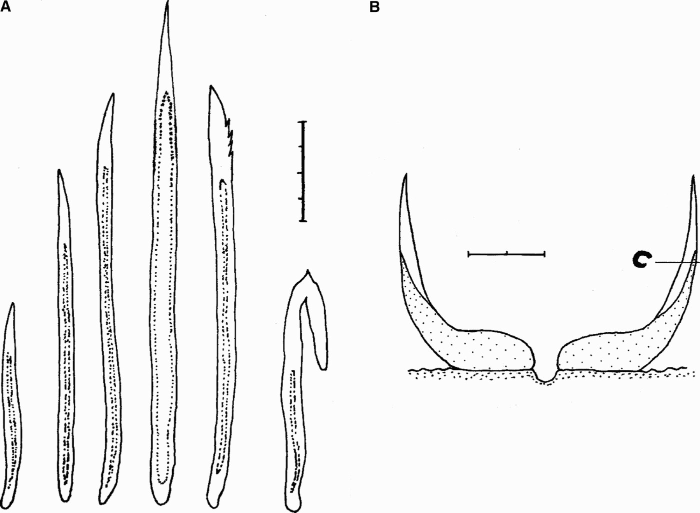
The single animal (Figure 1) is juvenile, 2.4 mm long and anteriorly 0.65 mm high, in cross-section high-triangular with a 4.5 mm wide main body and a tapering dorsal portion (Figures 5–7). This dorsal region is somewhat delimited; above the atrial sense organ it resembles a more separated carina or keel, elsewhere it is confluent. There are no epidermal papillae; the cuticle is 25–45 µm thick with cross-wise embedded hollow spicules (Figures 2–3A). These are slightly curved (70–250 µm long), interspersed by longer, straight and thin-walled elements (up to 250 µm). Hollow hook-shaped sclerites (80–110 µm) are present all along the dorsal body; anteriorly, spicules with a one-sided distal, moderate serration are interspersed (120–210 µm).
Gephyroherpia(?) triangulata sp. nov., holotype in preserved state (2.4 mm long, anterior end left).

Gephyroherpia(?) triangulata sp. nov., body sclerites: scale bar = 30 µm. (A) General type; (B) hook-shaped type; (C) distally serrated type.

Gephyroherpia(?) triangulata sp. nov. (A) Body sclerites (see text and Figure 2): scale bar = 40 µm; (B) one transverse row of the radula (reconstruction): scale bar = 10 µm.

The pedal pit extends as a pouch far dorso-posteriorly; the single longitudinal pedal fold is formed shortly behind the closure of the pit and ends at the mantle cavity opening. There are well-differentiated sole glands. The mantle or pallial cavity is simple, without folds; anteriorly it connects with the slender hindgut, whose ciliation continues for some distance along the roof of the cavity, and there is a paired anterior–lateral diverticle (primordia of spawning ducts?). A dorso-terminal sense organ is differentiated at the body end.
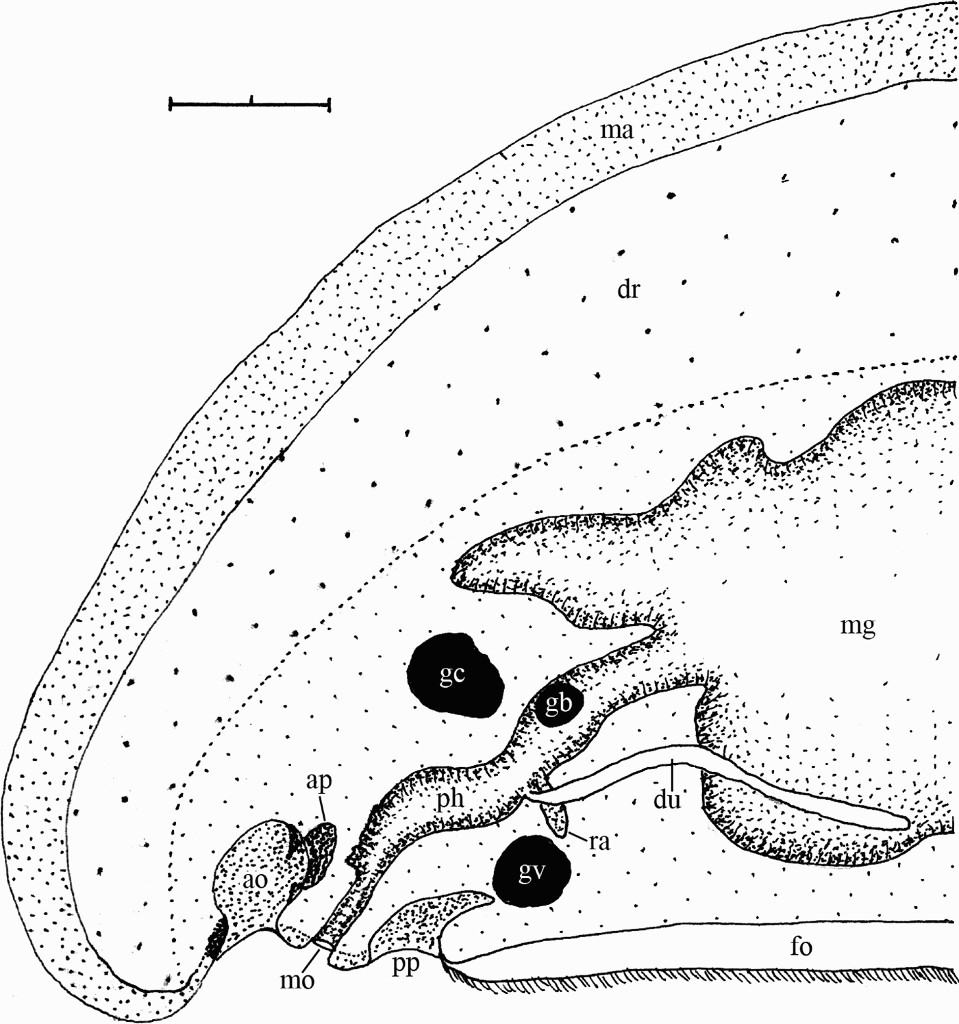
Just anterior to the beginning atrium, a knob-like, high epithelium is differentiated, possibly representing a sensory organ (see Pruvot, Reference Pruvot1891: figure 86 for Pararrhopalia pruvoti Simroth). The atrium or atrial sense organ itself is remarkable (Figures 4–5): posteriorly it shows a threefold configuration, the sensory area with papillae (and delimited by the horseshoe-like ciliary tract) extending bipartedly posteriorly; the open atrial cavity between the posterior curve of the ciliary tracts and the space behind them forms a central blind pouch extending beyond the lateral ends of the atrium (Figures 5–6). The atrial papillae are bulky, some with a common base for two. The narrow oral tube opens midventrally below the end of the atrial pouch (Figures 4 & 6); the mouth is thus distinctly separated from the atrial cavity.
Gephyroherpia(?) triangulata sp. nov., organization of the anterior body: scale bar = 100 µm. ao, atrial sense organ; ap, central blind atrial pouch; dr, differentiated dorsal region of body; du, outleading duct of ventral foregut glands; fo, pedal fold (foot); ma, mantle; gb, buccal ganglion; gc, cerebral ganglion; gv, ventral ganglion; mg, midgut; mo, mouth opening; ph, pharynx; pp, pedal pit; ra, radula sheath.

Gephyroherpia(?) triangulata sp. nov., cross-section through posterior end of bipartite atrial sense organ with blind pouch in between: scale bar = 50 µm. ao, atrial sense organ; ap, central blind atrial pouch; dr, differentiated dorsal region of body; ma, mantle; pg, pedal glands.

Gephyroherpia(?) triangulata sp. nov., cross-section through end of blind atrial pouch and mouth opening: scale bar = 50 µm. ap, central blind atrial pouch; dr, differentiated dorsal region of body; ma, mantle; mo, mouth opening; pg, pedal glands.

The oral tube opens ventrally into an irregularly widened, cuticularized anterior pharyngeal cavity, which is laterally and ventrally delimited by dorso-ventral musculature. From the cerebral ganglion to the radula region the foregut is somewhat narrowed and has its own circular musculature, which is distinct but not very strong. Peripherally, some scattered unicellular glands with granular contents are present, more densely arranged above the radula; there is no unpaired-dorsal papillous foregut glandular organ. The distichous radula is small and the delicate teeth (11 µm wide, 25 µm high) have no medial denticles; the distal portion is straight (not hook-like) and distinctly gutter-like formed at its outer edge (Figure 3B). The radula sheath is initially shortly divided, but with a common weak circular musculature; within the muscular radula support, some small turgescent are present. The buccal ganglia (diameter 30 µm) lie lateral to the foregut far above the radula sheath. The ventral foregut glandular organs open lateral of the anterior radula; they consist of long ducts surrounded by musculature and groups or bundles of intraepithelially opening glands (type A) whose granular contents are similar to the pharyngeal glands. There is a distinct postradular foregut (oesophagus; Figure 7) with poor circular musculature and without glands; it opens into the midgut, forming a short groove.
Gephyroherpia(?) triangulata sp. nov., cross-section through oesophagus: scale bar = 100 µm. ca, midgut caecum; dr, differentiated dorsal region of body; du, outleading duct of ventral foregut glands; dvm, dorso-ventral muscle bundle; gl, ventral foregut glands; ml, strengthened longitudinal musculature; nv, ventral nerve cord; oe, oesophagus; pg, pedal glands.

The midgut gives rise to an anterior caecum extending up to above the cerebral ganglion, where the caecum is paired. The midgut and part of its caecum are middorsally ciliated; the midgut is positioned some distance from the body wall and shows irregular sacculations, scarcely correlated to the dorso-ventral muscle bundles. The body wall musculature is dorsally diffuse and the longitudinal layer is ventrally distinctly strengthened.
The unpaired cerebral complex has two commissures: the anterior commissure interconnects the fused, true cerebral ganglia; these separately give rise dorso-laterally to the cerebro-lateral connectives, ventro-laterally first to the cerebro-buccal and then to the cerebro-ventral connectives. The posterior commissure traverses the posterior, unpaired portion of the cerebral complex region (without giving rise to nerves). The ventral ganglia (diameter 50 µm) lateral to the end of the pedal pit are interconnected by two commissures. Both pairs of body nerve cords are provided with nuclei (medullary) throughout, as is the supra-rectal commissure far anterior to the anus.
There are no gonads or anlagen of other genital apparatus.
MATERIAL CONSIDERED
- Present material:
SEA 6, Station 15#1/24.10.2004 (Ref.34940), Irish Sea, 53.46558°N 05.23011°W; no depth given. NMSZ: 2006.013.0002 (anterior body slides 1A–19A, posterior body slides 1B–19B; slide with mantle sclerites).
- Comparative material:
E. sierra (Pruvot) from Trondheim/Norway (cf. Salvini-Plawen, Reference Salvini-Plawen2003).
DIAGNOSIS
Probably an immature Eleutheromenia sp. of 4 mm length with middorsal midbody carina. Cuticle moderately thick, spicules in several layers, hollow hook-shaped sclerites present (Fig. 9), no epidermal papillae. One pedal fold, five bulky respiratory papillae. Foregut connected by a groove to the atrial sense organ; hooked radula teeth small without median denticles; outlet of ventral foregut glandular organs (type A) as paired long duct. Heart with paired atrium. Genital apparatus not yet fully differentiated.
DESCRIPTION
The present animal measured 4 mm in length and is anteriorly 0.65–0.75 mm high (Figure 8); in cross-section the body is high-triangular, distinctly higher than wide (Figure 10); in the midbody, the upper third (above the midgut) resembles a carina or keel. The mantle bears no epidermal papillae; the cuticle is 15–35 µm thick with cross-wise embedded hollow, slightly curved spicules (70–210 µm long) interspersed by straight ones. In the anterior body, some spicular elements (90–150 µm) have a one-sided distal serration (Figure 2B); hollow hook-shaped sclerites (60–90 µm; Figure 2A) are more abundant dorsally, where they are densely arranged. The pedal groove houses a single longitudinal fold that ends close to the mantle cavity; the sole glands are distinct throughout. The mantle or pallial cavity forms five bulky, echeloned respiratory papillae. No dorso-terminal sense organ could be discerned.
Eleutheromenia(?) sp. juv., animal in preserved state (4 mm long, anterior end left).

Eleutheromenia(?) sp. juv., sclerites: scale bar = 20µm. (A) Hook-shaped type; (B) distally serrated type.

Eleutheromenia(?) sp. juv., slightly oblique cross-section through posterior body: scale bar = 50 µm. at, paired atrium of heart; hg, hindgut; nl, lateral nerve cord; pc, pericardium; sd, spawning duct; sg, sole glands.

The longitudinal layer of the body wall musculature is distinct and ventrally strengthened.
In the investigated specimen, the anterior-most body is strongly contracted and ventrally curved: therefore, the configuration of the internal organs is atypical and difficult to recognize in the sections. The atrial sense organ is paired posteriorly; the atrial papillae are bulky, some with a common base for two. The central portion of the atrium continues as a furrow, at whose end the mouth opens as a narrow oral tube. It leads to a cuticularized, wide foregut. There is a distinct pre-radular circular musculature. The radula is distichous (basally 10 µm high fragments), the distal configuration of the teeth, however, are not accurately discernible in the sections. The radula sheath is proximally not bifid. There is no unpaired-dorsal papillous foregut glandular organ; the ventral foregut glandular organs consist of long, in part fairly wide ducts surrounded by groups or bundles of intraepithelially opening glands (type A). The post-radular foregut is short and narrowed with circular musculature; it opens from ventral into the midgut, which gives rise to a caecum anteriorly. The midgut is ciliated middorsally and shows invaginations and bulges, including regular paired dorsal sacculations; only lateroventrally, regular pouches are developed due to the dorso-ventral muscle bundles.
The animal is immature. The indistinct, short anlagen of the gonadial primordia continue with paired gonopericardioducts, which open frontally into the voluminous pericardium. The heart with paired atrium (Figure 10) is throughout connected by a double septum to the pericardial roof. The pericardioducts run in a curve anteriorly and open into the paired anlagen of the spawning ducts; these subsequently fuse and lead to the mantle cavity. Neither (anlagen of) copulatory stylets nor abdominal spicules were discernible.
SYSTEMATIC DISCUSSION
The characters of the mantle (cross-layered hollow needles and hooked sclerites), the distichous radula, and the ventral foregut glandular organs (type A) define both of the present ‘Pruvotina sp.’ specimens indeed as members of the Cavibelonia–Pruvotinidae; herein, due to the lack of an unpaired-dorsal papillous foregut glandular organ, they are members belonging to the Eleutheromeniinae (Salvini-Plawen, Reference Salvini-Plawen1978; García-Álvarez & Salvini-Plawen, Reference García-Álvarez and Salvini-Plawen2007). As concerns the Solenogastres fauna of the Irish Sea and the adjacent Celtic Sea, eight species are currently known to inhabit this geographical area (Hartley, Reference Hartley1979; Seaward, Reference Seaward1982; Salvini-Plawen, Reference Salvini-Plawen1985a, Reference Salvini-Plawen1997, Reference Salvini-Plawen2003; Caudwell et al., Reference Caudwell, Jones and Killeen1995; Mackie et al., Reference Mackie, Oliver and Rees1995). Among these, only two species of Pruvotinidae are included: although the specific identity of the records in Hartley (Reference Hartley1979) and Mackie et al. (1995a) is not ascertained, Eleutheromenia sierra (Pruvot, 1890) appears to belong to the biogegraphical area in question (cf. Salvini-Plawen, Reference Salvini-Plawen2003). Caudwell et al. (Reference Caudwell, Jones and Killeen1995) presented another record of an 8 mm long pruvotinid specimen from the same geographical area and tentatively referred it to the genus Pruvotina Cockerell. That individual, however, due to its rounded cross-section of the body, clearly is not conspecific with one of the two new specimens treated here.
With respect to the relation of the atrial sense organ and the mouth opening (distinctly separated in species 1, common atrio-buccal opening in species 2), as well as to the configuration of the foregut, both individuals cannot be classified as conspecific. Moreover, species 1 has to be placed into the genus Gephyroherpia Salvini-Plawen, the only genus within Eleutheromeniinae characterized by an independent mouth opening (cf. Salvini-Plawen, Reference Salvini-Plawen1978, Reference Salvini-Plawen2003; García-Álvarez & Salvini-Plawen, Reference García-Álvarez and Salvini-Plawen2007); the missing respiratory organs may be due to the juvenile state, and the lack of epidermal papillae possibly is not generic. (The idea, that the separate mouth opening in species 1 could be merely a developmental anomaly and the animal thus classified as an Eleutheromenia species, at least with respect to the sympatric E. sierra does not coincide with the configuration of the foregut and the radula; c.f. Salvini-Plawen, Reference Salvini-Plawen2003)
Within Gephyroherpia, only the type species G. antarctica Salvini-Plawen (Reference Salvini-Plawen1978) is known and characterized, among other features, by a circular body cross-section, by the radula teeth with 6–7 median denticles, by short and wide ducts of the ventral foregut glandular organs, as well as by the Antarctic provenance. There are thus clear specific differences of the species 1 from the Irish Sea, and this is therefore classified as a new species Gephyroherpia(?) triangulata sp. nov. Owing to our lack of knowledge of the configuration of the genital apparatus (e.g. with or without the generic character of copulatory stylets, etc.), however, the generic classification is tentative.
The present specimen 2, with a common atriobuccal opening and a distinct distichous radula, fits with the genus Eleutheromenia Salvini-Plawen. Both hitherto included species E. sierra (Pruvot, 1890) and E. carinata Salvini-Plawen & Öztürk, Reference Salvini-Plawen and Öztürk2006, are characterized by a striking middorsal carina (Pruvot, Reference Pruvot1891; Salvini-Plawen, Reference Salvini-Plawen2003; Salvini-Plawen & Öztürk, Reference Salvini-Plawen and Öztürk2006) which is met by the high-triangular cross-section. Though specimen 2, apart from the not exactly identical sclerites, roughly coincides with E. sierra (Pruvot) (e.g. atrial furrow with mouth, pre-radular sphincter, heart; cf. Salvini-Plawen, Reference Salvini-Plawen2003), it cannot more accurately be determined.
ACKNOWLEDGEMENTS
The author is very grateful to Mag Emanuel Redl (Vienna) for preparing the photographs and to Dr Michael Stachowitsch (Vienna) for improving the English text.