Implications
Early feed restriction during the suckling period of lambs promotes differences in both the gastrointestinal morphology and the liver proteome, thus impairing feed efficiency during the subsequent fattening period. This information will be useful to improve feed efficiency of milk-restricted lambs (e.g. due to udder problems, lack of lamb vitality and low milk production) by reducing the amount of feedstuff consumed by the animal during the fattening period to produce 1 kg of meat. This will enhance farm profitability and may have a positive environmental impact.
Introduction
The increasing world population has made the efficient use of resources progressively more important. In the livestock sector, this could be achieved by reducing the feed to gain ratio or ‘feed conversion rate’ (FCR, defined as the amount of feed needed to produce 1 unit of animal product) which would also reduce the environmental impact of animal farming. As far as lamb production is concerned, a decrease of up to 15% in dry matter intake (DMI) between efficient and inefficient animals to reach a similar target BW has been demonstrated (de Paula et al., Reference de Paula, de Souza, Monteiro, Santana, Gilaverte, Rossi and Dittrich2013).
Among the various factors that promote long-term effects on the efficiency and health of animals, prenatal (pregnancy) and early postnatal nutrition (milk consumption) have been postulated as key factors in ‘early nutritional programming’ (Gotoh, Reference Gotoh2015). It has been demonstrated in different species that nutrition of the pregnant dam modifies gene expression in several organs and tissues of the offspring, thus affecting both the digestive and metabolic utilisation of nutrients over the lifespan (Bell and Greenwood, Reference Bell and Greenwood2016). Studies of the effects of nutrition during the neonatal period of lambs are scarce but some (Davies and Owen, Reference Davies and Owen1967; Greenwood et al., Reference Greenwood, Hunt and Bell2004; Galvani et al., Reference Galvani, Pires, Hübner, Carbalho and Wommer2014) have revealed an open window during this early stage of life where the milk intake level generates long-term effects on feed efficiency. However, the underlying mechanisms responsible for the differences in feed efficiency, which might be multifactorial (e.g. differences in digestion, absorption or metabolic use of absorbed nutrients, as well as modifications in gene expression or activation of the immune response), are not completely understood (Meyer et al., Reference Meyer, Caton, Hess, Ford and Reynolds2012; Gotoh, Reference Gotoh2015).
Improving our understanding of these mechanisms will be necessary to produce more efficient animals. For example, the modern molecular – OMIC tools (i.e. epigenomics, transcriptomics, proteomics or metabolomics) are increasing our understanding of the cascade from genes to phenotypes and allow differences in gene expression to be detected (Tizioto et al., Reference Tizioto, Coutinho, Decker, Schnabel, Rosa, Oliveira, Souza, Mourão, Tullio, Chaves, Lanna, Zerlotini-Neto, Mudadu, Taylor and Regitano2015). Since the liver plays a major role in the metabolism of nutrients and other dietary substances, thus governing body energy and protein metabolism (Rui, Reference Rui2014; Santos et al., Reference Santos, Giráldez, Groenen, Madsen, Frutos, Valdés and Andrés2017), it can be considered a key metabolic organ to be studied with the use of transcriptomics and/or proteomic approaches to better understand the molecular mechanisms triggered by early feed restriction. In fact, some recent studies (Alexandre et al., Reference Alexandre, Kogelman, Santana, Passarelli, Pulz, Fantinato-Neto, Silva, Leme, Strefezzi, Coutinho, Ferraz, Eler, Kadarmideen and Fukumasu2015; Tizioto et al., Reference Tizioto, Coutinho, Decker, Schnabel, Rosa, Oliveira, Souza, Mourão, Tullio, Chaves, Lanna, Zerlotini-Neto, Mudadu, Taylor and Regitano2015) have identified that mitochondrial dysfunctions are at least partially responsible for differences in feed efficiency. However, the reasons for these differences, which might be partially related to early nutritional events and nutritional programming, were not ascertained.
The present study focuses on the effects of the early postnatal nutrition period of Merino lambs, and was designed to elicit the long-term effects promoted by feed restriction during the suckling period on feed efficiency during the fattening phase, in an attempt to clarify the underlying mechanisms related to digestion, absorption or metabolic use of nutrients, applying a liver proteomic approach for the final aspect.
Material and methods
All handling practices followed the recommendations of the Directive 2010/63/EU of the European Parliament and of the Council on the protection of animals used for scientific purposes and the IGM-CSIC Animal Experimentation Committee (protocol number 2015-04).
Animals and diets
In total, 24 male Merino lambs, penned individually with their corresponding ewe during the suckling period, were used in this experiment. The lambs were stratified based on live BW (LBW) at birth (average 4.81±0.256 kg) and randomly allocated to one of two experimental treatments (n=12 per dietary treatment) during the suckling period. In the first group, lambs (ad libitum (ADL)) were kept permanently with the dams, whereas the other 12 lambs (restricted (RES)) were separated from the dams from 0900 to 1800 h. At 1700 h, the dams of RES lambs were milked and injected with oxytocin to remove alveolar milk. All the lambs were weighed twice a week throughout the suckling period.
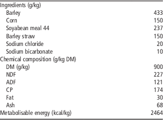
Each lamb was weaned progressively at a LBW of 13.5 kg, restricting the suckling time to 2 h with free access to a complete pelleted diet (CPD) and alfalfa until the LBW was 15 kg. At this weight (15 kg), each weaned lamb was penned individually and offered the same CPD at a restricted level (35 g/kg LBW per day) to ensure no variations in DMI during the fattening phase. Fresh drinking water was always available. All the lambs received the CPD once a day at 0900 h; the daily amount of feed offered was adjusted twice a week on the basis of LBW. Leftovers (if any) were collected daily, pooled in weekly composites for each animal, and analysed for dry matter (DM) content. Samples of the CPD offered were collected weekly and analysed for DM (ISO 6496:1999), ash (ISO 5984:2002), CP (ISO 5983:2009), NDF (Ankom Technology Corp., Macedon, NY, USA), ADF (Ankom Technology Corp.) and total fat (Acid Hydrolysis Filter Bag Technique using the AnkomHCl Hydrolysis System). Ingredients and chemical composition of the CPD administered are summarised in Table 1.
Ingredients and chemical composition (g/kg dry matter (DM) unless otherwise stated) of the complete pelleted diet administered during the fattening period of Merino lambs

Determination of digestibility
Digestibility was determined using acid insoluble ash (AIA) as an internal marker. Feed and faeces were sampled for 9 days starting when each animal reached a LBW of 20 kg. Samples of offered feed were taken daily from the 1st to the 7th day of the collection period; daily rectal grab samples were collected once a day before the morning meal for the last 7 days of the collection period. Samples of feed and faeces of each animal were weighed and pooled to form composite samples and stored at −30°C until AIA analysis (Van Keulen and Young, Reference Van Keulen and Young1977).
Slaughter procedure, ruminal sampling and histological examination of intestinal mucosa
All lambs were slaughtered after a fattening period of at least 50 days with a target LBW of 27 kg. Feed was withdrawn 6 h before slaughtering; the lambs were then weighed, stunned, slaughtered by exsanguination from the jugular vein, eviscerated and skinned. The weight of the organs of each animal was registered, fat depots being removed before weighing the different segments of the gut. A piece of liver was cut and kept at −80°C until fat content determination (AOAC, 2003). A second piece of liver was excised, stabilised with RNAlater (Ambion Inc., Austin, TX, USA) overnight at 4°C, and stored at −80°C for proteomic analysis.
Total rumen contents from each lamb were removed, mixed and sampled within 30 min of slaughter. Samples were collected in aseptic flasks, immediately frozen at −80°C for 48 h, freeze-dried, and stored at −20°C until DNA extraction for microbial diversity analysis as described in Andrés et al. (Reference Andrés, Bodas, Tejido, Giráldez, Valdés and López2016). In addition, about 200 g of ruminal contents were strained through two layers of cheesecloth and the pH was measured immediately. Subsequently, 40 ml of ruminal liquid was dispensed into a falcon tube with 1 ml of 20% sulphuric acid solution to acidify the medium and stop the fermentation. Fermentation end-products (volatile fatty acids, ammonia nitrogen and lactate) were determined according to Carro and Miller (Reference Carro and Miller1999).
A piece of the small intestine was removed from each animal, 20 cm from the pylorus, and placed in a 10% formaldehyde solution (Sigma-Aldrich, St. Louis, MO, USA) for histological examination (fixed by immersion in 10% buffered formalin for 1 week). Formalin-fixed samples from the ileum were trimmed and processed for paraffin embedding and histologic examination (haematoxylin–eosin staining). Slides were examined with a Leica microscope and 10 pictures from 10 different microscopic fields (selected at random from representative areas and damaged villi areas were rejected) per animal were taken at 40× magnification (eyepiece, 10×; objective lens, 4×). The heights of 40 villi were measured using the image processing and analysis software ImageJ v1.6.0_14 (National Institutes of Health, USA). The villus height was defined as the distance between the top of the villus and the crypt transition (Makovicky et al., Reference Makovicky, Tumova, Volek, Makovicky and Vodicka2014).
DNA extraction, quantitative real-time PCR and terminal restriction fragment length polymorphism of ruminal microbiota
Frozen samples of total rumen contents were freeze-dried and thoroughly homogenised before DNA extraction by physical disruption (1 min) using a bead beater (Mini-bead Beater; BioSpec Products, Bartlesville, OK, USA) and subsequent DNA purification with the QIAamp DNA Stool Mini Kit (Qiagen Inc., Valencia, CA, USA), with the modification of a greater temperature (95°C) to improve cell lysis. Deoxyribonucleic acid samples were used as templates for quantitative real-time PCR (qPCR) amplification and terminal restriction fragment length polymorphism (T-RFLP) analysis according to the procedures described by Andrés et al. (Reference Andrés, Bodas, Tejido, Giráldez, Valdés and López2016), except with the use of MspI as the restriction enzyme. Additionally, to quantify total bacteria, qPCR was performed as previously described (Andrés et al., Reference Andrés, Bodas, Tejido, Giráldez, Valdés and López2016), whereas the primers and qPCR details for Megasphaera elsdenii (Ouwerkerk et al., Reference Ouwerkerk, Klieve and Forster2002), Prevotella spp., Selenomonas ruminantium (Stevenson and Weimer, Reference Stevenson and Weimer2007) and methanogens (Denman et al., Reference Denman, Tomkins and McSweeney2007) can be found in the corresponding references. Results were expressed as fold change (log2 of number of copies per milligram of genomic DNA (RES/ADL)) of overrepresented species in ruminal microbial content (positive value if RES/ADL>1; negative value if ADL/RES>1).
Proteomic analysis of liver samples
For this procedure, four representative animals from each group were selected to cover the complete range of values of average daily gain during the fattening period in each group, and avoiding differences in BW between groups. Liver samples were homogenised in 0.01 M potassium phosphate buffer (pH 7.4, containing 0.07% 1,4-dithiothreitol (DTT) and supplemented with the cOmplete™ protease inhibitor cocktail; Roche Applied Science, Vilvoorde, Belgium) with the FastPrep® Instrument (MP Biomedicals, Illkirch, France) and LysisMatrix D and centrifuged (15 700×g, 4°C, 30 min) for clarification. An additional step for the removal of insoluble material by ultracentrifugation of samples at 157 000×g at 4°C for 4 h was applied. Protein supernatants were then mixed with an equal volume of pre-chilled 20% (w/v) 2,2,2-trichloroacetic acid/acetone containing 0.14% (w/v) DTT and incubated overnight at −20°C for protein precipitation. Precipitated proteins were separated by 3900×g centrifugation for 5 min at 4°C. The pellet was washed twice with pre-chilled acetone containing 0.07% (w/v) DTT and once with 80% (v/v) acetone/MilliQ water. Protein concentration was determined with the Bradford method (Bio-Rad Protein Assay, BioRad Laboratories, Hercules, CA, USA), which showed a high reproducibility for this protein extraction protocol. The final pellet was re-suspended in a rehydration solution containing 8 M urea, 2% (w/v) 3-[(3-cholamidopropyl)-dimethylammonio]-1-propanesulfonate, 0.5% (v/v) ampholytes, 25 mM DTT and 0.002% bromophenol blue. Samples were additionally processed using the 2-D Clean-Up Kit (GE Healthcare, Uppsala, Sweden) for salt removal, according to the manufacturer’s instructions, quantified again with Bradford assay, and stored at −80°C or applied directly to 18 cm immobilised pH gradient (IPG) strips before proteome resolution.
An active rehydration protocol for the first-dimension strips (450 µg of protein) was applied, followed by isoelectric focusing steps (non-linear gradient of pH 3 to 10) onto an Ettan IPGphor Isoelectric Focusing System (GE Healthcare) maintained at 20°C. Proteins resolved in the first-dimension strips were reduced and alkylated before second-dimensional electrophoresis. The second dimension was run in 12.5% to 15% SDS-PAGE in an Ettan Dalt Six apparatus (GE Healthcare) for 45 min at 3 W/gel and then for 4 h at 18 W/gel. Precision Plus Protein Standards (BioRad Laboratories) were used as markers. High sensitivity Colloidal Coomassie Blue G-250 Staining (blue silver) was used for protein visualisation.
All gels (two gels per sample, eight gels per condition) were scanned using an ImageScanner III (GE Healthcare), and images were analysed with the ImageMaster 2D Platinum software (GE Healthcare). The internal standard image with more spots was designated as the ‘Master,’ and was used as a template. Protein spots included in the rest of the images were matched to those in the Master to assure an accurate comparison. The intensity of individual spots was normalised and images (the best gel per sample, four gels per condition) were grouped. The intensity of individual spots was analysed and compared with other spots within and between image groups. Differential representation of protein spots was determined by Student’s t test. Spots showing a P-value⩽0.05 with a fold change ⩾1.5 were chosen for identification.
The protein spots of interest were manually excised, digested with trypsin and extracted for MALDI-TOF/TOF tandem MS analysis (4800 MALDI TOF/TOF™ Analyzer; Applied Biosystems, Foster City, CA, USA). Names of the proteins differentially accumulated in the liver of both groups of lambs were assigned according to the NCBI RefSeq annotation of the genes (GCF_000298735.2_Oar_v4.0), whereas the Kyoto Encyclopaedia of Genes and Genomes (KEGG) annotation (retrieved from the KEGG website) was used to assign proteins to specific metabolic pathways. Identification of a certain protein was considered correct if the score was higher than 93 (protein score >93; P<0.05). If more than one protein was identified in one spot, the protein with the highest score was selected.
Calculations and statistical analysis
Average daily gain (ADG, g/day) was estimated as the regression coefficient (slope) of BW against time using the REG procedure of the SAS statistical package (2011, SAS Institute Inc., Cary, NC, USA). The feed to gain ratio or ‘FCR’ was obtained by dividing the DMI per day by the previously estimated ADG during the fattening period (g DMI/g ADG). Growth performance data together with organ weights, fat depots, ruminal fermentation parameters, microbiota, digestibility and histology examination of the ileal mucosa were analysed by one-way ANOVA using the GLM procedure of SAS with feeding regime (ad libitum or restricted during the suckling period) as the only source of variation. The individual lamb was considered as the experimental unit. Significance was declared at P<0.05. The T-RFLP data obtained for each rumen sample were analysed by principal component analysis using SAS.
Results
Animal performance, digestibility, and ruminal fermentation and microbiota
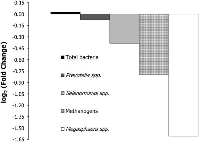
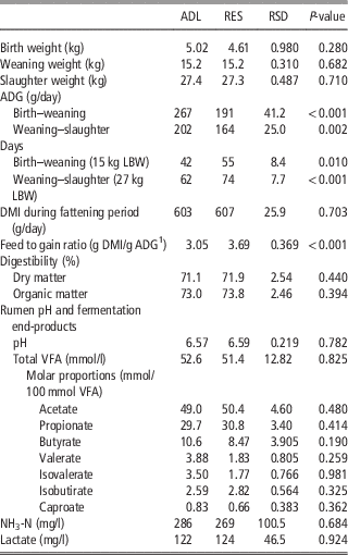
Body weight at birth, at weaning and at slaughter were unaffected (P>0.05) by dietary treatment (Table 2). As expected, the ADG of RES lambs was reduced during the suckling period (191 v. 267 g/day for RES and ADL lambs, respectively; P<0.010). Moreover, the ADG during fattening (from weaning to slaughter) was 164 v. 202 g/day for RES and ADL lambs, respectively (P<0.001), even though there were no differences in DMI between both groups (35 g/kg LBW per day) at this phase (P>0.05). Thus, the fattening period was longer for the RES lambs (62 v. 74 days, P<0.001) to reach target LBW at slaughter (27 kg LBW). As a consequence, the feed to gain ratio was increased in RES lambs compared with the ADL lambs (P<0.05). However, digestibility during the fattening period was unaffected (P>0.05) by early feed restriction. Neither fermentation end-products nor ruminal microbiota (T-RFLP, data not shown; qPCR, Figure 1) showed differences (P>0.05) between dietary treatments.
Relative quantitation compared with ad libitum lambs (ADL) of 16S ribosomal RNA copy numbers of microbiota groups in the ruminal contents of early fed restricted Merino lambs (RES) during the suckling period. Fold-changes for specific amplicon groups calculated as log2 ratio of normalised abundances. Positive value if RES/ADL>1; negative value if ADL/RES>1. No significant difference in copy number (P>0.05) calculated using Tukey’s test (SAS) were observed.

Effect of early feed restriction during the suckling period (ad libitum (ADL) v. early fed restricted lambs (RES)) on pre- and post-weaning growth performance of lambs, digestibility of the diet fed during the fattening period and rumen fermentation parameters

ADG=average daily gain; LBW=live BW; DMI=dry matter intake; AVG=Average daily gain; VFA=volatile fatty acids; NH3-N=ammonia nitrogen; RSD=residual standard deviation.
1 Average daily gain during the fattening period (weaning–slaughter).
Visceral organ weight, fat depots and histological structure of intestinal mucosa
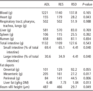
As shown in Table 3, visceral organ weights at slaughter were unaffected (P>0.05) by early feed restriction during the suckling period, with the exception of the heart, which was greater (P<0.05) in RES (179 g) compared with ADL lambs (155 g). On the other hand, although there were no differences (P>0.05) in small, large or total intestine weights, the small intestine represented a smaller (P<0.05) fraction of the total intestine in RES lambs (66.0%) compared with ADL lambs (69.3%). The average villus height in the ileal epithelium was shorter (P<0.05) in RES compared with ADL lambs (465.6 v. 487.3 µm). In addition, liver, omental and perirenal fat were significantly greater in RES compared with ADL lambs, whereas an opposite effect was observed for mesenteric fat.
Effect of early feed restriction during the suckling period (ad libitum (ADL) v. early fed restricted lambs (RES)) on visceral organ weights (g), fat depots (g) and ileum villi height (µm) of lambs at slaughter (27 kg)

DM=dry matter; RSD=residual standard deviation.
Liver proteomic profile
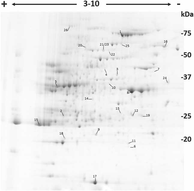
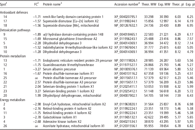
The proteomic profile of the liver identified 26 spots/proteins with significant differences (P<0.05) between both groups (Figure 2 and Table 4). In total, 18 of these proteins were down-regulated in the liver of RES lambs and most were involved in antioxidant defence (e.g. superoxide dismutase [Cu–Zn] isoform X2, superoxide dismutase [Mn], mitochondrial and nmrA-like family domain-containing protein 1) and detoxication pathways (e.g. α/β hydrolase domain-containing protein 14B, microsomal glutathione-S-transferase, dihydrodiol dehydrogenase 3 and indolethylamine N-methyltransferase-like isoform X2), protein synthesis (e.g. endoplasmic reticulum resident protein 29 precursor and guanidinoacetate N-methyltransferase) and glucose (e.g. retinol-binding protein 4 and galactokinase) or fatty acid metabolism (e.g. enoyl-CoA hydratase, mitochondrial). The spots increased in the liver of the RES lambs were primarily related to protein catabolic pathways (e.g. dihydrolipoyl dehydrogenase and selenium-binding protein), protein folding (e.g. protein disulfide-isomerase A3 precursor) or the tricarboxylic acid cycle (e.g. aconitase hydratase, mitochondrial). Spots 7 (actin, cytoplasmic 1) and 24 (lysozyme C) were not identified as Ovis aries proteins. The last spot (8) showed no correspondence with any known identified protein (protein score<93, P>0.05).
Representative colloidal Coomassie Blue stained 2D-E gel of a liver from early fed restricted lambs. A total of 450 μg of protein was loaded, and 2D-E was performed using a pH range of 3 to 10 in the first dimension, and SDS-PAGE in the second dimension. Marked spot numbers refer to numbers in Table 4.

List of spots/proteins identified by MALDI-TOF/TOF tandem MS analysis on the lamb liver 2D-E gel

FC=fold change; MW=molecular weight (Da); Theor.=theoretical; Exp.=experimental; pI=isoelectric point; RES=early fed restricted lambs; ADL=ad libitum.
1 Spots number labelled in Figure 2. Spot 8 showed no correspondence with any identified protein (protein score<93, P>0.05), whereas spot 7 (actin, cytoplasmic 1) and 24 (lysozyme C) were neither included in Table 4 because they were not identified as Ovis aries proteins.
2 positive value if RES/ADL>1.5; negative value if ADL/RES>1.5; ∞ if ADL=0.
3 Identified by results of MALDI-TOF/TOF tandem MS analysis.
4 From MASCOT database search (MatrixScience).
Discussion
The present study was designed to quantify the effect of early feed restriction on feed efficiency during the fattening period of Merino lambs and to elucidate the mechanisms responsible for the long-term effects promoted by such restriction. This is relevant as milk intake can be reduced during the suckling period in farm conditions due to unhealthy status of the lambs, reduction of milk production of the dam or pathologies in the udder, thus lowering profitability during post-weaning phases.
As expected, RES animals grew more slowly during the suckling period as a consequence of feed restriction. Likewise, and in accordance with previous studies (Davies and Owen, Reference Davies and Owen1967; Bhatt et al., Reference Bhatt, Tripathi, Verna and Karim2009; Galvani et al., Reference Galvani, Pires, Hübner, Carbalho and Wommer2014), RES lambs also showed lower ADG from weaning to slaughter than ADL lambs, even though DMI was identical for both groups (35 g/kg LBW per day). Consequently, the RES lambs showed reduced feed efficiency during the fattening period.
The forestomaches of ruminants host a great diversity of micro-organisms that break down and ferment most feed ingested by the animal; as a consequence, the ruminal microbiome diversity and functional capacity underpin digestive efficiency in the rumen (Weimer, Reference Wai, García-Prieto, Baker, Merkwirth, Benit, Rustin, Rupérez, Barbas, Ibañez and Langer2015). However, in the present study, there were no differences between treatments in the rumen microbiota (T-RFLP (not shown) or qPCR) or in the rumen fermentation end-products. In ruminants, the gastrointestinal tract is sterile at birth but is rapidly colonised by micro-organisms from adult animals and the surrounding environment within the 1st days of life (Yáñez-Ruiz et al., Reference Weimer2015). However, in the present study, the animals were ewe-reared, followed the same progressive weaning process, and were fed the same diet at a similar level of intake during the fattening period. Therefore, the effect of factors such as the introduction of solid feed or level of forage intake before weaning, which might affect rumen microbial population (Weimer, Reference Wai, García-Prieto, Baker, Merkwirth, Benit, Rustin, Rupérez, Barbas, Ibañez and Langer2015), was avoided. This would explain the lack of differences between groups in both ruminal microbiota and ruminal fermentation parameters.
Moreover, there is evidence that feed restriction and starvation induces changes in the morphology of the mucosa of the small intestine, and more precisely villus height and crypt depth, thus affecting absorption of nutrients in the gastrointestinal tract (Chappell et al., Reference Chappell, Thompson, Jeschke, Chung, Thompson and Wolf2003). In the present study, average villus height was shorter in the ileal epithelium of RES lambs, suggesting a reduced cellularity in the epithelia of this group (Chappell et al., Reference Chappell, Thompson, Jeschke, Chung, Thompson and Wolf2003). These results differ from those observed by other authors in lagomorphs, where an increase in size of the small intestine was observed when they were subjected to feed restriction, likely due to increased absorption of nutrients as an adaptation mechanism (Makovicky et al., Reference Makovicky, Tumova, Volek, Makovicky and Vodicka2014). Nevertheless, in our study the animals were slaughtered at the end of the fattening period, so differences in the weight of the intestine caused by early feed restriction during the suckling phase might have been modified throughout the fattening period, when DMI was similar for both groups. In any case, the smaller weight ratio of the small to large intestine, which is likely also related to the reduced mesenteric fat depot observed in the RES compared with the ADL lambs, together with the shorter villi height in the RES lambs, might have reduced the absorption of nutrients during the fattening period, thus contributing to the observed differences in ADG and feed efficiency of the lambs. However, lack of significant differences in total apparent digestibility of the diet led us to think about changes in feed efficiency promoted by the amount/proportion of nutrients digested in each part of the tract, which might have been modified according to the different proportions represented by the small/large intestine in the ADL and RES groups.
The greater weight of the heart observed in the RES lambs, which has also been described by Greenwood et al. (Reference Greenwood, Hunt and Bell2004) following postnatal feed restriction in sheep, could have been caused by an impairment of mitochondrial metabolism promoted by the lack of nutrients during early life (Wai et al., Reference Yáñez-Ruiz, Abecia and Newbold2015), as will be explained below for liver proteomic profiling. In fact, any deficiency in energy production might be critical for one of the highest energy demanding tissues, the cardiac muscle, so myocyte hypertrophy may be promoted as an adaptive response to increase myocyte contractile strengthening (Harvey and Leinwand, Reference Harvey and Leinwand2011; Meyers et al., Reference Meyers, Basha and Koenig2013).
Liver characteristics may also allow for better understanding of some of the mechanisms involved in the long-term effects of early feed restriction. The weight of this organ was similar for both groups, but important differences in the proteomic profile of the liver, which would be indicative of different metabolic uses of nutrients, were observed following early feed restriction. For example, fatty acid metabolism is one of the principal sources of energy for skeletal and cardiac muscle and is active in the liver (Quijano et al., Reference Quijano, Trujillo, Castro and Trostchansky2016). Short-, medium- and long-chain fatty acids are degraded to acetyl-CoA by mitochondrial β-oxidation, which allows acetyl-CoA to enter the tricarboxylic acid cycle. Energy released during this process is used for ATP generation by oxidative phosphorylation in the inner mitochondrial membrane. However, RES lambs showed more accumulation of fat in several depots including the liver (Table 2) and intramuscular fat (Santos et al., Reference Santos, Giráldez, Mateo, Frutos and Andrés2018), which might be related to inhibition of β-oxidation (e.g. down accumulation of enoyl-CoA hydratase, mitochondrial isoform X2, in RES lambs), accumulation of upstream metabolites, and hence impaired fatty acid catabolism for energy production (Alexandre et al., Reference Alexandre, Kogelman, Santana, Passarelli, Pulz, Fantinato-Neto, Silva, Leme, Strefezzi, Coutinho, Ferraz, Eler, Kadarmideen and Fukumasu2015; Fu et al., Reference Fu, Xu, Hou, Qi, Zhou, Liu, Luan, Jing, Miao, Zhao, Liu and Li2017; Santos et al., Reference Santos, Giráldez, Groenen, Madsen, Frutos, Valdés and Andrés2017). In agreement with this hypothesis, liver fat accretion (as occurred in the RES lambs) has also been related to severe impairment of mitochondrial lipid metabolism mainly promoted by a deficiency in adenosine kinase (Boison et al., Reference Boison, Scheurer, Zumsteg, Rülicke, Litynski, Fowler, Brandner and Mohler2002), an enzyme which was also reduced in the liver of the RES lambs in this study.
The lower levels of those proteins involved in antioxidant defences observed in the liver of RES lambs are also consistent with less active mitochondrial activity and reduced fatty acid catabolism for ATP production, which is tightly linked to the production of reactive oxygen species such as O2 ∙−and H2O2 (Quijano et al., Reference Quijano, Trujillo, Castro and Trostchansky2016). Additionally, the lower levels of serum albumin precursor (a carrier protein for a wide range of endogenous molecules including hormones, fatty acids, metabolites and Zn required by SOD2) in the RES lambs might have contributed to the decrease in lipid mobilisation and transport, leading to fat deposition. Furthermore, the increased levels of aconitate hydratase, mitochondrial isoform X1 in the livers of RES lambs also support the negative long-term effects of early feed restriction in the mitochondria. In fact, this enzyme is a moonlighting protein which binds directly to mitochondrial DNA under non-optimal conditions, contributing to its maintenance (Chen et al., Reference Chen, Wang, Kaufman and Butow2005). Accordingly, increased levels of this protein (which is also involved in the tricarboxylic acid cycle) in the livers of RES lambs do not contradict the mitochondrial dysfunction mentioned above. In fact, our results are in agreement with Vincent et al. (Reference Vincent, Louveau, Gondret, Tréfeu, Gilbert and Lefaucheur2015), who detected increments of this protein in the muscles of the least efficient pigs. It must be stated, however, that our results differ from those of Grubbs et al. (Reference Grubbs, Fritchen, Huff-Lonergan, Gabler and Lonergan2013), who described a decrease of aconitase in the muscles of the least efficient pigs. The apparent contradiction described by these studies might be explained by the fact that Grubbs et al. (Reference Grubbs, Fritchen, Huff-Lonergan, Gabler and Lonergan2013) studied only the intrinsic mitochondrial composition whose proteome might differ from the one obtained from muscle.
The up-regulation in RES lambs of mitochondrial dihydrolipoyl dehydrogenase, an enzyme involved in the pyruvate dehydrogenase complex, also seemed to contradict the mitochondrial dysfunction previously mentioned. In fact, again, our results differ from those described by Grubbs et al. (Reference Grubbs, Fritchen, Huff-Lonergan, Gabler and Lonergan2013), who showed a lower abundance of the dihydrolipoyl dehydrogenase in isolated mitochondria from the red portion of the semitendinosus muscle of the least efficient pigs. However, this protein also functions as a serine protease in non-optimal conditions (Babady et al., Reference Babady, Pang, Elpeleg and Isaya2007). Therefore, this protein might indicate the existence of long-term effects promoted by early feed restriction, with a shift towards increased protein catabolism in restricted lambs. In agreement with this idea, the selenium-binding protein, which may play a selenium-dependent role in ubiquitination/deubiquitination-mediated protein degradation, was also increased in the liver of RES lambs, likely to use amino acids as an energy source. This situation, together with reduced fatty acid catabolism, might help explain the reduced performance (in terms of decreased ADG) observed in the RES lambs (Santos et al., Reference Santos, Giráldez, Mateo, Frutos and Andrés2018). Long-term effects of early feed restriction were also observed in genes involved in glucose metabolism (e.g. galactokinase isoform X1 and retinol-binding protein 4 isoform X2). Furthermore, several proteins implicated in protein folding (e.g. protein disulfide-isomerases and endoplasmic reticulum protein 29) were differentially accumulated in both experimental groups. Finally, detoxification and excretion of xenobiotics and/or metabolites might have been affected by early feed restriction of lambs, as suggested by the down-regulation of several proteins (e.g. α/β hydrolase domain-containing protein 14B, microsomal glutathione-S-transferase, partial, dihydrodiol dehydrogenase 3 and indolethylamine N-methyltransferase-like isoform X2) likely involved in these functions. In contrast with our results, glutathione-S-transferase was up-regulated in the liver transcriptome of the least efficient bulls (Chen et al., Reference Chen, Gondro, Quinn, Herd, Parnell and Vanselow2011). However, unlike our study where early feed restriction was carried out, the reasons for different feed efficiency traits were not ascertained by Chen et al. (Reference Chen, Gondro, Quinn, Herd, Parnell and Vanselow2011), which could explain the differences observed in the two studies.
Conclusions
According to the results of the present study, early feed restriction impairs feed efficiency during the subsequent fattening phase of lambs. This effect might be largely related to differences in gastrointestinal morphology or metabolic use of nutrients, which may affect β-oxidation of fatty acids, leading to increased fat accretion in several depots. These effects, together with the increased catabolism of proteins indicated by the proteomic profile of the liver, suggest differences in the energy sources used for maintenance and growth, resulting in inferior post-weaning feed efficiency in early feed restricted lambs.
Acknowledgements
This work was funded by the Spanish Ministry of Economy, Industry and Competitiveness (MINECO, AGL2014-54124R). Alba Santos gratefully acknowledges receipt of a pre-doctoral grant from the Spanish Ministry of Education, Culture and Sport (MECD, FPU15/01630). The authors also thank Julio Benavides for providing histological sections of intestinal samples.
Declaration of interest
None.
Ethics statement
All handling practices followed the recommendations of the Directive 2010/63/EU of the European Parliament and of the Council on the protection of animals used for scientific purposes and the IGM-CSIC Animal Experimentation Committee (protocol number 2015-04).
Software and data repository resources
None.